Skip to main content Accessibility help
×
  • Cited by 12
Publisher:
Cambridge University Press
Online publication date:
March 2012
Print publication year:
2006
Online ISBN:
9780511545061

Book description

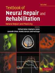
In two freestanding volumes, Textbook of Neural Repair and Rehabilitation provides comprehensive coverage of the science and practice of neurological rehabilitation. This volume, Neural Repair and Plasticity, covers the basic sciences relevant to recovery of function following injury to the nervous system, reviewing anatomical and physiological plasticity in the normal CNS, mechanisms of neuronal death, axonal regeneration, stem cell biology, and neuron replacement. Edited and written by leading international authorities, it is an essential resource for neuroscientists and provides a foundation for the work of clinical rehabilitation professionals.

Reviews

'In two freestanding but linked volumes, Textbook of Natural Repair and Rehabilitation provides comprehensive coverage of the science and practice of neurological rehabilitation. Edited and written by leading international authorities from the neurosciences and clinical neurorehabilitation, the two-volume set is an essential resource for rehabilitation professionals and a comprehensive reference for all scientists and clinicians in the field.'

Source: Advances in Clinical Neuroscience & Rehabilitation

Refine List

Actions for selected content:

Select all | Deselect all
  • View selected items
  • Export citations
  • Download PDF (zip)
  • Save to Kindle
  • Save to Dropbox
  • Save to Google Drive

Save Search

You can save your searches here and later view and run them again in "My saved searches".

Please provide a title, maximum of 40 characters.
×

Contents


Page 2 of 2


  • 19 - Axon guidance during development and regeneration
    pp 326-345
    • By Simon W. Moore, Department of Neurology and Neurosurgery, Center for Neuronal Survival, Montreal Neurological Institute, McGill University, Montreal, Quebec, Canada, Timothy E. Kennedy, Department of Neurology and Neurosurgery, Center for Neuronal Survival, Montreal Neurological Institute, McGill University, Montreal, Quebec, Canada
  • View abstract

    Summary

    This chapter provides an overview of molecular mechanisms that guide axon extension during neural development. It introduces the growth cone, a specialized motile structure at the tip of the axon responsible for sensing and responding to guidance cues. Growth cone morphology is a direct consequence of the organization of the two main components of its cytoskeleton, microtubules, and filamentous actin. The chapter describes the trajectory of embryonic spinal commissural axons, and reviews axonal guidance cues. It presents a description of the growing understanding of the cellular and molecular mechanisms that transduce extracellular guidance cues into directed axon growth. As an axon extends along its trajectory, its growth cone has the capacity to change its response to local guidance cues. The chapter concludes with a discussion on the possibility that cues now known to regulate axon guidance during development may subsequently influence axon regeneration in the adult central nervous system (CNS).
  • 20 - Synaptogenesis
    pp 346-362
    • By Matthew S. Kayser, Department of Neuroscience, University of Pennsylvania School of Medicine, Philadelphia, PA, USA, Matthew B. Dalva, Department of Neuroscience, University of Pennsylvania School of Medicine, Philadelphia, PA, USA
  • View abstract

    Summary

    This chapter describes the process of synapse development, focusing on the signaling and molecular cues involved in the formation of mammalian central excitatory synapses. It provides an overview of central nervous system (CNS) synapse structure and describes the initial contact between axon and dendrite. Contact between axon and dendrite occurs both en passent along the shaft of the axon and at its growing tip, just as synapses form both on dendritic protrusions and the dendritic shaft. The chapter discusses the differentiation of those pre- and post-synaptic compartments, and the large number of molecules implicated in the regulation of synaptogenesis. It addresses how activity might be involved in the formation and/or maturation of synaptic contacts, and how control of this intricate process might differ in young animals compared to adults or following neural injury. Glutamate release from presynaptic terminals may control axonal filopodia motility and selection of postsynaptic targets.
  • 21 - Inhibitors of axonal regeneration
    pp 365-389
    • By Tim Spencer, Department of Biological Sciences, Hunter College of the City, University of New York, New York, NY, USA, Marco Domeniconi, Department of Biological Sciences, Hunter College of the City, University of New York, New York, NY, USA, Marie T. Filbin, Department of Biological Sciences, Hunter College of the City, University of New York, New York, NY, USA
  • View abstract

    Summary

    This chapter outlines some of the various components which may contribute to the observed lack of regeneration which occurs after injury to the adult mammalian central nervous system (CNS). To date, three major inhibitors of axonal regeneration associated with myelin have been identified: myelin-associated glycoprotein (MAG), Nogo, and oligodendrocyte-myelin glycoprotein (OMgp). The myelin-associated inhibitors all appear to be present in undamaged myelin and may have roles other than the block of regeneration or inadvertent sprouting. Following the binding of each of the myelin-associated inhibitors to the NgR-p75NTR-LINGO receptor complex, there is an induction of a signaling pathway which eventually leads to the blockage of neurite extension from damaged or naïve adult neurons. If the binding or signaling of a single receptor complex can be compromised, it may be possible to permit sufficient regeneration in the adult mammalian CNS after injury, particularly prior to formation of the glial scar.
  • 22 - Effects of the glial scar and extracellular matrix molecules on axon regeneration
    pp 390-404
    • By Jared H. Miller, Department of Neurosciences, School of Medicine, Case Western Reserve University, Cleveland, OH, USA, Jerry Silver, Department of Neurosciences, School of Medicine, Case Western Reserve University, Cleveland, OH, USA
  • View abstract

    Summary

    This chapter discusses the environment of the glial scar, with particular focus on the role of chondroitin sulfate proteoglycans (CSPGs) in regeneration failure. Many injuries of the central nervous system (CNS) occur with an accompanying opening of the blood-brain barrier. Non-CNS molecules entering the brain parenchyma through the disrupted blood-brain barrier have significant effects on the immune system and subsequent development of the glial scar. It should be reiterated that following injury in the vicinity of blood-brain barrier extravasation, much of the glial scar forms without astrocyte proliferation, but rather with a switch to the reactive state followed by inhibitory extracellular matrix (ECM) production and then hypertrophy. The growth inhibitory and growth promoting molecules exist in a balance that favors stalled regeneration of axons, but it is important to reiterate that non-regenerating axons still need to be supported if they are to remain indefinitely in the vicinity of the lesion.
  • 23 - Trophic factors and their influence on regeneration
    pp 405-420
    • By Joel M. Levine, Department of Neurobiology and Behavior, State University of New York at Stony Brook, Stony Brook, NY, USA, Lorne M. Mendell, Department of Neurobiology and Behavior, State University of New York at Stony Brook, Stony Brook, NY, USA
  • View abstract

    Summary

    This chapter reviews the biology of neurotrophins (NTs) and their receptors with an emphasis on their use to encourage nerve regeneration after damage to both the central and peripheral nervous system. NTs bind to a receptor complex comprised of two components: p75 and a tropomyosin-related kinase (Trk) transmembrane receptor protein tyrosine Kinase. Ligand binding induces Trk subunit dimerization and the autophosphorylation of multiple tyrosine residues. This autophosphorylation initiates several different intracellular signaling cascades that are described in the chapter. p75 can also play an important role as a negative regulator of axon growth by virtue of its interactions with the rho GTPase. Damaged peripheral axons undergo a characteristic sequence of morphological and physiological changes. The specificity of different NTs in promoting regeneration of different functional classes of dorsal root (DR) fibers suggests that spinal axons should also be selectively responsive to different NTs.
  • 24 - Intraneuronal determinants of regeneration
    pp 421-442
    • By Lisa J. McKerracher, Département de Pathologie et Biologie Cellulaire, Université de Montréal, Montréal, Québec, Canada, Michael E. Selzer, Department of Neurology, University of Pennsylvania Medical Center, Philadelphia, PA, USA
  • View abstract

    Summary

    This chapter focuses on the translation of extracellular cues to intracellular programs that are determinants of regenerative capacity. While the extracellular environment in the adult central nervous system (CNS) contains molecules that act as growth inhibitors, both in vitro and in vivo, co-culture experiments suggest that much of the failure of axon regeneration seen in the adult CNS can be attributed to a developmental reduction in the intrinsic regenerative ability of neurons. An irreversible loss of regenerative ability occurs at birth in rat retinal ganglion cell (RGC). Another indication of the importance of neuronintrinsic factors in determining the regenerative ability of axons is the heterogeneity in regenerative ability expressed by axons of different neurons growing through the same environment. The intrinsic growth capacity of an injured neuron is influenced by its external environment. Finally, the chapter presents some of the important extrinsic signals, and considers the intrinsic drivers of regeneration.
  • 25 - Cell replacement in spinal cord injury
    pp 445-467
    • By Itzhak Fischer, Department of Neurobiology and Anatomy, Drexel University College of Medicine, Angelo C. Lepne, Department of Neurobiology and Anatomy, Drexel University College of Medicine, Steve Sang Woo Han, Department of Neurobiology and Anatomy, Drexel University College of Medicine, Alan R. Tessler, Department of Neurobiology and Anatomy, Drexel University College of Medicine and Department of Veterans Affairs Hospital, Philadelphia, PA, USA
  • View abstract

    Summary

    This chapter summarizes pioneering work on neural tissue transplantation that showed the feasibility of cell replacement as a treatment for spinal cord injury (SCI). It focuses on neural transplants with emphasis on the potential for neural stem cells (NSCs) and lineage-restricted precursors to replace damaged neurons and glia and to enhance regeneration. NSC has been identified not only in the fetal central nervous system (CNS), but also at later stages of CNS development and in select regions of the brain and spinal cord throughout adult life. The chapter present results of their transplantation into the spinal cord with respect to issues of fate, potential therapeutic properties and problems that need to be solved. It considers strategies for activating endogenous stem cells in the adult spinal cord for repair. Finally, the chapter reviews experimental strategies for cellular replacement in traumatic injury.
  • 26 - Dysfunction and recovery in demyelinated and dysmyelinated axons
    pp 468-486
    • By Stephen G. Waxman, Department of Neurology and Center for Neuroscience Research, Yale University School of Medicine, New Haven and Rehabilitation Research Center, VA Connecticut Healthcare System, West Haven, CT, USA
  • View abstract

    Summary

    This chapter discusses the organization and function of normal myelinated axons and demyelinated and dysmyelinated axons. The myelinated fiber consists of an axon and its surrounding myelin sheaths. As a result of its high electrical resistance and low capacitance, the myelin functions as an insulator which prevents current loss during action potential conduction. Action potential conduction is rapid, and occurs in a unidirectional manner because sodium channels close soon after activation and remain refractory for a short time. Occasional reports have purported that neuroelectric blocking factors or sodium channel blocking factors may contribute to axonal conduction block in neuro-inflammatory disorders. Oligodendrocyte and Schwann cell-mediated remyelination of central nervous system (CNS) axons can both enhance conduction along CNS axons. Although the relationship of axonal degeneration to demyelination is not yet clear, neuroprotection of axons has emerged as a major theme in recent MS research.
  • 27 - Role of Schwann cells in peripheral nerve regeneration
    pp 487-512
    • By Wesley J. Thompson, Section of Neurobiology, School of Biological Sciences, Institutes for Cell and Molecular Biology and Neuroscience, University of Texas, Austin, TX 78712, USA
  • View abstract

    Summary

    This chapter considers evidence that Schwann cells, the glial cells of the peripheral nervous system, play a crucial role in guiding and supporting the regeneration of peripheral axons. A role of the endoneurial tubes in guiding regenerating axons to synaptic sites in muscles was indicated by earlier vital imaging experiments. The observations that suggested regenerating axons grew to adjacent synaptic sites by following the processes extended by Schwann cells raised the issue of whether Schwann cells played a similar role in another type of nerve growth that had been extensively described and studied, the phenomenon of terminal sprouting. Several laboratories have examined muscle reinnervation by collecting repeated, vital images of axons, postsynaptic acetylcholine receptors, and Schwann cells. Observations of the reinnervation of frog neuromuscular junctions ultimately lead to the discovery of the crucial synapse-organizing-protein agrin.
  • 28 - Transplantation of Schwann cells and olfactory ensheathing cells to promote regeneration in the CNS
    pp 513-531
    • By Mary Bartlett Bunge, The Miami Project to Cure Paralysis, University of Miami Miller School of Medicine, Miami, FL, USA, Patrick M. Wood, The Miami Project to Cure Paralysis, University of Miami Miller School of Medicine, Miami, FL, USA
  • View abstract

    Summary

    This chapter provides an overview of the efficacy of Schwann cell (SC) and olfactory ensheathing cell (OEC) transplantation to repair the central nervous system (CNS). Remyelination of CNS axons by SCs has been observed in many studies of experimentally induced and naturally occurring pathologic processes. Axons are remyelinated by either endogenous SCs that have migrated into the demyelinated site or by transplanted SCs. Restoration of conduction properties has been studied following remyelination by transplanted SCs. Using the microtransplantation technique to minimize disturbance of the tract architecture, cultured SCs placed into either the cervical corticospinal or ascending dorsal column tracts cause sprouting of both types of axons. A combination strategy involved the transplantation of OECs into the stumps beside the SC bridge after complete transaction. Although OECs do not normally form myelin in the olfactory system, numerous studies have demonstrated that SC-like myelin is produced following the transplantation of OECs.
  • 29 - Trophic factor delivery by gene therapy
    pp 532-547
    • By Ken Nakamura, Department of Neurology, University of California, San Francisco, CA, Un Jung Kang, Department of Neurology, University of Chicago, Chicago, IL, USA
  • View abstract

    Summary

    This chapter examines the rational, therapeutic potential, strategies, and obstacles to the delivery of neurotrophic factors using gene therapy for the treatment of neurologic diseases. Two general approaches are used for gene therapy. In ex vivo approaches, cells are genetically modified in vitro to express relevant genes, and then delivered to target areas. The chapter reviews the application of neurotrophic factor gene therapy to animal models of selected neurologic diseases, in which clinical applications are being investigated. In order to maximize the effectiveness of neurotrophic factors, other components of growth factor signaling pathways such as receptor expression can also be targeted. Even if neurotrophic factors are found to protect against degenerative processes in humans, these protective effects may be unrelated to underlying disease pathophysiology. As a result, there may be continued degeneration that negates any protective effects over time.
  • 30 - Assessment of sensorimotor function after spinal cord injury and repair
    pp 548-562
  • View abstract

    Summary

    This chapter focuses on methods to assess postural and locomotor performance in laboratory animals. It identifies the specific neuromotor deficits resulting from spinal cord injury (SCI) and those interventions that may be used to improve the level of recovery. An assessment of fine motor control of the forelimbs would be more appropriate to test following a corticospinal tract (CST) lesion. The basic locomotor pattern in mammals can be elicited from the brainstem and largely from the mesencephalic locomotor region (MLR). Rating scales have been used clinically for many years to assess motor performance in humans. One established electrophysiological test for sensory function is the stimulation of the tibial nerve in the popliteal fossa and recording somatosensory-evoked potentials (SSEPs) in the sensorimotor cortex via needle electrodes in the scalp. A wide variety of monosynaptic and polysynaptic reflexes have been used to test some aspects of neuromotor connectivity after SCI.
  • 31 - Alzheimer's disease, model systems and experimental therapeutics
    pp 565-586
    • By Donald L. Price, Departments of Pathology, Neurology, Neuroscience and the Division of Neuropathology, The Johns Hopkins University School of Medicine, Baltimore, Tong Li, Departments of the Division of Neuropathology, The Johns Hopkins University School of Medicine, Baltimore, Huaibin Cai, Departments of The National Institute on Aging, Laboratory of Neurogenetics, Bethesda, MD, USA, Philip C. Wong, Departments of Pathology, Neuroscience and the Division of Neuropathology, The Johns Hopkins University School of Medicine, Baltimore
  • View abstract

    Summary

    This chapter focuses on important research relevant to Alzheimer's disease (AD) including: the diagnosis of clinical syndrome; value of laboratory studies, particularly new imaging efforts; and advances in genetics and neuropathology/biochemistry. It discusses the pathogenesis of AD and development of experimental models of value for understanding disease mechanisms and for developing experimental therapeutics. Transgenic approaches have been used to model features of autosomal dominant neurodegenerative diseases in mice. In mice, expression of APPswe or APP717 minigenes leads to an Aβ amyloidosis in the murine central nervous system (CNS). To better understand the functions of some of the proteins playing roles in AD, investigators have targeted genes encoding these proteins, including: APP; amyloid precursor-like protein genes (APLPs); BACE1; PS1; Nct; and Aph-1. Both β and γ-secretase activities represent therapeutic targets for the development of novel protease inhibitors.
  • 32 - Biomimetic design of neural prostheses
    pp 587-601
    • By Gerald E. Loeb, Department of Biomedical Engineering and the A.E. Mann Institute for Biomedical Engineering, University of Southern California, Los Angeles, CA, USA, Cesar E. Blanco, Department of Biomedical Engineering and the A.E. Mann Institute for Biomedical Engineering, University of Southern California, Los Angeles, CA, USA
  • View abstract

    Summary

    This chapter deals with the neural prosthetic devices that integrate directly with the nervous system. The individual computational elements of the nervous system, neurons, are physically small in diameter, allowing them to be packed together into dense nerve tracts and nuclei. In order to achieve biomimetic function, it is desirable to exchange information with neurons on a similar spatial scale. Improving the biomimetic function of a neural prosthesis generally depends on packing yet more electrodes and signal processing functionality into ever-smaller places in the body from which they are not easily retrieved. The seemingly mundane requirements for packaging are likely to remain limiting factors in the clinical performance of neural prostheses. Many neurological deficits involve loss of function in central rather than peripheral pathways, such as inability to store or access information in various forms of dementia.
  • 33 - Brain–computer interfaces for communication and control
    pp 602-614
    • By Jonathan R. Wolpaw, Laboratory of Nervous System Disorders, Wadsworth Center, NYS Department of Health, Albany, NY, USA, Niels Birbaumer, Institute Behavioural Neuroscience, Eberhard-Karls-University, Tubingen, Germany
  • View abstract

    Summary

    As a communication and control system, a brain-computer interface (BCI) establishes a real-time interaction between the user and the outside world. Human BCI experience to date has been confined almost entirely to electroencephalographic (EEG) studies and short-term electrocorticographic activity (EcoG) studies. A BCI records brain signals and processes them to produce device commands. This signal processing has two stages. The first stage is feature extraction, the calculation of the values of specific features of the signals. The second stage is a translation algorithm that translates these features into device commands. The eventual extent and impact of BCI applications depend on the speed and precision of the control that can be achieved and on the reliability and convenience of their use. Simple BCI applications appear to have a secure future in their potential to make a difference in the lives of extremely disabled people.
  • 34 - Status of neural repair clinical trials in brain diseases
    pp 615-632
    • By Olle Lindvall, Section of Restorative Neurology, Wallenberg Neuroscience Center, University Hospital, Peter Hagell, Section of Restorative Neurology, Wallenberg Neuroscience Center, University Hospital and Department of Nursing, Lund University, Lund, Sweden
  • View abstract

    Summary

    This chapter reviews the clinical observations following neural transplantation in three major brain disorders, namely Parkinson's disease (PD), Huntington's disease (HD), and stroke. It discusses the scientific advancements that are needed for the further development of cell-based therapies. The clinical trials with embryonic mesencephalic grafts in PD patients have provided proof-of-principle that cell replacement can restore function in the parkinsonian brain. Cell-based approaches give rise to long-lasting, major improvements of mobility and suppression of dyskinesias without the need for further therapeutic interventions. The optimum strategy for neuronal replacement in stroke will probably be to combine transplantation of neural stem cells (NSCs) close to the damaged area with stimulation of neurogenesis from endogenous NSCs. Recent progress shows that specific types of neurons and glia cells suitable for transplantation can be generated from stem cells in culture.

Page 2 of 2


Metrics

Full text views

Total number of HTML views: 0
Total number of PDF views: 0 *
Loading metrics...

Book summary page views

Total views: 0 *
Loading metrics...

* Views captured on Cambridge Core between #date#. This data will be updated every 24 hours.

Usage data cannot currently be displayed.