Skip to main content Accessibility help
×
  • Cited by 12
Publisher:
Cambridge University Press
Online publication date:
March 2012
Print publication year:
2006
Online ISBN:
9780511545061

Book description

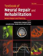
In two freestanding volumes, Textbook of Neural Repair and Rehabilitation provides comprehensive coverage of the science and practice of neurological rehabilitation. This volume, Neural Repair and Plasticity, covers the basic sciences relevant to recovery of function following injury to the nervous system, reviewing anatomical and physiological plasticity in the normal CNS, mechanisms of neuronal death, axonal regeneration, stem cell biology, and neuron replacement. Edited and written by leading international authorities, it is an essential resource for neuroscientists and provides a foundation for the work of clinical rehabilitation professionals.

Reviews

'In two freestanding but linked volumes, Textbook of Natural Repair and Rehabilitation provides comprehensive coverage of the science and practice of neurological rehabilitation. Edited and written by leading international authorities from the neurosciences and clinical neurorehabilitation, the two-volume set is an essential resource for rehabilitation professionals and a comprehensive reference for all scientists and clinicians in the field.'

Source: Advances in Clinical Neuroscience & Rehabilitation

Refine List

Actions for selected content:

Select all | Deselect all
  • View selected items
  • Export citations
  • Download PDF (zip)
  • Save to Kindle
  • Save to Dropbox
  • Save to Google Drive

Save Search

You can save your searches here and later view and run them again in "My saved searches".

Please provide a title, maximum of 40 characters.
×

Contents


Page 1 of 2


  • Neural repair and rehabilitation: an introduction
    pp xxvii-xxxvi
  • View abstract

    Summary

    Neurorehabilitation is the clinical subspecialty that is devoted to the restoration and maximization of functions that have been lost due to impairments caused by injury or disease of the nervous system. The concepts of neural plasticity have been accepted as important elements in the scientific understanding of functional recovery. As practiced in most countries, rehabilitation is a multidisciplinary process, involving combinations of treatment modalities administered by multiple therapists. Moreover, the most important outcome of the rehabilitation process is the degree of reintegration of the patient in society, in terms of roles in work, family, and community. In order to catch up to other fields in the practice of evidence-based medicine, the rehabilitation field has been forced to become extremely resourceful in designing outcome measures to evaluate the efficacy of its treatments. The combination of rehabilitation and either neuroplasticity or regeneration did not appear until after the term neurorehabilitation became current.
  • 1 - Anatomical and biochemical plasticity of neurons: regenerative growth of axons, sprouting, pruning, and denervation supersensitivity
    pp 5-25
    • By Oswald Steward, Departments of Anatomy and Neurobiology, Neurobiology and Behavior, and Neurosurgery, Reeve-Irvine Research Center, University of California, Irvine, CA, USA
  • View abstract

    Summary

    This chapter describes the reactive changes that central nervous system (CNS) neurons do exhibit following injury, both degenerative responses that occur following denervation and axotomy, and reactive growth that may contribute to recovery of function. Damage to the CNS affects all cell populations in the brain including neurons, glia, ependymal cells, and vascular elements. The chapter explores what happens to neurons and their interconnections. Trauma can cause physical transection of axons (axotomy), causing the portion distal to the injury to degenerate (Wallerian Degeneration). The term "specific regeneration" indicates a specific re-growth of an interrupted axon to its normal target. Developing neurons that have not received their full complement of innervation would be available to the aberrant axons. In mature animals, the prediction is that regenerative sprouting and pruning-related sprouting might not result in the formation of new connections unless sites were made available by denervation.
  • 2 - Learning and memory: basic principles and model systems
    pp 26-43
    • By Kimberly M. Christian, Neuroscience Program, University of Southern California, Los Angeles, CA, USA, Andrew M. Poulos, Neuroscience Program, University of Southern California, Los Angeles, CA, USA, Richard F. Thompson, Neuroscience Program, University of Southern California, Los Angeles, CA, USA
  • View abstract

    Summary

    This chapter provides a taxonomic overview of different forms of learning and memory at the behavioral and neural system levels. Long-term memory can be divided roughly into two categories, namely, declarative and non-declarative memory. Non-declarative memories encompass a wide range of phenomena from priming to skill learning. Some of the most basic forms of memory result from non-associative learning processes. Non-declarative priming memory is a form of memory that results from exposure to stimuli prior to a testing session. Damage to medial temporal structures including the hippocampus in human studies is associated with marked impairments in trace eyeblink conditioning. Many researchers have looked to the neocortex as the prime candidate for the permanent storage of declarative memories but there is limited evidence at this point to demonstrate this with certainty. Specificity of neocortical sites for memory storage and retrieval has been observed in imaging studies of healthy patients.
  • 3 - Short-term plasticity: facilitation and post-tetanic potentiation
    pp 44-59
    • By Ralf Schneggenburger, AG Synaptische Dynamik und Modulation, Abteilung Membranbiophysik, Max-Planck-Institut für Biophysikalische Chemie, Göttingen, Germany and Laboratory of Synaptic Mechanisms, Ecole Polytechnique Fédérale de Lausanne, Brain Mind Institute, 1015 Lausanne, Switzerland
  • View abstract

    Summary

    This chapter discusses the presynaptic, Ca2+-dependent mechanisms of short-term enhancement (STE) of synaptic transmission. Synaptic transmission takes place at specialized contact sites, at which the active zone of the presynaptic neuron approaches the postsynaptic density of a postsynaptic neuron. During prolonged action potential (AP) trains, STE of synaptic transmission, like augmentation, and post-tetanic potentiation (PTP) are also observed at many synapses. A framework for understanding in which ways synaptic strength can be modified is the quantal hypothesis of transmitter release, which originated from electrophysiological studies at the neuromuscular junction. Following longer-lasting trains of presynaptic APs, other forms of STE besides facilitation occur. These can be separated from one another by their time course of decay following the trains. Thus, synapses have a wide range of possibilities to fine-tune their signaling properties and short-term plasticity, according to the needs of the neuronal networks in which they operate.
  • 4 - Long-term potentiation and long-term depression
    pp 60-78
    • By Zafir I. Bashir, Department of Anatomy, MRC Centre for Synaptic Plasticity, University of Bristol, Bristol, UK, Peter V. Massey, Department of Anatomy, MRC Centre for Synaptic Plasticity, University of Bristol, Bristol, UK
  • View abstract

    Summary

    This chapter concentrates on the basic mechanisms that are thought to underlie induction and expression of long-term plasticity (LTP). LTP has been demonstrated at all of the major synapses in the hippocampus. Activity-dependent LTP is a commonly observed feature of the neocortex and does not differ from the hippocampus in its induction or expression mechanisms. The induction and/or the magnitude of LTP and long-term depression (LTD) can be affected by prior activity that in itself does not produce observable changes in synaptic efficacy. Thus there is some plastic change of a different, or meta, form that influences traditional synaptic plasticity. N-methyl-D-aspartate receptors (NMDAR)-mediated synaptic transmission can undergo potentiation and depression, and given the critical role for NMDARs in LTP and LTD induction it is apparent that any change in NMDAR synaptic transmission brought about by a priming stimulus could have dramatic consequences for induction of LTP and LTD.
  • 5 - Cellular and molecular mechanisms of associative and nonassociative learning
    pp 79-94
    • By John H. Byrne, Department of Neurobiology and Anatomy, University of Texas Health Science Center at Houston, Houston, TX, USA, Diasinou Fioravante, Department of Neurobiology and Anatomy, University of Texas Health Science Center at Houston, Houston, TX, USA, Evangelos G. Antzoulatos, Department of Neurobiology and Anatomy, University of Texas Health Science Center at Houston, Houston, TX, USA
  • View abstract

    Summary

    The neural mechanisms that contribute to the adaptation of an organism to environmental changes through learning are likely to contribute to the adaptation of the organism to physical changes (e.g., trauma) through repair and rehabilitation. Learning can be distinguished depending on whether it is associative or nonassociative. The examination of learning in simple model systems, such as the marine mollusc Aplysia californica, has proven to be very fruitful. The simplicity and tractability of the neural circuits mediating some behaviors in Aplysia have allowed the cellular/molecular dissection of the underlying neural mechanisms. Indeed a number of critical cells, synapses, and molecules have been identified in Aplysia to mediate basic forms of learning. One way to recruit the processes of neuronal plasticity in repair and rehabilitation is through behavioral learning paradigms, which provide the basis for current therapeutic strategies.
  • 6 - Plasticity of mature and developing somatosensory systems
    pp 97-108
    • By Jon H. Kaas, Department of Psychology, Vanderbilt University, Nashville, TN, Tim P. Pons, Department of Neurosurgery, Wake Forest University School of Medicine, Winston-Salem, NC, USA
  • View abstract

    Summary

    The somatosensory system of adult primates is capable of considerable reactivation after the loss of some of the activating connections This chapter examines what happens when damage occurs at each of four levels of the system: the receptor or primary afferent level, the level of the brain stem relay to the dorsal column-trigeminal complex, the thalamic ventroposterior nucleus, and primary somatosensory cortex. It summarizes several important features that relate to plasticity. The chapter discusses the consequences of a nerve crush with regeneration, a nerve cut and repair with regeneration, a nerve cut without regeneration, and transplanting and regenerating a nerve to a new skin location. The clear evidence for extensive reorganization came with the opportunity to study the somatosensory cortex of monkeys with a longstanding loss of all afferents from a forelimb. One mechanism for the extensive reactivation appears to be the growth of new connections in the brain stem.
  • 7 - Activity-dependent plasticity in the intact spinal cord
    pp 109-125
    • By Jonathan R. Wolpaw, Laboratory of Nervous System Disorders, Wadsworth Center, NYS Department of Health, Albany, NY, USA
  • View abstract

    Summary

    Interest in activity-dependent spinal cord plasticity is increasing with the growing recognition that the acquisition and maintenance of normal motor performances reflect activity-dependent plasticity at multiple sites throughout the central nervous system (CNS), including the spinal cord. This chapter addresses the range of activity-dependent plasticity during normal life. The behavioral effects associated with spinal cord plasticity appear to reflect the interaction of plasticity at both spinal and supraspinal sites. The substantial capacity for activity-dependent plasticity in the spinal cord has important theoretical and practical implications. The substantial capacity suggests that most motor skills that are acquired gradually through prolonged practice involve spinal cord plasticity. The ubiquity of activity-dependent plasticity and the inevitable interaction between primary, compensatory, and reactive types, implies that functional effects may change over time. Early gains will not always evolve into long-term improvements, while deleterious early effects may give way to long-term benefits.
  • 8 - Plasticity of cerebral motor functions: implications for repair and rehabilitation
    pp 126-146
  • View abstract

    Summary

    This chapter focuses on the plasticity now known to be possible in the motor regions of the brain. It explores the recent findings regarding motor cortex plasticity and reorganization. The most common approach taken to investigate the potential for cortical plasticity has been to evaluate the reorganization of sensory and motor maps following peripheral or central lesions and compare them to normal animals. The chapter also explores the structure of the motor cortex and how it relates to plasticity. The motor cortex contains a neural circuitry conducive to motor plasticity, which includes both intrinsic and extrinsic components. The chapter presents an overview of the development in a new field of rehabilitation, neural prostheses, also called brain-machine interfaces (BMIs). Neural prostheses open an important window into plasticity by allowing detailed research into how the brain changes with practice and learning and the extent to which the brain is able to adapt.
  • 9 - Plasticity in visual connections: retinal ganglion cell axonal development and regeneration
    pp 147-161
    • By Kurt Haas, Department of Cellular and Physiological Sciences, Brain Research Centre, University of British Columbia, Vancouver, BC, Canada, Hollis T. Cline, Cold Spring Harbor Laboratory, Cold Spring Harbor, NY, USA
  • View abstract

    Summary

    This chapter discusses current knowledge of how precisely ordered afferent synaptogenesis occurs during development. It also explains the potential for reforming functional circuits by correct rewiring during regeneration. Most of the knowledge of the mechanisms involved in establishing circuits between distant central nervous system (CNS) neuronal populations comes from studies of the axonal projection from the eye to central brain targets. The output neurons of the eye are the retinal ganglion cells (RGCs), whose axons exit the eye as the optic nerve, cross the midline at the optic chiasm, and innervate central brain structures. RGCs in fish and frogs survive optic nerve lesion and sprout new axonal extensions that correctly navigate to the tectum, reform the retinotectal map, and demonstrate visual responsivity. Regeneration recapitulates a critical period of heightened plasticity during which activity-dependent mechanisms mediate map refinement through pruning of ectopic axonal branches.
  • 10 - Plasticity in auditory functions
    pp 162-179
    • By Josef P. Rauschecker, Department of Physiology and Biophysics, Georgetown University School of Medicine, Washington, DC, USA
  • View abstract

    Summary

    This chapter covers plasticity in the central auditory system, most notably in the auditory cortex, from a variety of viewpoints. Plasticity of the auditory system has an important function in the compensation of early loss of vision and hearing. The chapter considers neuroanatomical and neurophysiological studies in animals as well as behavioral and functional imaging studies in humans. Auditory cortex is involved in the identification and recognition of auditory objects including the decoding and interpretation of speech sounds and the appreciation of music. Plasticity in most cases can be seen as an adaptive process, but sometimes has consequences that expose the vulnerability of the brain for deprivation during critical periods of sensory development. Sensory deprivation in one modality can also have beneficial effects on other modalities. Deleterious consequences of auditory plasticity are revealed in phenomena such as tinnitus.
  • 11 - Cross-modal plasticity in sensory systems
    pp 180-193
    • By Krishnankutty Sathian, Departments of Neurology and Rehabilitation Medicine, Emory University School of Medicine, Atlanta, GA, USA
  • View abstract

    Summary

    This chapter reviews a large body of work that has demonstrated the cross-modal involvement of visual cortical areas in non-visual tasks, both in the sighted and in the blind. According to common belief, blindness is associated with superior non-visual perception. Rats deprived of vision at birth are able to navigate a maze for a food reward faster than normal, and also show altered somatosensory receptive fields in the whisker barrel representation in somatosensory cortex. Paralleling the changes in performance and somatosensory cortex, neonatal visual deprivation in rats results in the appearance of somatosensory responsiveness in the anterior parts of occipital cortex, as shown by both electrophysiology and autoradiography. The effects of blindness on non-visual perceptual abilities and on cerebral cortical function might be attributed to long-term neural plasticity. However, the same cannot apply to similar changes noted, amazingly, after short-term visual deprivation of normally sighted subjects.
  • 12 - Attentional modulation of cortical plasticity
    pp 194-206
    • By Bharathi Jagadeesh, Department of Physiology and Biophysics, University of Washington, Seattle, WA, USA
  • View abstract

    Summary

    This chapter develops a possible model for the role of attention in cortical plasticity. It presents the definition of attention, its behavioral implications, and its neural and pharmacological basis. Attention is most frequently studied in the visual domain, where attention is known to improve the processing of certain visual information at the expense of other information that is presented simultaneously. The chapter also presents evidence that attention or behavioral interaction is required. It outlines some examples of plasticity that appears to occur in the absence of behavioral engagement. The chapter considers studies where attentional modulation or awareness of stimuli was required for learning to occur. The concept of a common pathway for attention and learning is supported by psychophysical studies that show that attentional enhancement of perception and learning enhancement of perception operate on the same substrates.
  • 13 - Plasticity in the injured spinal cord
    pp 209-227
    • By Serge Rossignol, Department of Physiology, Centre for Research in Neurological Sciences, Universite de Montreal, Montreal, Quebec, Canada
  • View abstract

    Summary

    This chapter discusses the mechanisms of spinal cord plasticity in animal models as revealed by the recovery of motor functions after a spinal lesion. It shows that in cats, rats and mice, motor programs such as locomotion are re-expressed after a complete spinal transection at the low-thoracic level. The pharmacological work with intrathecal cannula suggested that important effects on locomotion could be observed when the intrathecal injections were localized to the rostral spinal segments. Clinically, the notion that certain spinal segments may play a critical role in the control of spinal locomotion may help to somewhat simplify where to target pharmacological stimulation, cell grafts or electrical stimulation. With the advent of genetic characterization and the potential for genetic manipulations, the mouse is becoming an increasingly important model for spinal cord injury research. The characteristics of motor patterns result from an intricate dynamic sensori-motor interaction between the spinal and supraspinal levels.
  • 14 - Plasticity after brain lesions
    pp 228-247
    • By Randolph J. Nudo, Department of Molecular and Integrative Physiology, University of Kansas Medical Center, Kansas City, KS, USA, Ines Eisner-Janowicz, Department of Molecular and Integrative Physiology, University of Kansas Medical Center, Kansas City, KS, USA, Ann M. Stowe, Department of Molecular and Integrative Physiology, University of Kansas Medical Center, Kansas City, KS, USA
  • View abstract

    Summary

    Animal models have been especially useful in understanding the role of neuroplasticity in recovery of sensorimotor skills after brain injury. The type of injury and the method of induction vary with the specific purposes of the experiment. Typically, injuries are of two types: those designed to mimic traumatic brain injury (TBI) and those designed to mimic cerebral ischemia (or stroke). Studies conducted in cortical sensory areas over the past several years have revealed that representational maps are alterable as a function of the integrity of their sensory inputs, and as a function of experience. Changes in two neurotransmitter systems, gammaaminobutyric acid (GABA) and glutamate, have been implicated to play a role in functional recovery. New strategies for promoting recovery that were derived from basic studies in preclinical models are being tested in clinical trials. Approaches employing neurotrophins, neuromodulators, stem cells, magnetic stimulation, and electrical stimulation are currently under development.
  • 15 - From bench to bedside: influence of theories of plasticity on human neurorehabilitation
    pp 248-266
    • By Agnes Floel, Human Coritical Physiology Section, National Institute of Neurological Disorders and Stroke, National Institutes of Health, Bethesda, MD, USA, Leonardo G. Cohen, Human Coritical Physiology Section, National Institute of Neurological Disorders and Stroke, National Institutes of Health, Bethesda, MD, USA
  • View abstract

    Summary

    The primary vehicle for acquiring knowledge on plasticity in the human central nervous system (CNS) has been animal research. Understanding of mechanisms, development of strategies to purposefully modulate these mechanisms, and translation into rational strategies to promote recovery of function are the goals of modern neurorehabilitation. Training leads to specific changes in brain organization in the motor, somatosensory, auditory, and visual domain. Acute and chronic alterations in neurotransmitter regulation after injury affect plasticity, and may thus provide a basis for new pharmacologic targets for stroke recovery. One of the strategies proposed to enhance functional recovery and sensory substitution is to use mechanical devices interfaced with human sensory afferents or interacting with the CNS. Intravenous human umbilical cored blood cells (HUCB) infusion may have a beneficial effect on recovery processes. Growth factors seem to exert their effects by local processes including autocrine, paracrine, and juxtacrine stimulation.
  • 16 - Neuronal death and rescue: neurotrophic factors and anti-apoptotic mechanisms
    pp 271-292
    • By Thomas W. Gould, Department of Neurobiology and Anatomy, and the Neuroscience Program, Wake Forest University School of Medicine, NC, USA, Ronald W. Oppenheim, Department of Neurobiology and Anatomy, and the Neuroscience Program, Wake Forest University School of Medicine, NC, USA
  • View abstract

    Summary

    Developmental cell death is an integral part of the many adaptive strategies employed during ontogeny for generating the mature nervous system. There is increasing evidence that some of the mechanisms that regulate the death and survival of developing neurons and that of mature neurons following injury or in neurologic disease are similar. This chapter discusses several hypotheses of the etiology of amyotrophic lateral sclerosis (ALS). Neuronal cell death observed in brains of Huntington's disease (HD) patients, mutant Htt-expressing transgenic mice or neurotoxin-treated rats occurs through apoptosis. Recent studies reveal continuity in the molecular mechanisms by which apoptosis occurs in the various animal models of Parkinson's disease (PD). The chapter describes the cellular and molecular events of spinal cord injury (SCI) and suggests potential strategies for therapeutic rescue. Anti-apoptotic agents, including neurotrophic factors (NTFs), are necessary but not exclusive components of any effective therapy for neurodegenerative disease and neuronal injury.
  • 17 - Axon degeneration and rescue
    pp 293-302
    • By John W. Griffin, Departments of Neurology and Neuroscience, Johns Hopkins University, School of Medicine, Baltimore, MD, USA, Ahmet Höke, Departments of Neurology and Neuroscience, Johns Hopkins University, School of Medicine, Baltimore, MD, USA, Thien T. Nguyen, Departments of Neurology and Neuroscience, Johns Hopkins University, School of Medicine, Baltimore, MD, USA
  • View abstract

    Summary

    This chapter reviews the sequence of changes in Wallerian degeneration after transection, and suggests that the late stages are similar in the disorders of axonal transport. The axons develop swellings containing densely packed accumulations of mitochondria, dense bodies and multivesicular bodies. In settings where interrupted axons survive for long periods, such as the Wallerian-like degeneration slow (Wlds) mouse, there is also an accumulation of neurofilaments. Neurofilament proteins were initially recognized to move in the slow anterograde phase of axonal transport. The importance of defects in axonal transport have become especially clear in genetic disorders where molecules involved in axonal transport have been responsible for human length-dependent neuropathies. In multiple sclerosis (MS) and other human and experimental settings with inflammatory demyelination, axonal degeneration and loss can result from focal axonal interruption consequent to the presence of nearby inflammatory cells and inflammatory mediators.
  • 18 - Adult neurogenesis and neural precursors, progenitors, and stem cells in the adult CNS
    pp 303-325
    • By Jeffrey D. MacKlis, Department of Neurology, MGH-HMS Center for Nervous System Repair, Harvard Medical School, Boston, MA, USA, Gerd Kempermann, Max Delbruck Center for Molecular Medicine (MDC), Berlin-Buch, Germany
  • View abstract

    Summary

    This chapter deals with adult neurogenesis and examines what is known about the behavior and function of precursor cells in the adult brain. It outlines few examples of normally occurring neurogenesis in the mammalian central nervous system (CNS), and describes adult neural precursors. Functional adult neurogenesis occurs in many non-mammalian vertebrates. The chapter reviews a few lines of recent research demonstrating that endogenous neural precursors can be induced to differentiate into neurons in regions of the adult brain that do not normally undergo neurogenesis. In the adult mammalian brain, neurogenesis normally occurs only in the olfactory bulb and the dentate gyrus (DG) of the hippocampus. Transplantation studies support the concept of neurogenic and non-neurogenic regions, and provide evidence about the role of the microenvironment in realizing the potential of neuronal stem or progenitor cells. Neuronal replacement therapies based on manipulation of endogenous precursors may be possible in the future.

Page 1 of 2


Metrics

Full text views

Total number of HTML views: 0
Total number of PDF views: 0 *
Loading metrics...

Book summary page views

Total views: 0 *
Loading metrics...

* Views captured on Cambridge Core between #date#. This data will be updated every 24 hours.

Usage data cannot currently be displayed.