CLINICIAN'S CAPSULE
What is known about the topic?
Absence of cardiac activity on an electrocardiogram (ECG) and point-of-care ultrasound (POCUS) during cardiac arrest independently predicts poor outcomes.
What did this study ask?
Is there a benefit to combining findings on ECG and POCUS when predicting outcome in patients arriving at the emergency department with cardiac arrest?
What did this study find?
Negative POCUS findings and negative combined POCUS and ECG findings performed better than ECG alone for predicting negative clinical outcomes.
Why does this study matter to clinicians?
Adding POCUS to ECG improves the accuracy of predicting which patients will not benefit from continued resuscitation during cardiac arrest.
INTRODUCTION
Advanced cardiac life support (ACLS) protocols guide the management of patients in cardiac arrest. Although these algorithms do not currently mandate the use of echocardiography, cardiac point-of-care ultrasound (POCUS) is recognized in resuscitation guidelines and has become standard practice in many emergency departments (EDs) and critical care settings.1–Reference Atkinson, Bowra and Milne5 Cardiac POCUS during resuscitation has been shown to be feasible, and, although it can be performed without interrupting cardiopulmonary resuscitation (CPR) beyond the standard rhythm check,Reference Hayhurst, Lebus and Atkinson6 when incorporated routinely, it may also prolong interruptions in CPR.Reference Allison, Bostick and Fisher7 Physicians use POCUS to identify potentially reversible conditions, as well as to identify mechanical cardiac activity.Reference Hernandez, Shuler and Hannan8,Reference Andrus and Dean9
Rules for the termination of out-of-hospital cardiac arrest have been proposed.Reference Morrison, Visentin and Kiss10 Several studies also report that an absence of cardiac activity on POCUS during cardiac arrest is associated with a significantly lower (but not zero) likelihood that a patient will experience return of spontaneous circulation (ROSC).Reference Blyth, Atkinson, Gadd and Lang11–Reference Salen, Melniker and Chooljian14 A large prospective study suggested that cardiac activity on POCUS could be used as a reliable predictor of favourable outcomes.Reference Gaspari, Weekes and Adhikari15 A recent systematic review suggests that POCUS can aid in decision-making for resuscitation termination but is not the absolute determining factor.Reference Tsou, Kurbedin and Chen12 Patients presenting with no cardiac activity on POCUS, as well as an electrocardiogram (ECG) rhythm of asystole have been shown to have lower rates of survival, whereas increased survival has been reported in patients with cardiac activity on POCUS and with pulseless electrical activity (PEA) on ECG.Reference Gaspari, Weekes and Adhikari15
Our primary enquiry was to assess whether the combined use of POCUS and ECG rhythm improves outcome prediction during ED cardiac arrest in patients without prior advanced life support, who had been selected for a transfer to hospital.
METHODS
A health records review was completed for patients who presented in cardiac arrest to a single tertiary Canadian ED between 2010 and 2014. All adult non-trauma cardiac arrest patients presenting during the study period were considered for inclusion. Prehospital basic life support had been administered. A prehospital resuscitation termination protocol was in place. Patients were excluded if under age 19 years, if their initial cardiac rhythm was shockable, if resuscitation was halted due to end-of-life decisions, or if no cardiac POCUS was performed. Standardized data extraction was completed.Reference Gaspari, Weekes and Adhikari15,Reference Gilbert, Lowenstein, Koziol-McLain, Barta and Steiner16 Initial ECG rhythm and cardiac activity on the initial POCUS exam were recorded. We report the diagnostic test performance of initial ECG rhythm alone, initial POCUS alone, and initial ECG and POCUS combined, for our primary outcome of failure to achieve ROSC, as well as secondary outcomes of failure to survive to hospital admission and to hospital discharge. Further details on delivery of resuscitation, definitions of outcomes, and data extraction methods are provided in Supplemental Appendix 1.
Research Ethics Board approval was obtained (Horizon Health Network 2015–2132).
Point estimates and proportions are reported with appropriate confidence intervals. Categorical data were analysed using Fisher's exact test and continuous data using unpaired student's t-test, using GraphPad Prism software (2012) (GraphPad Software Inc., La Jolla, CA).
RESULTS
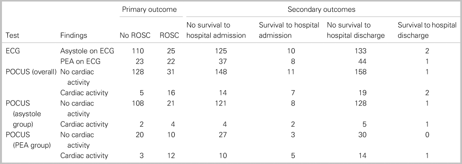
Of the 264 cardiac arrest cases reviewed, 84 were excluded, leaving a total of 180 cases meeting the inclusion criteria (Supplemental Figure 1). A median of 2 (range 1 to 9) POCUS exams was performed; 45 (25.0%; 19.2% to 31.8%) patients demonstrated cardiac activity on the initial ECG, and 21 (11.7%; 7.7% to 17.2%) had cardiac activity on the initial POCUS exam; 15 (8.3%; 5.0% to 13.4%) patients demonstrated activity on both ECG and POCUS, and 129 (71.7%; 64.7% to 77.7%) had no activity on either ECG or POCUS; 47 patients (26.1%; 20.2% to 33.0%) achieved ROSC, 18 (10.0%; 6.3% to 15.3%) survived to admission, and 3 (1.7%; 0.3% to 5.0%) survived to hospital discharge (Table 1). Baseline population characteristics are shown in Supplemental Appendix 2.
Table 1. Contingency tables showing patient numbers achieving no ROSC (primary outcome) and no survival to hospital admission and discharge (secondary outcomes) for initial ECG rhythm, initial POCUS findings overall and POCUS findings in patients with ECG asystole, and PEA

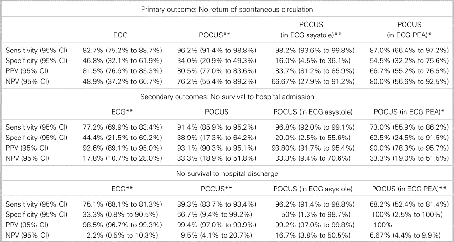
As a predictor of failure to achieve ROSC, ECG rhythm alone had a sensitivity of 82.7% (95% CI 75.2% to 88.7%) and a specificity of 46.8% (32.1% to 61.9%). Overall, POCUS alone had a higher sensitivity of 96.2% (91.4% to 98.8%) but a similar specificity of 34.0% (20.9% to 49.3%). In patients with ECG asystole, POCUS had a sensitivity of 98.2% (93.6% to 99.8%) and a specificity of 16.0% (4.5% to 36.1%). In patients with PEA, POCUS had a sensitivity of 86.96% (66.41% to 97.22%) and a specificity of 54.55% (32.2% to 75.6%).
The diagnostic performance of ECG, POCUS, and both combinations for the secondary outcomes of survival to hospital admission and discharge along with further details on diagnostic performance are shown in Table 2 and in Appendix 2. Only 0.8% (0.0 to 4.7%) of patients with asystole on ECG and absence of cardiac activity on POCUS survived to hospital discharge.
Table 2. Diagnostic test performance of initial ECG rhythm, initial POCUS findings overall and POCUS findings in patients with ECG asystole, and PEA, for the predication of no ROSC (primary outcome) and no survival to hospital admission and discharge (secondary outcomes)

CI = Confidence interval; NPV = negative predictive value; PPV = positive predictive value; McNemar's chi-squared test (P-value), **P < 0.01, *P < 0.05.
DISCUSSION
Our findings show that both ECG rhythm and POCUS findings, either independently or in combination, perform better as predictors of negative outcomes (death) than they do for positive outcomes (ROSC, survival to hospital admission and discharge) in adult ED cardiac arrest patients with non-shockable rhythms. Moreover, ECG and POCUS findings are not independent variables, and, as such, any direct statistical comparison of these tests would be inappropriate. However, describing the relative utility of each, either independently or when used in combination, is useful for understanding how they can contribute to decision-making during resuscitation.
In patients with non-shockable rhythms, we found ECG alone to be a poor predictor of outcome in cardiac arrest, with a sensitivity of 82.7% and a specificity of 46.8% for failure to achieve ROSC. POCUS alone was a better predictor of futility, with a higher sensitivity of 96.2%, as was the combination of asystole on ECG and absence of activity on POCUS with a sensitivity of 98.2%, demonstrating the utility of adding POCUS to ECG for the identification of patients who are unlikely to survive. Unfortunately, neither ECG nor POCUS, alone, or in combination, performed well as reliable tests to identify patients likely to survive, with specificities maximizing at just over 50%. A similar pattern was also seen when assessing prediction for long-term outcomes with POCUS alone, and negative POCUS findings in patients with asystole on ECG, providing reasonable sensitivities for failure to survive to hospital discharge, but again with poor specificity.
Our findings are consistent with results reported by Gaspari et al., where the absence of activity on POCUS in patients with asystole on ECG was associated with poorer outcomes than in patients with PEA and activity on POCUS.Reference Gaspari, Weekes and Adhikari15 Future research should examine, among PEA patients, whether those with cardiac activity on POCUS have improved clinical outcomes. Previous studies have suggested that an absence of cardiac activity on POCUS can aid in resuscitation termination decisions. We found that POCUS, and especially combined ECG and POCUS, demonstrated high sensitivity and positive predictive values for our primary and secondary outcomes, suggesting that POCUS could add value for termination of resuscitation decisions if incorporated into ACLS guidelines for adult cardiac arrest.Reference Tsou, Kurbedin and Chen12
There are a number of limitations with this project. The database was formed by a health records review in a single centre. Although standardized methodology was used (see Appendix 1), not all population data could be acquired for each patient, and charting quality could not be controlled. Variable delays until the initial POCUS may explain the inconsistency of recording cardiac activity on POCUS in a small number of patients who had asystole on their initial ECG. We did not include any functional assessment as a formal outcome in our analysis. However, all of the survivors (1.7%) required home care or rehabilitation support.
CONCLUSIONS
Our findings suggest that for adult patients arriving to the ED in non-shockable cardiac arrest, the use of POCUS independently or in combination with ECG rhythm more accurately predicted death than the use of ECG rhythm alone. No combination reliably predicted survival.
Supplementary material
The supplementary material for this article can be found at https://doi.org/10.1017/cem.2019.397
Acknowledgements
The authors acknowledge the clinical staff at Saint John Regional Hospital Emergency Department for their help in identifying patients for this study and for their participation in performing the required ultrasonographic examinations and clinical assessments.


