Introduction
Strongyloidiasis, which is caused by Strongyloides stercoralis, is one of the most difficult-to-diagnose soil-transmitted helminth (STH) infections. It occurs worldwide and is endemic in tropical and temperate climates (Schär et al., Reference Schär, Trostdorf, Giardina, Khieu, Muth, Marti, Vounatsou and Odermatt2013; Jourdan et al., Reference Jourdan, Lamberton, Fenwick and Addiss2018). It is considered the most neglected of the neglected tropical diseases and its prevalence is largely underestimated (Olsen et al., Reference Olsen, van Lieshout, Marti, Polderman, Polman, Steinmann, Stothard, Thybo, Verweij and Magnussen2009; Viney, Reference Viney2017). Moreover, several aspects of the epidemiology of S. stercoralis infection remain poorly documented (Bisoffi et al., Reference Bisoffi, Buonfrate, Montresor, Requena-Méndez, Muñoz, Krolewiecki, Gotuzzo, Mena, Chiodini, Anselmi, Moreira and Albonico2013; Nutman, Reference Nutman2017). Based on available information; it is estimated that about 370 million people are infected with S. stercoralis worldwide, with the prevalence rate ranging from 10 to 70% in tropical and subtropical countries where conducive conditions for transmission such as moist soil and inadequate sanitation coexist. It is particularly evident in underprivileged communities in Latin America, West Africa and Southeast Asia (Schär et al., Reference Schär, Trostdorf, Giardina, Khieu, Muth, Marti, Vounatsou and Odermatt2013; Bisoffi et al., Reference Bisoffi, Buonfrate, Sequi, Mejia, Cimino, Krolewiecki, Albonico, Gobbo, Bonafini, Angheben, Requena-Mendez, Muñoz and Nutman2014; Puthiyakunnon et al., Reference Puthiyakunnon, Boddu, Li, Zhou, Wang, Li and Chen2014).
Humans acquire S. stercoralis infection generally through skin penetration of filariform larvae that exist in contaminated soil. Moreover, donor-derived strongyloidiasis infection in solid organ transplant recipients is increasingly being reported (Kim et al., Reference Kim, Kim, Yoon, Sohn and Kim2016; Winnicki et al., Reference Winnicki, Eder, Mazal, Mayer, Sengölge and Wagner2018). Strongyloidiasis is commonly chronic and long lasting with a majority of infected individuals remaining asymptomatic, and infections could be sustained in individuals for more than 75 years (Prendki et al., Reference Prendki, Fenaux, Durand, Thellier and Bouchaud2011; Junior et al., Reference Junior, Zaman, Zaman and Zaman2017). Pulmonary migration of larvae results in transient eosinophilia with cough, dyspnoea (shortness of breath), wheezing and haemoptysis; known as Loeffler's syndrome which is seen in heavy infections (Al Hadidi et al., Reference Al Hadidi, Shaaban, Jumean and Peralta2018). Chronic S. stercoralis infection may present with fatigue, anorexia, vomiting, abdominal pain, diarrhoea and urticaria (Khieu et al., Reference Khieu, Srey, Schär, Muth, Marti and Odermatt2013; Nutman, Reference Nutman2017). In immunocompetent individuals, infection produces negligible symptoms, but in immunocompromised patients the autoinfection cycle of this parasite can be life-threatening because it can lead to Strongyloides hyperinfection syndrome that has a mortality rate of nearly 100% if left untreated and typically presents as intestinal or pulmonary failure (Kassalik and Mönkemüller, Reference Kassalik and Mönkemüller2011; Nutman, Reference Nutman2017). In immunocompromised patients, disseminated strongyloidiasis can occur, with large numbers of parasites spreading to affect various organs, including the liver, kidneys, brain, cutaneous and subcutaneous tissue and heart, with occasional gut translocation of bacteria causing bacteraemia (Bisoffi et al., Reference Bisoffi, Buonfrate, Montresor, Requena-Méndez, Muñoz, Krolewiecki, Gotuzzo, Mena, Chiodini, Anselmi, Moreira and Albonico2013; Shimasaki et al., Reference Shimasaki, Chung and Shiiki2015).
Strongyloides stercoralis diagnosis remains a challenge because individuals carrying the infection often remain asymptomatic and there is no gold-standard reference method for its diagnosis (Requena-Méndez et al., Reference Requena-Méndez, Chiodini, Bisoffi, Buonfrate, Gotuzzo and Muñoz2013; Nutman, Reference Nutman2017). The most common diagnostic techniques that are used to detect most intestinal parasites are the direct fecal smear, formalin-ether sedimentation (FES) and Kato-Katz, but these methods often failed to detect S. stercoralis larvae or they have low sensitivity, particularly in the case of chronic infections (Requena-Méndez et al., Reference Requena-Méndez, Chiodini, Bisoffi, Buonfrate, Gotuzzo and Muñoz2013; Buonfrate et al., Reference Buonfrate, Perandin, Formenti and Bisoffi2017). Hence, more-sensitive and Strongyloides-specific methods such as the Baermann method, Koga agar plate culture (APC) and molecular assays are now being used to detect this parasite and to update the epidemiology of strongyloidiasis worldwide (Amor et al., Reference Amor, Rodriguez, Saugar, Arroyo, López-Quintana, Abera, Yimer, Yizengaw, Zewdie, Ayehubizu, Hailu, Mulu, Echazú, Krolewieki, Aparicio, Herrador, Anegagrie and Benito2016). In reality, however, these methods are not routinely used in parasitological examinations because they are time consuming and require more resources than the most commonly applied methods, particularly in the potentially endemic settings of resource-poor countries (Agrawal et al., Reference Agrawal, Agarwal and Ghoshal2009; Schär et al., Reference Schär, Trostdorf, Giardina, Khieu, Muth, Marti, Vounatsou and Odermatt2013). Although serological methods are very useful in the diagnosis of strongyloidiasis and have been validated for clinical and epidemiological use, their specificity is variable and they show reduced sensitivity in the case of low-intensity infections (Bisoffi et al., Reference Bisoffi, Buonfrate, Sequi, Mejia, Cimino, Krolewiecki, Albonico, Gobbo, Bonafini, Angheben, Requena-Mendez, Muñoz and Nutman2014; Buonfrate et al., Reference Buonfrate, Perandin, Formenti and Bisoffi2017; Fradejas et al., Reference Fradejas, Herrero-Martínez, Lizasoaín, Rodríguez de Las Parras and Pérez-Ayala2018).
In Southeast Asia, high prevalence rates of S. stercoralis infection have been reported in Lao PDR, Cambodia and Thailand (Puthiyakunnon et al., Reference Puthiyakunnon, Boddu, Li, Zhou, Wang, Li and Chen2014; Schär et al., Reference Schär, Giardina, Khieu, Muth, Vounatsou, Marti and Odermatt2016; Senephansiri et al., Reference Senephansiri, Laummaunwai, Laymanivong and Boonmar2017; Forrer et al., Reference Forrer, Khieu, Schär, Vounatsou, Chammartin, Marti, Muth and Odermatt2018). In Malaysia, several previous studies are available on the prevalence of intestinal parasitic infections (IPIs) throughout the country; however, data on S. stercoralis infection are still very limited. The first study on S. stercoralis in Malaysia reported in a single case among Orang Asli in the Kelantan state; however, this study applied low-sensitivity diagnostic methods (Rahmah et al., Reference Rahmah, Ariff, Abdullah, Shariman, Nazli and Rizal1997). In addition, despite the high seropositive rates reported among indigenous populations in Selangor state, West Malaysia (31.5%; 17/54) and Sarawak state, East Malaysia (11%; 26/236), parasitological methods failed to detect S. stercoralis larvae in fecal samples among the studied participants (Ahmad et al., Reference Ahmad, Hadip, Ngui, Lim and Mahmud2013; Ngui et al., Reference Ngui, Halim, Rajoo, Lim, Ambu, Rajoo, Chang, Woon and Mahmud2016).
In light of the above status, the present study used multiple diagnostic methods to investigate the prevalence and risk factors of S. stercoralis infection among Orang Asli schoolchildren in Peninsular Malaysia. This study is crucial because it will not only provide reliable community-based evidence on the presence of S. stercoralis in Orang Asli communities to enable a better understanding of the epidemiology of human strongyloidiasis in Malaysia, it will also update the available information on the global distribution of strongyloidiasis.
Materials and methods
Study design and study area
A cross-sectional, community-based study was carried out among Orang Asli (aboriginal) schoolchildren in six states of Peninsular Malaysia: (Selangor, Pahang, Negeri Sembilan, Kelantan, Johor and Perak) from January to April 2017.
The Orang Asli communities that were involved in this study were residents in 11 districts as follows: Hulu Selangor (3°35′N latitude, 101°35′E longitude) and Petaling (3°05′N latitude, 101°35′E longitude), which are districts in Selangor state; Raub (3°47′N latitude, 101°51′E longitude) and Lipis (4°15′N latitude, 101°50′E longitude) in Pahang state; Jelebu (3°0′N latitude, 102°05′E longitude) and Kuala Pilah (2°44′N latitude, 102°14′E longitude)in Negeri Sembilan state; Gua Musang (4°53′N latitude, 101°58′E longitude) in Kelantan state; Segamat (2°30′N latitude, 102°55′E longitude) in Johor state; and Hulu Perak (5°20′N latitude, 101°15′E longitude), Muallim (3°50′N latitude, 101°30′E longitude) and Batang Padang (4°05′N latitude, 101°20′E longitude) in Perak state (Fig. 1). These districts were randomly selected from a list describing the distribution of Orang Asli communities in Peninsular Malaysia that was provided by the Department of Orang Asli Development (also known as JAKOA). In these Orang Asli communities, primary schools that had an enrolment of more than 100 schoolchildren were selected for this study.
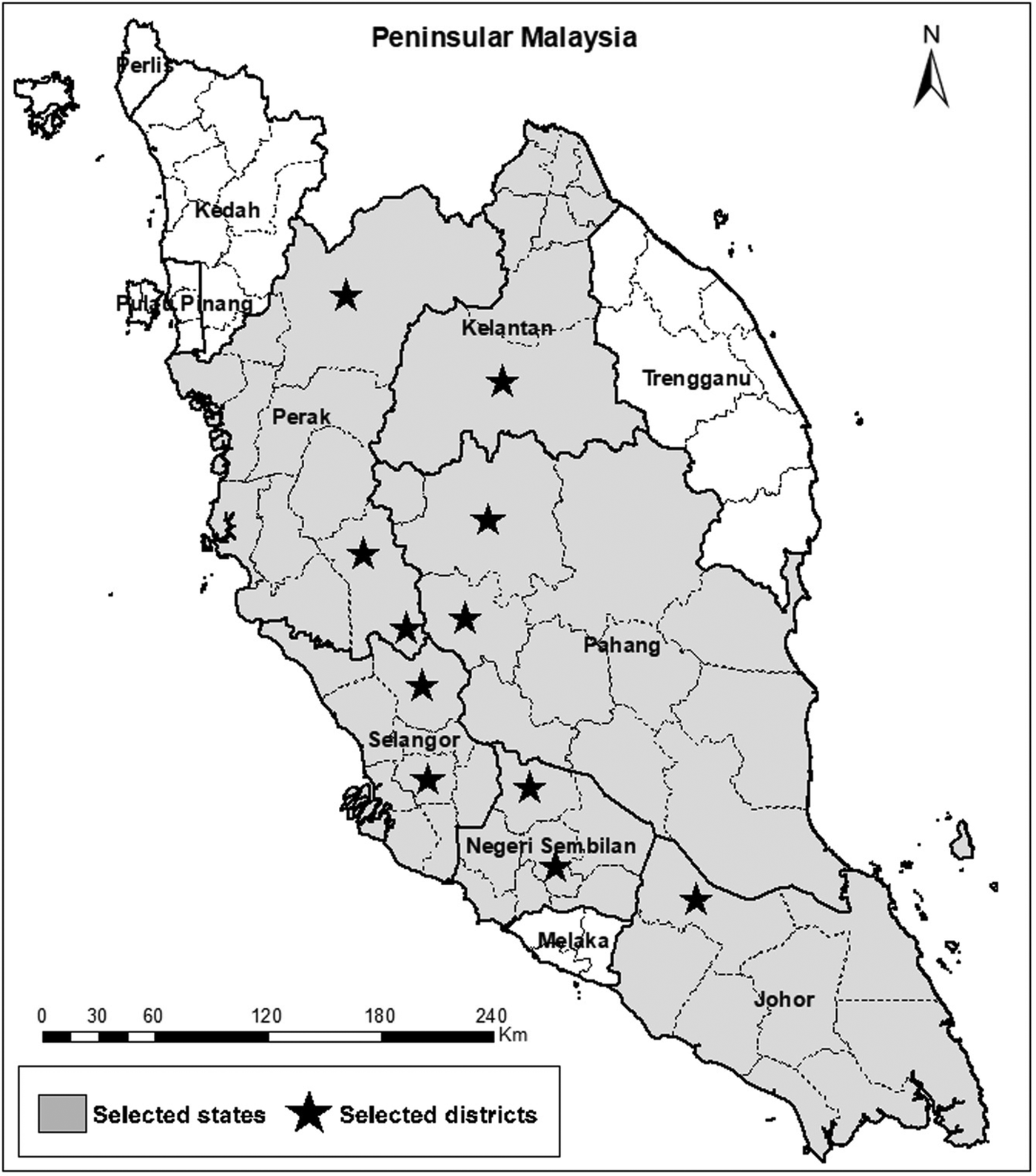
Fig. 1. A geographic map showing Peninsular Malaysia and the districts involved in the study (11 districts within six states). The map was created using the Esri ArcGIS 10.7 software.
Peninsular Malaysia, also known as West Malaysia, covers a land area of 1 31 587 km2 that extends 740 km from north (bordering Thailand) to south (bordering Singapore) and its maximum width is 322 km. It boasts a tropical climate; warm and humid all year round with temperatures ranging from 21 to 32 °C and average humidity of 90%. Moreover, rainfall is plentiful with an average of 2300 mm per year, with thick rainforest (Wong et al., Reference Wong, Venneker, Uhlenbrook, Jamil and Zhou2009).
Study population
This study was carried out among Orang Asli schoolchildren who were residents in the above-mentioned districts. Orang Asli (a Malay term that can be translated as ‘original people’ or ‘the first people’) are the aboriginal minority people of Peninsular Malaysia who accounts for 0.6% of the country's total population (i.e. approximately 180 000 people) (Department of Statistics Malaysia, 2010). They comprised of three main tribal groups: Senoi, constituting the largest group (54.9%), Proto-Malay (42.3%) and Negrito (2.8%). Within these three tribal groups, there are a total of 19 ethnic groups (SyedHussain et al., Reference SyedHussain, Krishnasamy and Hassan2017). The largest Orang Asli population reside in the states of Pahang and Perak, while the smallest can be found in the states of Penang and Perlis. Most Orang Asli communities live in remote forest areas but some live in the vicinity of suburban areas. A majority are marginalized socioeconomically and culturally, with a significant population below the poverty line, and about half of them are classified as living in hard-core poverty (SyedHussain et al., Reference SyedHussain, Krishnasamy and Hassan2017). Moreover, Orang Asli have an average life expectancy of 53 years, compared to the national average of 73 years, and an infant mortality rate of 51.7 per 1000 live births compared to the national rate of 8.9 (Masron et al., Reference Masron, Masami and Ismail2013; Odani, Reference Odani2017). Some Orang Asli work for wages in rubber tapping, in oil palm plantations as labourers or as factory workers, while many other Orang Asli are engaged in other kinds of daily activities to earn a living by selling jungle products such as fruit, rattan and bamboo (Masron et al., Reference Masron, Masami and Ismail2013).
Sample size
A randomized, open-label controlled intervention trial (Trial Registration: clinicaltrials.gov; identifier: NCT03930901) was designed with the aim of investigating the impact of an improved health education learning package in controlling IPIs among Orang Asli schoolchildren. The cross-sectional study discussed herein represents the baseline assessment before randomization and intervention.
The minimum sample size required for the main interventional study was calculated as 924 children; 462 per intervention arm (154 children from each tribe, including 10% to avoid the effects of dropouts and potential losses). It was estimated that this number would give the study at least 80% power at 5% significance to detect at least 10% difference in the prevalence and intensity of IPIs between the intervention group and the control group. This calculation assumed that 70% of Orang Asli children have IPIs (Nasr et al., Reference Nasr, Al-Mekhlafi, Ahmed, Roslan and Bulgiba2013; Al-Delaimy et al., Reference Al-Delaimy, Al-Mekhlafi, Lim, Nasr, Sady, Atroosh and Mahmud2014; Elyana et al., Reference Elyana, Al-Mekhlafi, Ithoi, Abdulsalam, Dawaki, Nasr, Atroosh, Abd-Basher, Al-Areeqi, Sady, Subramaniam, Anuar, Lau, Moktar and Surin2016). This sample size was judged safe enough to achieve a final sample size of 646, a figure that was calculated separately for the cross-sectional baseline survey at a 5% level of significance, a 95% confidence level, and a design effect of 2 (Lwanga and Lemeshow, Reference Lwanga and Lemeshow1991). All of the eligible children who were present during our visit to each of the selected schools were invited to participate in the baseline study (universal sampling).
According to the official rolls, a total of 2763 children were enrolled in the 13 selected primary schools. However, at the time of our visits, there were 1914 children in the targeted schools all of whom were invited to participate in this study. Of these, 96 refused to participate and 676 did not deliver stool samples in the next 2 days following our visits. |Hence, 1142 children (49.4% boys and 50.6% girls) who delivered suitable fecal samples for examination and complete questionnaire data were included in this study.
Questionnaire survey
A pre-tested structured questionnaire was used to collect information about the demographic (age, sex and family size) and socioeconomic (e.g. parents' educational and occupational status, and family monthly income) background of the participants. Moreover, information about personal hygiene (e.g. washing hands before eating and after using the toilet, wearing shoes when outside the house, cutting nails periodically and washing vegetables/fruit before eating), health (e.g. history of IPIs and presence of any signs and symptoms particularly those related to gastrointestinal infections) and household characteristics (e.g. type of house, availability and type of toilets, sources of drinking water and presence of domestic animals) was also obtained. The types of toilet facility in the study area were categorized into two groups; improved (pour flush toilet) and unimproved (pit latrine without a slab) in line with the WHO/UNICEF criteria (WHO and UNICEF, 2015). Similarly, types of drinking water source were categorized into improved water sources (i.e. piped water supply) and unimproved water sources (e.g. wells, streams and rain).
The children were interviewed (face to face) by two assistants, one from the children's respective school and one from the Department of Parasitology, University of Malaya. Both assistants received proper training on the purpose of the study and on the administration of the questionnaire. A separate sheet containing questions to obtain demographic information about the children's parents was given to the children to hand to their parents to complete and was collected back the next day.
Fecal examination
Each child received a clean wide-mouth 100 mL screw-capped container clearly labelled with child's name and unique reference number for the collection of fecal samples. A clear explanation of how to collect the stool sample, the suitable amount of sample, and how to avoid possible contamination in the course of collection at home was provided to the participants in their classrooms. The children were instructed to bring their early morning stool samples the next day. Upon collection, the samples were placed into zipped plastic bags, kept in suitable cool boxes, and transported (within 2–8 h of collection) to the Department of Parasitology, University of Malaya, Kuala Lumpur for parasitological examination.
To detect S. stercoralis, the fecal samples were examined by using four different methods. First, the samples were examined using a direct smear or wet mount, using normal saline and iodine. Second, the samples were examined using the FES method (Cheesbrough, Reference Cheesbrough2005). Third, the Koga APC method, which is one of the most sensitive techniques, was used to detect S. stercoralis and/or hookworm larvae (Koga et al., Reference Koga, Kasuya, Khamboonruang, Sukhavat, Ieda, Takatsuka, Kita and Ohtomo1991; Pocaterra et al., Reference Pocaterra, Ferrara, Peñaranda, Rojas, Pérez-Chacón, Hernán, Certad, Goldstein and Núñez2017). Strongyloides stercoralis larvae were identified based on morphology (i.e. size of the buccal cavity, presence of genital primordium in rhabditiform larvae, and presence of forked tail-end in filariform larvae). Fourth, an aliquot of each fecal sample was preserved in 70% ethanol alcohol and kept at 4 °C until subsequent DNA extraction, and conventional polymerase chain reaction (PCR) assay to detect S. stercoralis. The samples were considered positive via the detection of S. stercoralis larvae DNA using PCR or by using any of the parasitological methods with confirmation by PCR. For those fecal samples that were found to be negative by PCR but positive by agar culture, the larvae collected from the positive agar cultures were examined by PCR to confirm the presence of S. stercoralis.
DNA extraction
About 500 mg of each ethanol-alcohol-preserved fecal sample was shaken vigorously and centrifuged at 1000 rpm for 2 min. The resulting pellet was washed twice with phosphate-buffered saline (0.01 M, pH 7.2) and then used for genomic DNA extraction using the commercially available QIAamp® DNA stool Mini Kit (QIAGEN, Hilden, Germany), according to the manufacturer's instructions. The extracted DNA was then eluted with 100 µl of the AE elution buffer (included in the kit) and quantified using a Micro UV-Vis fluorescence spectrophotometer (Malcom e-spect, Tokyo, Japan). The extracted DNA was then kept at −20 °C until it was subjected to PCR amplification.
DNA amplification and sequencing
Strongyloides stercoralis-specific primer Stro18S-1530 (5′-GAATTCCAAGTAAACGTAAGTCATTAGC-3′) and Stro18S-1630 (5′-TGCCTCTGGATATTGCTCAGTTC-3′) were used as an upstream and downstream oligonucleotide pair, respectively, to amplify a 101 base pair (bp) region of S. stercoralis 18S rRNA (GenBank accession number AF279916), as described by Verweij et al. (Reference Verweij, Canales, Polman, Ziem, Brienen, Polderman and van Lieshout2009). The PCR was optimized and run using MyCycler thermal cycler (Bio-Rad, CA, USA) in a final volume of 25 µL of the reaction mixture that contained 1X of ExPrime Taq Master Mix (Genet Bio, Korea), 200 mm of each primer and 1 µl of DNA. The PCR thermal conditions included an initial denaturing step at 94 °C for 5 min followed by 40 cycles of 30 s at 94 °C, 60 s at 60.5 °C and 60 s at 72 °C, and a final extension step at 72 °C for 10 min. The PCR products were then visualized in 2.5% agarose gel stained with Sybr® safe DNA gel stain (Invitrogen, CA, USA) using a UV documenting system (Bio-Rad, Hercules, CA, USA).
The DNA that was extracted from the filariform larvae of S. stercoralis obtained from positive APC was subjected to PCR amplification and DNA sequencing in both directions. The resulting sequences were checked in NCBI BLAST to confirm the S. stercoralis species, and those with 100% specificity were used as a positive control.
Statistical analysis
Data analysis was performed using the IBM SPSS Statistics, version 18.0 (IBM Corporation, New York, USA). The dependent variable (i.e. S. stercoralis infection status) and the independent variables (i.e. demographic, socioeconomic, household and behavioural variables) were treated as categorical variables and presented as frequencies and percentages. All the variables were coded as binary dummy variables (i.e. 0 and 1). For instance, S. stercoralis infection (yes = 1, no = 0); age (>10 years = 1, ⩽10 years = 0); gender (male = 1, female = 0); wearing shoes when outside (no = 1, yes = 0); and type of drinking water source (unimproved = 1, improved = 0). Pearson's χ 2 test was used to test the association between S. stercoralis infection prevalence and each of the explanatory variables. The odds ratio (OR) and 95% CI were calculated. Also, in order to identify the risk factors that were significantly associated with S. stercoralis infection, all the variables that showed associations with P ⩽ 0.25 in the univariate analysis were included in a multivariate logistic regression analysis, as suggested in the literature (Bendel and Afifi, Reference Bendel and Afifi1977). In addition, the population attributable risk fraction (PARF) was calculated for the significantly associated risk factors (Rockhill et al., Reference Rockhill, Newman and Weinberg1998). A P value of <0.05 was considered as the level of significance.
Results
General characteristics of the participants
A total of 1142 schoolchildren aged 8–12 years, with a mean age of 10.19 years (s.d. = 1.36), attending 13 primary schools in six states of Peninsular Malaysia participated in this study. Of these, 49.4% were male and 50.6% were female. The highest percentage of children (49%) belonged to the Senoi tribe, while the lowest percentage belonged to the Negrito tribe (13%). About half of the mothers (54.8%) and fathers (52.8%) had completed their primary education (i.e. at least 6 years of formal education). Approximately two thirds of the participants had a low household monthly income [Malaysian Ringgit (RM) 500; US$1 = RM4.05]. Most of the houses were made of wood or bamboo and some houses were small, single-storey, concrete terraced houses built by the government to replace the old bamboo and wooden houses. Almost half of the houses had an improved (piped, gravity-fed) water supply and electricity. Over one-third (40.1%) of the fathers worked as labourers in oil palm or rubber plantations. Full details of the general characteristics of the participants are presented in Table 1.
Table 1. General characteristics of Orang Asli schoolchildren who participated in the study (n = 1142)
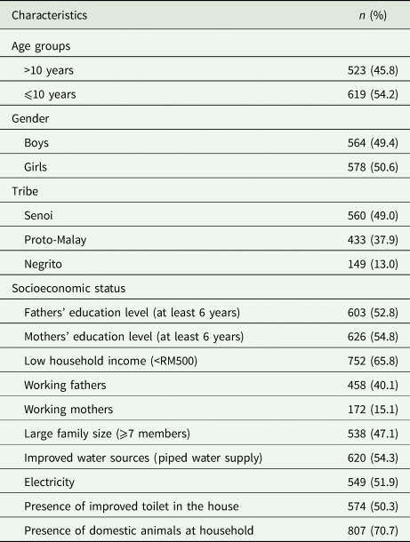
All values are number (%). RM, Malaysian Ringgit; US$1 = RM4.05.
Prevalence and distribution of S. stercoralis infections
Based on the above-described examination of the fecal samples for the presence of S. stercoralis larvae, 15.8% (180/1142) of the children were found to be infected with S. stercoralis. As shown in Fig. 2, the results indicated that the highest detection rate was achieved by using the Koga APC method (174/1142; 15.2%; 95% CI = 13.12–17.28), followed by PCR assay, which detected S. stercoralis in 157 samples (13.7%; 95% CI = 11.71–15.69); however, the difference in rates by these methods as well as the combined rate was not statistically significant (P > 0.05). On the other hand, S. stercoralis was detected in only 15 (1.3%; 95% CI = 0.64–1.96) and 2 (0.2%; 95% CI = −0.06–0.46) samples by FES and direct smear, respectively. Six samples that were found to be negative by agar culture, FES and direct smear were identified as positive by PCR assay.
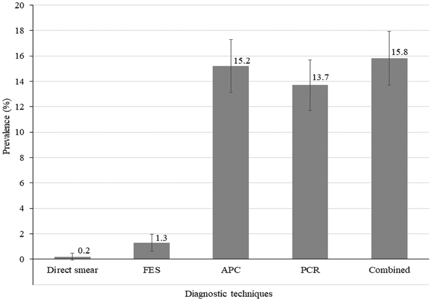
Fig. 2. Prevalence (%) of Strongyloides stercoralis identified by each technique, and by the combination of the four techniques among the participants (n = 1142). FES, formalin-ether sedimentation; APC, agar plate culture. Error bars represent 95% confidence interval of the proportion.
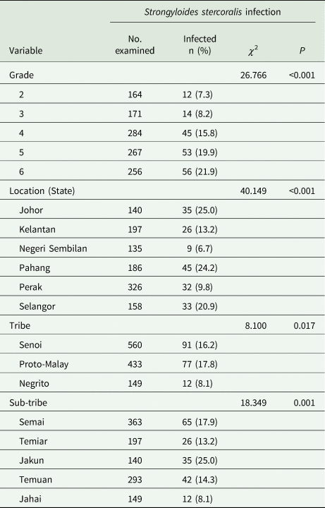
Table 2 shows the distribution of S. stercoralis among the participants. The prevalence of S. stercoralis infection increased significantly with age (χ 2 = 26.766; P < 0.001), with the highest prevalence reported among children in Grade 6, aged 12 years (21.9%), while the lowest prevalence was found among children in Grade 2, aged 8 years (7.3%). Also, the prevalence of S. stercoralis infection among boys was significantly higher than among girls (18.8 vs 12.8%; χ 2 = 7.718; P = 0.005). In addition, the prevalence of S. stercoralis infection differed significantly among the six states (χ 2 = 40.149; P < 0.001), with the highest prevalence of S. stercoralis infection found among children in the states of Johor (25%) and Pahang (24.2%) and the lowest found among children in Negeri Sembilan (6.7%). Moreover, the prevalence of S. stercoralis infection was significantly higher among children belonging to the Proto-Malay (17.8%) and Senoi (16.2%) tribes compared to those belonging to the Negrito tribe (8.1%) (χ 2 = 8.100; P = 0.017). Furthermore, children belonging to the Jakun sub-tribe of the Proto-Malay tribal group had the highest prevalence of infection (25.0%) compared to children of other sub-tribes (χ 2 = 18.349; P = 0.001).
Table 2. Distribution of Strongyloides stercoralis infection among Orang Asli schoolchildren in Malaysia (n = 1142)
Factors associated with S. stercoralis infection
Table 3 shows the result of the univariate analysis conducted to determine the association of S. stercoralis infection with the demographic, socioeconomic, housing and behavioural variables. The results showed that children aged >10 years had a significantly higher prevalence of strongyloidiasis than younger children (21.0 vs 11.3%; P < 0.001). Also, children of the Proto-Malay (17.8 vs 8.1%; P = 0.004) and Senoi (16.2 vs 8.1%; P = 0.012) tribes had a significantly higher strongyloidiasis prevalence when compared with children of the Negrito tribe. Similarly, a higher prevalence of strongyloidiasis was found among children who lived in houses without improved toilet facilities (19.7 vs 11.8%; P < 0.001) and children who used unimproved sources of drinking water (19.5 vs 12.6%; P = 0.001) when compared to their counterparts with improved toilet facilities and improved sources of drinking water. With regard to personal hygiene variables, it was found that children who practised indiscriminate/open defecation had a significantly higher prevalence of strongyloidiasis than those who always used toilets (18.5 vs 8.1%; P < 0.001). Similarly, the prevalence was significantly higher among children who did not wear shoes when outside their house (22.2 vs 12.3%; P < 0.001), and children who did not wash their hands after defecation (19.8 vs 14.2%; P = 0.020) compared to those who wore shoes or slippers and those who practised proper hand washing after defecation.
Table 3. Univariate analysis of factors associated with Strongyloides stercoralis infection among Orang Asli schoolchildren in Malaysia (n = 1142)
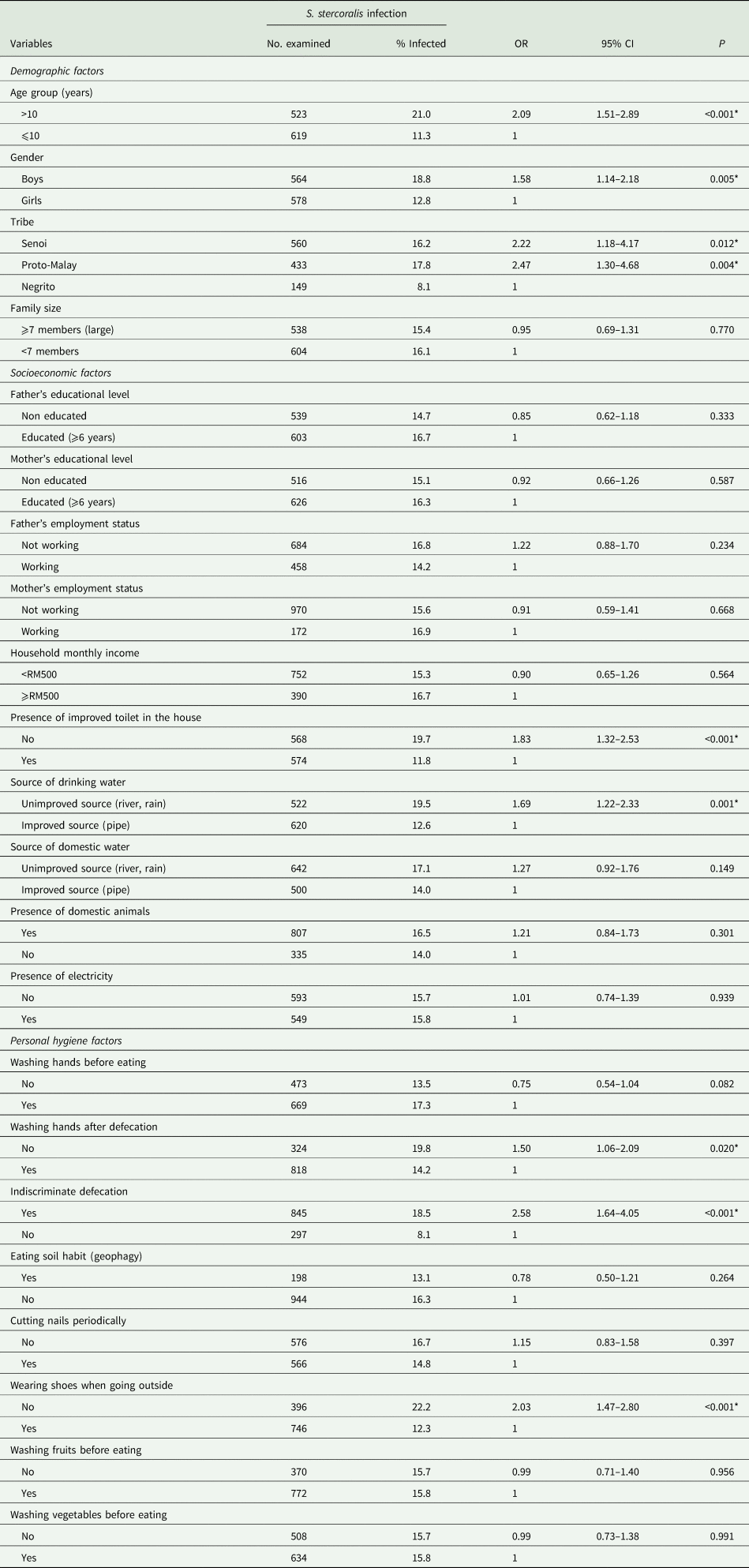
RM, Malaysian Ringgit; US$1 = RM4.05. OR, odds ratio. CI, confidence interval.
*Significant association (P < 0.05).
In the multivariate logistic regression analysis, six variables were identified as significant risk factors of S. stercoralis infection among Orang Asli schoolchildren (Table 4). The Hosmer–Lemeshow test showed that the model fit the data well [χ 2 = 6.789 (8 degrees of freedom); P = 0.560]. Children aged > 10 years and boys had higher odds of being infected by strongyloidiasis compared to younger children [adjusted odds ratio (aOR) = 1.91; 95% CI = 1.36–2.68] and girls (aOR = 1.52; 95% CI = 1.09–2.13), respectively. Also, children belonging to the Proto-Malay and Senoi tribes had 2.50 (95% CI = 1.28–4.85) and 1.93 (95% CI = 1.01–3.70) higher odds of contracting strongyloidiasis compared to children belonging to the Negrito tribe. Moreover, children who did not have improved sources of drinking water in their houses had 2.91 odds (95% CI = 1.50–5.66) of having strongyloidiasis compared to those who had safe piped sources of drinking water. In addition, practising indiscriminate/open defecation and not wearing shoes when outside the house increased the children's odds of having strongyloidiasis by 2.81 (95% CI = 1.75–4.50) and 1.91 times (95% CI = 1.37–2.67), respectively, compared to their counterparts.
Table 4. Multivariate analysis of factors associated with Strongyloides stercoralis infection among Orang Asli schoolchildren in Malaysia (n = 1142)
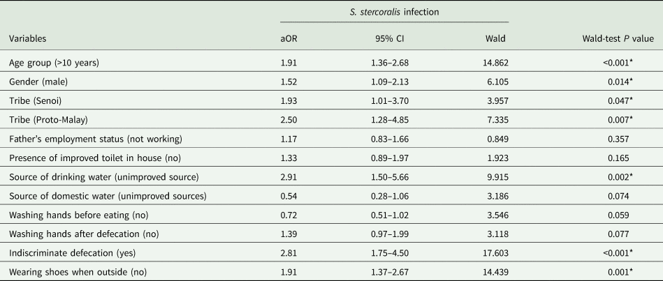
aOR, adjusted odds ratio. CI, confidence interval.
*Significant risk factors of S. stercoralis infection (P < 0.05).
Finally, the results of the PARF analysis showed that almost half (48.7%) of the strongyloidiasis cases among Orang Asli schoolchildren could be reduced if all children avoided indiscriminate/open defecation in rivers and surrounding areas. Moreover, the prevalence of strongyloidiasis could be reduced by 21.8% if the children in this population practised good standards of personal hygiene; particularly, wearing shoes or slippers when going or playing outside the house. In addition, 20.2% of the cases could be avoided if these children were provided with improved sources of drinking water in their houses.
Discussion
This study revealed a relatively high prevalence (15.8%; 180/1142) of strongyloidiasis among the study population, which consisted of Orang Asli schoolchildren in Peninsular Malaysia. The prevalence of this infection was detected by using different diagnostic techniques, namely the direct smear, FES, Koga APC and PCR. Hence, the current study provides reliable and comprehensive evidence on the occurrence of S. stercoralis among Orang Asli communities. Moreover, it is the first in Malaysia to use the Koga APC method on fecal samples.
The prevalence of strongyloidiasis reported by the current study is significantly higher than that reported in previous studies conducted in Malaysia. For instance, a recent community-based study that attempted to screen 236 participants in indigenous communities in Sarawak (East Malaysia) for S. stercoralis infection showed that 26 participants (11%) were seropositive for strongyloidiasis by ELISA assay, while none of the collected fecal samples was positive for S. stercoralis larvae by direct smear and FES (Ngui et al., Reference Ngui, Halim, Rajoo, Lim, Ambu, Rajoo, Chang, Woon and Mahmud2016). On the other hand, a recent study among Negrito communities reported that only seven out of 416 (1.7%) fecal samples were found positive for S. stercoralis; however, the methods used (direct smear, FES and Kato-Katz techniques) have a low sensitivity for S. stercoralis (Muslim et al., Reference Muslim, Mohd Sofian, Shaari, Hoh and Lim2019). By using PCR, the current study found 157 S. stercoralis-positive cases (equating to 13.7% of the participants), whereas only five and three cases were reported by previous studies in Sarawak and Selangor, respectively (Ahmad et al., Reference Ahmad, Hadip, Ngui, Lim and Mahmud2013; Ngui et al., Reference Ngui, Halim, Rajoo, Lim, Ambu, Rajoo, Chang, Woon and Mahmud2016). Using a pentaplex real-time PCR analysis, Basuni et al. revealed that 30 out of 77 (39%) individuals presenting with abdominal symptoms in two district hospitals in Sarawak, East Malaysia were positive for S. stercoralis (Basuni et al., Reference Basuni, Muhi, Othman, Verweij, Ahmad, Miswan, Rahumatullah, Aziz, Zainudin and Noordin2011).
In neighbouring Southeast Asian countries, higher prevalence rates of S. stercoralis infection have been reported in Cambodia (Forrer et al., Reference Forrer, Khieu, Schär, Vounatsou, Chammartin, Marti, Muth and Odermatt2018), Lao PDR (Vonghachack et al., Reference Vonghachack, Sayasone, Bouakhasith, Taisayavong, Akkavong and Odermatt2015) and Thailand (Laoraksawong et al., Reference Laoraksawong, Sanpool, Rodpai, Thanchomnang, Kanarkard, Maleewong, Kraiklang and Intapan2018), while much lower prevalence rates were reported in Indonesia (Wiria et al., Reference Wiria, Wammes, Hamid, Dekkers, Prasetyani, May, Kaisar, Verweij, Tamsma, Partono, Sartono, Supali, Yazdanbakhsh and Smit2013) and Myanmar (Aung et al., Reference Aung, Hino, Oo, Win, Maruyama, Htike and Nagayasu2018). Nevertheless, it is believed that strongyloidiasis is likely to have a much higher burden in Southeast Asia than currently indicated in the available literature (Schär et al., Reference Schär, Giardina, Khieu, Muth, Vounatsou, Marti and Odermatt2016) not least because the region exhibits conditions that favour high transmission of strongyloidiasis, such as humid and wet climates, inadequate sanitary conditions and poverty (Al-Mekhlafi et al., Reference Al-Mekhlafi, Lim, Moktar, Ngui, Lim and Vythilingam2013; Schär et al., Reference Schär, Trostdorf, Giardina, Khieu, Muth, Marti, Vounatsou and Odermatt2013).
Elsewhere, strongyloidiasis is hyperendemic among aboriginal populations in Australia, with a reported prevalence ranging from 35 to 60% (Kearns et al., Reference Kearns, Currie, Cheng, McCarthy, Carapetis, Holt, Page, Shield, Gundjirryirr, Mulholland, Ward and Andrews2017). In addition, endemic foci for S. stercoralis have been reported in Europe and North America (Asundi et al., Reference Asundi, Beliavsky, Liu, Akaberi, Schwarzer, Bisoffi, Requena-Méndez, Shrier and Greenaway2019). For instance, until 2018, strongyloidiasis was reported in a total of 1083 Spanish-born individuals without a history of travel to endemic areas (Barroso et al., Reference Barroso, Salvador, Sánchez-Montalvá, Bosch-Nicolau and Molina2019). Also, an earlier study in Canada revealed a prevalence rate of 76.6 and 11.8% among Cambodian and Vietnamese refugees, respectively (Gyorkos et al., Reference Gyorkos, Genta, Viens and MacLean1990).
In the current study, the APC method showed superior sensitivity (174 cases) in detecting S. stercoralis larvae in fecal samples compared to direct smear (two cases), FES (15 cases) and conventional PCR (157 cases), with six samples found positive only by PCR. These results are consistent with those reported elsewhere (Requena-Méndez et al., Reference Requena-Méndez, Chiodini, Bisoffi, Buonfrate, Gotuzzo and Muñoz2013; Amor et al., Reference Amor, Rodriguez, Saugar, Arroyo, López-Quintana, Abera, Yimer, Yizengaw, Zewdie, Ayehubizu, Hailu, Mulu, Echazú, Krolewieki, Aparicio, Herrador, Anegagrie and Benito2016; Aung et al., Reference Aung, Hino, Oo, Win, Maruyama, Htike and Nagayasu2018; Tuyizere et al., Reference Tuyizere, Ndayambaje, Walker, Bayingana, Ntirenganya, Dusabejambo and Hale2018). It is also interesting to note that the number of positive cases detected by the combined methods was not significantly different from using APC. Although 17 agar culture-positive fecal samples were found to be negative by PCR, these samples were confirmed by amplification of S. stercoralis larvae collected from the agar culture. The differing results by APC and PCR could be explained by the low intensity of the larvae in those samples, as shown by the agar culture, as well as the presence of fecal inhibitors that might not have been completely removed prior to PCR (Knopp et al., Reference Knopp, Salim, Schindler, Karagiannis Voules, Rothen, Lweno, Mohammed, Singo, Benninghoff, Nsojo, Genton and Daubenberger2014; Requena-Méndez et al., Reference Requena-Mendez, Buonfrate, Bisoffi and Gutiérrez2014).
The current study investigated the possible risk factors of S. stercoralis infection among the study participants and revealed three sets of key risk factors; (1) demographic: the age of >10 years, tribe (Senoi and Proto-Malay) and male gender; (2) socioeconomic: using an unimproved water source for drinking water; and (3) behavioural: practising indiscriminate defecation and not wearing shoes when outside the house. The age-related findings of the current study showed that children aged >10 years are generally at a higher risk of S. stercoralis infection compared to younger children, which is consistent with the general conclusion drawn in prior research (Schär et al., Reference Schär, Trostdorf, Giardina, Khieu, Muth, Marti, Vounatsou and Odermatt2013). This finding is also in agreement with many previous studies that have linked this infection to increasing age because S. stercoralis can be sustained in infected individuals for decades by means of autoinfection (Prendki et al., Reference Prendki, Fenaux, Durand, Thellier and Bouchaud2011; Conlan et al., Reference Conlan, Khamlome, Vongxay, Elliot, Pallant, Sripa, Blacksell, Fenwick and Thompson2012; Khieu et al., Reference Khieu, Schär, Marti, Bless, Char, Muth and Odermatt2014; Aung et al., Reference Aung, Hino, Oo, Win, Maruyama, Htike and Nagayasu2018). Hence, a higher prevalence of S. stercoralis infection is expected among adult individuals in Orang Asli communities. However, further studies are required to confirm this conjecture.
The results of the current study also showed that boys were more prone to carry the infection than girls, which is consistent with previous studies in different countries (Steinmann et al., Reference Steinmann, Zhou, Du, Jiang, Wang, Wang, Li, Marti and Utzinger2007; Conlan et al., Reference Conlan, Khamlome, Vongxay, Elliot, Pallant, Sripa, Blacksell, Fenwick and Thompson2012; Khieu et al., Reference Khieu, Schär, Marti, Bless, Char, Muth and Odermatt2014; Tuyizere et al., Reference Tuyizere, Ndayambaje, Walker, Bayingana, Ntirenganya, Dusabejambo and Hale2018; Gétaz et al., Reference Gétaz, Castro, Zamora, Kramer, Gareca, Torrico-Espinoza, Macias, Lisarazu-Velásquez, Rodriguez, Valencia-Rivero, Perneger and Chappuis2019). Interestingly, previous studies have reported a consistent male-bias in helminth infections, particularly hookworm infection, and have suggested sex-related differences in susceptibility to infection arising from immunosuppression associated with male hormones (Poulin, Reference Poulin1996; Moore and Wilson, Reference Moore and Wilson2002; Brooker et al., Reference Brooker, Bethony and Hotez2004). However, the reported sex-dependent difference could be also attributed to higher exposure among males who are more involved in outdoor activities such as playing football, swimming in streams or ponds as well as in helping their parents in farming activities.
The findings also showed that children belonging to the Proto-Malay (17.8%) and Senoi (16.2%) tribes were more likely to be infected with S. stercoralis compared to those in the Negrito tribe (8.1%). Indeed, previous studies have shown that IPIs other than S. stercoralis are prevalent to varying degrees in all Orang Asli communities throughout Peninsular Malaysia. For instance, the prevalence of Trichuris trichiura infection is significantly higher among the Negrito tribe and that of the Ascaris lumbricoides infection is significantly higher among the Senoi tribe, whereas the prevalence of hookworm infection is comparable among the tribes (Anuar et al., Reference Anuar, Salleh and Moktar2014). A similar profile of infections was also found according to location, with Pahang and Selangor states having the highest prevalence. As S. stercoralis was reported in all targeted states, the significant differences could be explained by the different socioeconomic status and behavioural factors among the tribes rather than climatic or environmental factors. Most of the Senoi and Proto-Malay tribes including the Jakun, Semai, Temiar and Temuan communities practise permanent agriculture and manage their own rubber, oil palm or cocoa farms or engage in ‘shifting cultivation’ (hill rice cultivation), in which human/animal fecal materials are used as fertilizer (night soil). They also prefer to live in remote areas located deep in the jungle, which have inadequate sanitary facilities and are far away from healthcare facilities (Masron et al., Reference Masron, Masami and Ismail2013; Choy et al., Reference Choy, Al-Mekhlafi, Mahdy, Nasr, Sulaiman, Lim and Surin2014). These conditions provide the ideal ecological and economic setting for a high burden of S. stercoralis infection, and this may explain the significantly higher prevalence among these tribes compared to the Negrito tribe.
With regards to the socioeconomic and behavioural risk factors of S. stercoralis infection, this study found that children who practised indiscriminate defecation used an unimproved water source for drinking water, and walked barefooted when outside the house were more likely to be infected than their counterparts. These findings are consistent with those of previous studies in other Asian countries (Khieu et al., Reference Khieu, Schär, Marti, Bless, Char, Muth and Odermatt2014; Senephansiri et al., Reference Senephansiri, Laummaunwai, Laymanivong and Boonmar2017; Aung et al., Reference Aung, Hino, Oo, Win, Maruyama, Htike and Nagayasu2018). Moreover, several studies have identified poor personal hygienic practices as significant predictors of IPIs among Orang Asli populations (Al-Mekhlafi et al., Reference Al-Mekhlafi, Azlin, Nor Aini, Shaikh, Sa'iah, Fatmah, Ismail, Firdaus, Aisah, Rozlida and Norhayati2006; Ahmed et al., Reference Ahmed, Al-Mekhlafi, Al-Adhroey, Ithoi, Abdulsalam and Surin2012; Anuar et al., Reference Anuar, Al-Mekhlafi, Abdul Ghani, Abu Bakar, Azreen, Salleh, Ghazali, Bernadus and Moktar2012; Elyana et al., Reference Elyana, Al-Mekhlafi, Ithoi, Abdulsalam, Dawaki, Nasr, Atroosh, Abd-Basher, Al-Areeqi, Sady, Subramaniam, Anuar, Lau, Moktar and Surin2016; Muslim et al., Reference Muslim, Mohd Sofian, Shaari, Hoh and Lim2019).
Given the fact that the filariform infective larvae of S. stercoralis are found in soil and mainly infect humans through skin penetration, it is not particularly surprising that the result of the PARF analysis showed that wearing shoes when outside the house will help in preventing strongyloidiasis. However, this study showed that using an unimproved water source for drinking water (aOR = 2.91) and indiscriminate defecation (aOR = 2.81) was the most significant risk factors of S. stercoralis infection. Based on the PARF results, about half (48.7%) and one fifth (21.7%) of the strongyloidiasis cases could be prevented if all children frequently used improved toilets for defecation and had an improved source for drinking water in their households, respectively. In Malaysia, rivers are considered the lifeblood of Orang Asli populations and are still the main water source for drinking water and for domestic use (e.g. bathing, washing and swimming). Unfortunately, rivers are also the preferred site for defecation, particularly among Orang Asli children who have also been noted to defecate indiscriminately close to their houses or within the village confines (Al-Mekhlafi et al., Reference Al-Mekhlafi, Surin, Atiya, Ariffin, Mahdy and Abdullah2008; Elyana et al., Reference Elyana, Al-Mekhlafi, Ithoi, Abdulsalam, Dawaki, Nasr, Atroosh, Abd-Basher, Al-Areeqi, Sady, Subramaniam, Anuar, Lau, Moktar and Surin2016). Young children commonly select shallow areas of rivers and streams where the water flows more slowly so they can sit, individually or in small groups, to defecate and then cleanse themselves after defecation. Thus, this untreated water is likely to be highly contaminated with intestinal parasitic ova, cysts, oocysts and/or larvae (Lim and Ahmad, Reference Lim and Ahmad2004; Lee et al., Reference Lee, Ngui, Tan, Roslan, Ithoi and Lim2014). In such epidemiological situation, water, sanitation and hygiene may have a crucial role in the prevention and control of STH infections, including strongyloidiasis.
Although not found to be significant in this study, contact with domestic animals (mainly dogs) has also been identified as a significant risk factor of S. stercoralis infection, and thus zoonotic strongyloidiasis, with dogs as reservoirs, has been suggested (Thamsborg et al., Reference Thamsborg, Ketzis, Horii and Matthews2017). Moreover, clinical and subclinical cases of S. stercoralis infection have been increasingly reported among dogs in Europe (Paradies et al., Reference Paradies, Iarussi, Sasanelli, Capogna, Lia, Zucca, Greco, Cantacessi and Otranto2017; Iatta et al., Reference Iatta, Buonfrate, Paradies, Cavalera, Capogna, Iarussi, Šlapeta, Giorli, Trerotoli, Bisoffi and Otranto2019). Similarly, dogs in rural Cambodia have been found to carry two populations of S. stercoralis, one of which is shared with humans (Jaleta et al., Reference Jaleta, Zhou, Bemm, Schär, Khieu, Muth, Odermatt, Lok and Streit2017). In Malaysia, S. stercoralis larvae have been detected in fecal samples collected from dogs in rural Orang Asli communities and in soil samples collected from an urban area in Kuala Lumpur (Azian et al., Reference Azian, Sakhone, Hakim, Yusri, Nurulsyamzawaty, Zuhaizam, Rodi and Maslawaty2008). Interestingly, larvae of Strongyloides spp. have been detected in domestic pig-tailed macaques working in the harvesting of coconuts in Kelantan state (Choong et al., Reference Choong, Mimi Armiladiana, Ruhil and Peng2019). Furthermore, S. stercoralis larvae have been detected in common vegetables and herbs in the city of Kota Bharu in Kelantan (Zeehaida et al., Reference Zeehaida, Zairi, Rahmah, Maimunah and Madihah2011).
Based on current and previous findings, it is possible to suggest a number of different scenarios for the transmission of S. stercoralis infection among the studied Orang Asli children. First, filariform larvae in the soil can penetrate the intact skin of the feet when children walk or play barefooted or help their parents in farms fertilized by human and animal excreta. Second, while defecating in the river, filariform larvae in the water may penetrate the exposed skin of the feet, legs, anal or buttock area and hands. Third, recalling some valiant self-experimentation in the early 1900s to induce S. stercoralis infection by oral ingestion of larvae in water (Grove, Reference Grove1996), water-borne transmission by ingesting the larvae in untreated drinking water collected from rivers may occur among these children. Fourth, food-borne transmission through the ingestion of larvae in contaminated vegetables or fruit may also take place. However, except for the first scenario, which has been documented as the exclusive mode of transmission, further studies are required to confirm the other suggested scenarios.
The strengths of this study include the screening of a large number of children (n = 1142) from the main three Orang Asli tribes and the use of a combination of screening methods to increase the sensitivity of S. stercoralis diagnosis. However, we also acknowledge some limitations that should be considered when interpreting the results of this study. For instance, the study design (i.e. cross-sectional) limited our ability to confirm the existence of a causal association between S. stercoralis infection and the reported significant risk factors. Moreover, only one fecal sample from each participant was examined for S. stercoralis instead of the three repeated samples due to the level of cooperation of the children and the cultural beliefs of the Orang Asli that oppose the giving of fecal samples. Previous studies have shown that there is a high risk that a single-sample examination will miss an infection because of the day-to-day variation in larval excretion, particularly in asymptomatic light S. stercoralis infections (Dreyer et al., Reference Dreyer, Fernandes-Silva, Alves, Rocha, Albuquerque and Addiss1996; Uparanukraw et al., Reference Uparanukraw, Phongsri and Morakote1999). Thus, the overall prevalence of S. stercoralis infection reported by the current study is likely to have been significantly underestimated.
In conclusion, this study revealed that S. stercoralis infection is prevalent among Orang Asli schoolchildren in Malaysia, particularly among Proto-Malay and Senoi tribes, and is thus a matter of serious concern. An age of >10 years, the male gender, indiscriminate defecation, using unimproved sources for drinking water and not wearing shoes when outside were identified as the significant risk factors of infection in the studied population. Orang Asli populations in Peninsular Malaysia share similar epidemiological characteristics. Thus, the findings reported herein can be generalized to Orang Asli schoolchildren in other communities that were not included in this study. However, there is still a need for further studies among preschool children and adults.
Orang Asli children are vulnerable to many infectious diseases and can be considered partially immunocompromised as a result of a high level of malnutrition (Al-Mekhlafi et al., Reference Al-Mekhlafi, Azlin, Aini, Shaikh, Sa'iah, Fatmah, Ismail, Ahmad, Aisah, Rozlida and Norhayati2005; Khor and Zalilah, Reference Khor and Zalilah2008; Ahmed et al., Reference Ahmed, Al-Mekhlafi, Al-Adhroey, Ithoi, Abdulsalam and Surin2012). In such a situation, S. stercoralis infection may have severe consequences. Therefore, access to adequate diagnosis and treatment of strongyloidiasis is urgently needed and should be a public health priority for Orang Asli population. Moreover, specific measures to control this infection should be implemented in any efforts to improve the quality of life of the Orang Asli population as a whole. Proper health education regarding good personal hygiene, good sanitary practices, provision of improved water supply and adequate sanitation, as well as the implementation of periodic mass drug administration, will help in reducing the prevalence of S. stercoralis infection in these communities.
Acknowledgements
The authors wish to acknowledge the Department of Orang Asli Development (JAKOA), Ministry of Rural and Regional Development, Kuala Lumpur, Malaysia and the Departments of Education in the targeted states for their generous collaboration during this study. The authors also would like to express their appreciation to the support and cooperation of the headmasters, teachers and staff of the targeted schools. Special thanks also go to the children and their parents/guardians for their voluntary participation in this study.
Financial support
The work presented in this paper was funded by the University of Malaya High Impact Research Fund, HIR-MOHE (H-20001-00-E00051), and also by the University of Malaya Research Grants (RG331-15AFR). The funders had no role in study design, data collection and analysis, decision to publish, or preparation of the manuscript.
Conflict of interest
None.
Ethical standards
This study was carried out according to the guidelines laid down in the Declaration of Helsinki. The study protocol was approved by the Medical Ethics Committee of the University of Malaya Medical Centre, Malaysia (Reference number: 201731-4985). Before recruiting participants, meetings were held with the headmasters and teachers of schools to provide important information about the aims and methods of this study and their consents were obtained. In addition, the research team visited Orang Asli communities in the targeted areas and met the heads of the villages and the parents of schoolchildren to inform them about the objectives and to request the involvement of their children in the study. They were informed that the involvement of their children in this study was totally voluntary and that they have the right to withdraw from the study at any point without having to give a reason. Written and signed or thumb-printed informed consents were collected from parents or guardians on behalf of their children before the commencement of the survey.


