Articles
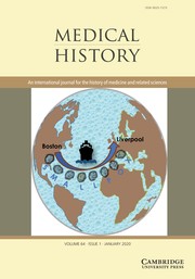
An Analysis of the United States and United Kingdom Smallpox Epidemics (1901–5) – The Special Relationship that Tested Public Health Strategies for Disease Control
-
- Published online by Cambridge University Press:
- 19 December 2019, pp. 1-31
-
- Article
-
- You have access
- Open access
- HTML
- Export citation
‘To Awaken the Medical and Hygienic Conscience of the People’: Cultivating Enlightened Citizenship through Free Public Healthcare in Haiti from 1915–34
-
- Published online by Cambridge University Press:
- 19 December 2019, pp. 32-51
-
- Article
-
- You have access
- HTML
- Export citation
Still Controversial: Early Detection and Screening for Breast Cancer in Brazil, 1950–2010s
-
- Published online by Cambridge University Press:
- 19 December 2019, pp. 52-70
-
- Article
-
- You have access
- HTML
- Export citation
Re-assessing the Foundations: Worldwide Smallpox Eradication, 1957–67
-
- Published online by Cambridge University Press:
- 19 December 2019, pp. 71-93
-
- Article
-
- You have access
- Open access
- HTML
- Export citation
Between Donor Interest, Global Models and Local Conditions: Treatment and Decision-Making in the Somalia-Finland Tuberculosis Control Project, 1981–3
-
- Published online by Cambridge University Press:
- 19 December 2019, pp. 94-115
-
- Article
-
- You have access
- HTML
- Export citation
Photograph as Skin, Skin as Wax: Indexicality and the Visualisation of Syphilis in Fin-de-Siècle France The William Bynum Prize Essay
- Part of:
-
- Published online by Cambridge University Press:
- 19 December 2019, pp. 116-141
-
- Article
-
- You have access
- HTML
- Export citation
Book Review
Cynthia Carson Bisbee, Paul Bisbee, Erika Dyck, Patrick Farrell, James Sexton and James W. Spisak (eds), Psychedelic Prophets: The Letters of Aldous Huxley and Humphry Osmond (Montreal and Kingston: McGill-Queen’s University Press, 2018), pp. lxxix + 644, $75.00 CAD, hardback, ISBN: 9780773555068.
-
- Published online by Cambridge University Press:
- 19 December 2019, pp. 142-143
-
- Article
-
- You have access
- HTML
- Export citation
Christopher E. Forth, Fat: A Cultural History of the Stuff of Life (London: Reaktion Books, 2019), pp. 360, £25, hardback, ISBN: 9781789140620.
-
- Published online by Cambridge University Press:
- 19 December 2019, pp. 144-145
-
- Article
-
- You have access
- HTML
- Export citation
María Jesús Santesmases, The Circulation of Penicillin in Spain: Health, Wealth and Authority (Cham, Switzerland: Palgrave McMillan, 2018), pp. xi + 239, €78, ebook, ISBN: 9783319697185.
-
- Published online by Cambridge University Press:
- 19 December 2019, pp. 145-147
-
- Article
-
- You have access
- HTML
- Export citation
Lucas Richert, Strange Trips: Science, Culture and the Regulation of Drugs (Montreal and Kingston: McGill-Queen’s University Press, 2019), pp. xii + 247, $34.95 CAD, paperback, ISBN: 978773556379.
-
- Published online by Cambridge University Press:
- 19 December 2019, pp. 147-148
-
- Article
-
- You have access
- HTML
- Export citation
Katerina Gardikas, Landscapes of Disease: Malaria in Modern Greece (Budapest: Central European University Press, 2018), pp. 348, £48, hardback, ISBN: 9786155211980.
-
- Published online by Cambridge University Press:
- 19 December 2019, pp. 148-150
-
- Article
-
- You have access
- HTML
- Export citation
Hugh Cagle, Assembling the Tropics: Science and Medicine in Portugal’s Empire, 1450–1700 (Cambridge: Cambridge University Press, 2018), pp. 382, £35.99, hardback, ISBN: 9781107196636.
-
- Published online by Cambridge University Press:
- 19 December 2019, pp. 150-152
-
- Article
-
- You have access
- HTML
- Export citation
Keith Andrew Stewart, Galen’s Theory of Black Bile: Hippocratic Tradition, Manipulation, Innovation, Studies in Ancient Medicine, vol. 51 (Leiden: Brill, 2018), pp. x + 178, € 94/US$113, hardback, ISBN: 9789004382787.
-
- Published online by Cambridge University Press:
- 19 December 2019, pp. 152-154
-
- Article
-
- You have access
- HTML
- Export citation
Jim Kristofic, Medicine Women: The Story of the First Native American Nursing School (Albuquerque, New Mexico: University of New Mexico Press, 2019), pp. xvi + 396, $36.95, paperback, ISBN: 9780826360670.
-
- Published online by Cambridge University Press:
- 19 December 2019, pp. 154-155
-
- Article
-
- You have access
- HTML
- Export citation
Wendee M. Wechsberg (ed.), HIV Pioneers: Lives Lost, Careers Changed and Survival (Baltimore, MD: Johns Hopkins University Press, 2018), pp. 272, $32.95, paperback, ISBN: 9781421425726. - Lukas Engelmann, Mapping Aids: Visual Histories of an Enduring Epidemic (Cambridge: Cambridge University Press, 2018), pp. 266, £75, hardback, ISBN: 9781108425773.
-
- Published online by Cambridge University Press:
- 19 December 2019, pp. 156-158
-
- Article
-
- You have access
- HTML
- Export citation
Books also Received
Books also Received
-
- Published online by Cambridge University Press:
- 19 December 2019, p. 159
-
- Article
-
- You have access
- HTML
- Export citation
Media Review
Bilingual Exhibition Auf Messers Schneide. Der Chirurg Ferdinand Sauerbruch zwischen Medizin und Mythos [On a Knife-edge. Surgeon Ferdinand Sauerbruch between Medicine and Myth] (22.3.2019–2.2.2020) + accompanying programme + same titled exhibition catalogue (German), Berlin, Medizinhistorisches Museum der Charité, Germany
-
- Published online by Cambridge University Press:
- 19 December 2019, pp. 160-162
-
- Article
-
- You have access
- HTML
- Export citation
Front Cover (OFC, IFC) and matter
MDH volume 64 Issue 1 Cover and Front matter
-
- Published online by Cambridge University Press:
- 19 December 2019, pp. f1-f2
-
- Article
-
- You have access
- Export citation
Back Cover (OBC, IBC) and matter
MDH volume 64 Issue 1 Cover and Back matter
-
- Published online by Cambridge University Press:
- 19 December 2019, pp. b1-b4
-
- Article
-
- You have access
- Export citation

