Hostname: page-component-586b7cd67f-2plfb Total loading time: 0 Render date: 2024-11-28T13:14:21.499Z Has data issue: false hasContentIssue false

Stimuli-responsive liquid crystal elastomers for dynamic cell culture

Published online by Cambridge University Press:  10 February 2015

Aditya Agrawal
Affiliation:
Department of Chemical and Biomolecular Engineering, William Marsh Rice University, Houston, Texas 77005, USA
Oluwatomiyin Adetiba
Affiliation:
Department of Bioengineering, William Marsh Rice University, Houston, Texas 77005, USA
Hojin Kim
Affiliation:
Department of Chemical and Biomolecular Engineering, William Marsh Rice University, Houston, Texas 77005, USA
Huiying Chen
Affiliation:
Department of Bioengineering, William Marsh Rice University, Houston, Texas 77005, USA
Jeffrey G. Jacot*
Affiliation:
Department of Bioengineering, William Marsh Rice University, Houston, Texas 77005, USA; and Division of Congenital Heart Surgery, Texas Children's Hospital, Congenital Heart Surgery Services, Houston, Texas 77030, USA
Rafael Verduzco*
Affiliation:
Department of Chemical and Biomolecular Engineering, William Marsh Rice University, Houston, Texas 77005, USA
*
a)Address all correspondence to these authors. e-mail: [email protected]
Get access

Abstract

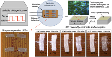
Responsive, biocompatible substrates are of interest for directing the maturation and function of cells in vitro during cell culture. This can potentially provide cells and tissues with desirable properties for regenerative therapies. Here, we demonstrate a straightforward and scalable approach to attach, align, and dynamically load cardiomyocytes on responsive liquid crystal elastomer (LCE) substrates. Monodomain LCEs exhibit reversible shape changes in response to cyclic heating, and when immersed in an aqueous medium on top of resistive heaters, shape changes are fast, reversible, and produce minimal temperature changes in the surroundings. We systematically characterized the strain response of LCEs in water and demonstrated the attachment and alignment of neonatal rat ventricular myocytes on LCE substrates. Cardiomyocytes attached to both static and stimulated LCE substrates, and under cyclic stimulation, cardiomyocytes aligned along the primary direction of strain. This work demonstrates the potential of LCEs as stimuli-responsive substrates for dynamic cell culture.

Type
Invited Feature Papers
Copyright
Copyright © Materials Research Society 2015 

Access options

Get access to the full version of this content by using one of the access options below. (Log in options will check for institutional or personal access. Content may require purchase if you do not have access.)

References

REFERENCES

Li, Y., Huang, G., Zhang, X., Wang, L., Du, Y., Lu, T.J., and Xu, F.: Engineering cell alignment in vitro. Biotechnol. Adv. 32, 347 (2014).CrossRefGoogle ScholarPubMed
Helm, P., Beg, M.F., Miller, M.I., and Winslow, R.L.: Measuring and mapping cardiac fiber and laminar architecture using diffusion tensor MR imaging. Ann. N. Y. Acad. Sci. 1047, 296 (2005).Google Scholar
Nikkhah, M., Edalat, F., Manoucheri, S., and Khademhosseini, A.: Engineering microscale topographies to control the cell–substrate interface. Biomaterials 33, 5230 (2012).Google Scholar
Neidlinger-Wilke, C., Grood, E.S., Wang, J.H-C., Brand, R.A., and Claes, L.: Cell alignment is induced by cyclic changes in cell length: Studies of cells grown in cyclically stretched substrates. J. Orthop. Res. 19, 286 (2001).Google Scholar
Berthiaume, F., Maguire, T.J., and Yarmush, M.L.: Tissue engineering and regenerative medicine: History, progress, and challenges. Annu. Rev. Chem. Biomol. Eng. 2, 403 (2011).CrossRefGoogle ScholarPubMed
Eschenhagen, T. and Zimmermann, W.H.: Engineering myocardial tissue. Circ. Res. 97, 1220 (2005).CrossRefGoogle ScholarPubMed
Zimmermann, W-H., Schneiderbanger, K., Schubert, P., Didié, M., Münzel, F., Heubach, J.F., Kostin, S., Neuhuber, W.L., and Eschenhagen, T.: Tissue engineering of a differentiated cardiac muscle construct. Circ. Res. 90, 223 (2002).CrossRefGoogle ScholarPubMed
Black, L.D., Meyers, J.D., Weinbaum, J.S., Shvelidze, Y.A., and Tranquillo, R.T.: Cell-induced alignment augments twitch force in fibrin gel-based engineered myocardium via gap junction modification. Tissue Eng., Part A 15, 3099 (2009).CrossRefGoogle ScholarPubMed
Lee, A.A., Delhaas, T., Waldman, L.K., MacKenna, D.A., Villarreal, F.J., and McCulloch, A.D.: An equibiaxial strain system for cultured cells. Am. J. Physiol.: Cell Physiol. 271, C1400 (1996).Google Scholar
Gopalan, S.M., Flaim, C., Bhatia, S.N., Hoshijima, M., Knoell, R., Chien, K.R., Omens, J.H., and McCulloch, A.D.: Anisotropic stretch-induced hypertrophy in neonatal ventricular myocytes micropatterned on deformable elastomers. Biotechnol. Bioeng. 81, 578 (2003).Google Scholar
Garvin, J., Qi, J., Maloney, M., and Banes, A.J.: Novel system for engineering bioartificial tendons and application of mechanical load. Tissue Eng. 9, 967 (2003).CrossRefGoogle ScholarPubMed
Pang, Y., Wang, X., Lee, D., and Greisler, H.P.: Dynamic quantitative visualization of single cell alignment and migration and matrix remodeling in 3-D collagen hydrogels under mechanical force. Biomaterials 32, 3776 (2011).Google Scholar
Guan, J., Wang, F., Li, Z., Chen, J., Guo, X., Liao, J., and Moldovan, N.I.: The stimulation of the cardiac differentiation of mesenchymal stem cells in tissue constructs that mimic myocardium structure and biomechanics. Biomaterials 32, 5568 (2011).CrossRefGoogle ScholarPubMed
Emmert, M.Y., Hitchcock, R.W., and Hoerstrup, S.P.: Cell therapy, 3D culture systems and tissue engineering for cardiac regeneration. Adv. Drug Delivery Rev. 6970, 254 (2014).CrossRefGoogle ScholarPubMed
Furuta, A., Miyoshi, S., Itabashi, Y., Shimizu, T., Kira, S., Hayakawa, K., Nishiyama, N., Tanimoto, K., Hagiwara, Y., Satoh, T., Fukuda, K., Okano, T., and Ogawa, S.: Pulsatile cardiac tissue grafts using a novel three-dimensional cell sheet manipulation technique functionally integrates with the host heart, in vivo. Circ. Res. 98, 705 (2006).CrossRefGoogle ScholarPubMed
Sawa, Y., Miyagawa, S., Sakaguchi, T., Fujita, T., Matsuyama, A., Saito, A., Shimizu, T., and Okano, T.: Tissue engineered myoblast sheets improved cardiac function sufficiently to discontinue LVAS in a patient with DCM: Report of a case. Surg. Today 42, 181 (2012).Google Scholar
Davis, K.A., Burke, K.A., Mather, P.T., and Henderson, J.H.: Dynamic cell behavior on shape memory polymer substrates. Biomaterials 32, 2285 (2011).CrossRefGoogle ScholarPubMed
Yang, P., Baker, R.M., Henderson, J.H., and Mather, P.T.: In vitro wrinkle formation via shape memory dynamically aligns adherent cells. Soft Matter 9, 4705 (2013).CrossRefGoogle Scholar
Mayer, M., Rabindranath, R., Börner, J., Hörner, E., Bentz, A., Salgado, J., Han, H., Böse, H., Probst, J., Shamonin, M., Monkman, G.J., and Schlunck, G.: Ultra-soft PDMS-based magnetoactive elastomers as dynamic cell culture substrata. PLoS One 8, e76196 (2013).CrossRefGoogle ScholarPubMed
Mather, P.T., Luo, X., and Rousseau, I.A.: Shape memory polymer research. Annu. Rev. Mater. Res. 39, 445 (2009).CrossRefGoogle Scholar
Small, W. IV, Singhal, P., Wilson, T.S., and Maitland, D.J.: Biomedical applications of thermally activated shape memory polymers. J. Mater. Chem. 20, 3356 (2010).Google Scholar
Rickert, D., Lendlein, A., Peters, I., Moses, M.A., and Franke, R-P.: Biocompatibility testing of novel multifunctional polymeric biomaterials for tissue engineering applications in head and neck surgery: An overview. Eur. Arch. Oto-Rhino-Laryngol Head Neck 263, 215 (2006).Google Scholar
Chen, Q., Liang, S., and Thouas, G.A.: Elastomeric biomaterials for tissue engineering. Prog. Polym. Sci. 38, 584 (2013).CrossRefGoogle Scholar
Mano, J.F.: Stimuli-responsive polymeric systems for biomedical applications. Adv. Eng. Mater. 10, 515 (2008).Google Scholar
Ratna, D. and Karger-Kocsis, J.: Recent advances in shape memory polymers and composites: A review. J. Mater. Sci. 43, 254 (2008).Google Scholar
Biggs, J., Danielmeier, K., Hitzbleck, J., Krause, J., Kridl, T., Nowak, S., Orselli, E., Quan, X., Schapeler, D., Sutherland, W., and Wagner, J.: Electroactive polymers: Developments of and perspectives for dielectric elastomers. Angew. Chem., Int. Ed. 52, 9409 (2013).Google Scholar
Warner, M. and Terentjev, E.M.: Liquid Crystal Elastomers (Oxford University Press, Oxford, England, 2003).Google Scholar
Jiang, H., Li, C., and Huang, X.: Actuators based on liquid crystalline elastomer materials. Nanoscale 5, 5225 (2013).Google Scholar
Ohm, C., Brehmer, M., and Zentel, R.: Liquid crystalline elastomers as actuators and sensors. Adv. Mater. 22, 3366 (2010).CrossRefGoogle ScholarPubMed
Ali, S.A., Al-Muallem, H.A., Rahman, S.U., and Saeed, M.T.: Bis-isoxazolidines: A new class of corrosion inhibitors of mild steel in acidic media. Corros. Sci. 50, 3070 (2008).Google Scholar
Agrawal, A., Luchette, P., Palffy-Muhoray, P., Biswal, S.L., Chapman, W.G., and Verduzco, R.: Surface wrinkling in liquid crystal elastomers. Soft Matter 8, 7138 (2012).Google Scholar
Küpfer, J. and Finkelmann, H.: Nematic liquid single crystal elastomers. Macromol. Chem. Rapid Commun. 12, 717 (1991).Google Scholar
Mark, J.E.: Polymer Data Handbook (Oxford University Press, New York, USA, 1999).Google Scholar
Agrawal, A., Yun, T., Pesek, S.L., Chapman, W.G., and Verduzco, R.: Shape-responsive liquid crystal elastomer bilayers. Soft Matter 10, 1411 (2014).Google Scholar
Komuro, I., Kaida, T., Shibazaki, Y., Kurabayashi, M., Katoh, Y., Hoh, E., Takaku, F., and Yazaki, Y.: Stretching cardiac myocytes stimulates protooncogene expression. J. Biol. Chem. 265, 3595 (1990).CrossRefGoogle ScholarPubMed
Yamazaki, T., Komuro, I., Kudoh, S., Zou, Y., Shiojima, I., Mizuno, T., Takano, H., Hiroi, Y., Ueki, K., and Tobe, K.: Mechanical stress activates protein kinase cascade of phosphorylation in neonatal rat cardiac myocytes. J. Clin. Invest. 96, 438 (1995).Google Scholar
Fink, C., Ergün, S., Kralisch, D., Remmers, U., Weil, J., and Eschenhagen, T.: Chronic stretch of engineered heart tissue induces hypertrophy and functional improvement. FASEB J. 14, 669 (2000).Google Scholar
Mansour, H., de Tombe, P.P., Samarel, A.M., and Russell, B.: Restoration of resting sarcomere length after uniaxial static strain is regulated by protein kinase C epsilon and focal adhesion kinase. Circ. Res. 94, 642 (2004).Google Scholar
Yu, J-G. and Russell, B.: Cardiomyocyte remodeling and sarcomere addition after uniaxial static strain in vitro. J. Histochem. Cytochem. 53, 839 (2005).Google Scholar
Chambers, M., Finkelmann, H., Remškar, M., Sánchez-Ferrer, A., Zalar, B., and Žumer, S.: Liquid crystal elastomer–nanoparticle systems for actuation. J. Mater. Chem. 19, 1524 (2009).Google Scholar
Chambers, M., Zalar, B., Remskar, M., Zumer, S., and Finkelmann, H.: Actuation of liquid crystal elastomers reprocessed with carbon nanoparticles. Appl. Phys. Lett. 89, 243116 (2006).CrossRefGoogle Scholar
Greco, F., Domenici, V., Desii, A., Sinibaldi, E., Zupančič, B., Zalar, B., Mazzolai, B., and Mattoli, V.: Liquid single crystal elastomer/conducting polymer bilayer composite actuator: Modelling and experiments. Soft Matter 9, 11405 (2013).Google Scholar
Kohlmeyer, R.R. and Chen, J.: Wavelength-selective, IR light-driven hinges based on liquid crystalline elastomer composites. Angew. Chem., Int. Ed. 52, 9234 (2013).Google Scholar
Ahir, S.V. and Terentjev, E.M.: Photomechanical actuation in polymer-nanotube composites. Nat. Mater. 4, 491 (2005).Google Scholar
Ahir, S.V., Squires, A.M., Tajbakhsh, A.R., and Terentjev, E.M.: Infrared actuation in aligned polymer-nanotube composites. Phys Rev. B 73, 085420 (2006).CrossRefGoogle Scholar
Marshall, J.E. and Terentjev, E.M.: Photo-sensitivity of dye-doped liquid crystal elastomers. Soft Matter 9, 8547 (2013).Google Scholar
File 4.9 MB
File 19.5 MB

Agrawal supplementary movie

Supplementary movie 1

Download Agrawal supplementary movie(Video)
Video 2.2 MB

Agrawal et al. supplementary movie

Supplementary movie

Download Agrawal et al. supplementary movie(Video)
Video 10.9 MB
Supplementary material: File

Agrawal et al. supplementary material

Supplementary figure

Download Agrawal et al. supplementary material(File)
File 11.1 MB