Hostname: page-component-586b7cd67f-l7hp2 Total loading time: 0 Render date: 2024-11-24T15:47:07.889Z Has data issue: false hasContentIssue false

Spatial localization of Mms6 during biomineralization of Fe3O4 nanocrystals in Magnetospirillum magneticum AMB-1

Published online by Cambridge University Press:  16 February 2016

Zachery Oestreicher
Affiliation:
School of Natural System, College of Science and Engineering, Kanazawa University, Kakuma-machi, Kanazawa 920-1192, Japan; and School of Environment and Natural Resources, The Ohio State University, Columbus, Ohio 43210, USA
Eric Mumper
Affiliation:
School of Earth Sciences, The Ohio State University, Columbus, Ohio 43210, USA
Carol Gassman
Affiliation:
Department of Chemistry, Columbia Basin College, Pasco, Washington 99301, USA
Dennis A. Bazylinski
Affiliation:
School of Life Sciences, University of Nevada, Las Vegas, Las Vegas, Nevada 89154, USA
Steven K. Lower*
Affiliation:
School of Earth Sciences, The Ohio State University, Columbus, Ohio 43210, USA; and School of Environment and Natural Resources, The Ohio State University, Columbus, Ohio 43210, USA
Brian H. Lower*
Affiliation:
School of Environment and Natural Resources, The Ohio State University, Columbus, Ohio 43210, USA
*
a) Address all correspondence to this author. e-mail: [email protected]
Get access

Abstract

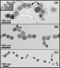
Magnetotactic bacteria mineralize nanometer-size crystals of magnetite (Fe3O4) through a series of protein-mediated reactions that occur inside of organelles called magnetosomes. Mms6 is a transmembrane protein thought to play a key role in magnetite mineralization. We used both electron and fluorescent microscopy to examine the subcellular location of Mms6 protein within single cells of Magnetospirillum magneticum AMB-1 using Mms6-specific antibodies. We also purified magnetosomes from M. magneticum to determine if Mms6 was physically attached to magnetite crystals. Our results show that Mms6 proteins are present during crystal growth, and Mms6 is found in direct contact with the magnetite crystals or within the lipid/protein membrane surrounding the magnetite crystals. Mms6 was not detected at other subcellular locations within the bacteria or isolated fractions. Because Mms6 was found to completely surround the magnetosomes rather than being localized to one specific area of the magnetosome, it appears that this protein could act on the entire magnetite crystal during the biomineralization process. This supports a model in which Mms6 functions to regulate Fe3O4 crystal morphology. This knowledge is important for future in vitro experiments utilizing Mms6 to synthesize tailored nanomagnets with specific physical or magnetic properties.

Type
Biomineralization and Biomimetics Articles
Copyright
Copyright © Materials Research Society 2016 

Access options

Get access to the full version of this content by using one of the access options below. (Log in options will check for institutional or personal access. Content may require purchase if you do not have access.)

References

REFERENCES

Tartaj, P., Morales, M.P., Gonzalez-Carreno, T., Veintemillas-Verdaguer, S., and Serna, C.J.: Advances in magnetic nanoparticles for biotechnology applications. J. Magn. Magn. Mater. 290, 28 (2005).Google Scholar
Matsunaga, T. and Arakaki, A.: Molecular bioengineering of bacterial magnetic particles for biotechnological applications. In Magnetoreception and Magnetosomes in Bacteria, Schuler, D. ed.; Springer: New York, 2007; p. 227254.Google Scholar
Prozorov, T., Palo, P., Wang, L., Nilsen-Hamilton, M., Jones, D., Orr, D., Mallapragada, S.K., Narasimhan, B., Canfield, P.C., and Prozorov, R.: Cobalt ferrite nanocrystals: Out-performing magnetotactic bacteria. ACS Nano 1, 228 (2007).Google Scholar
Deng, Y., Qi, D., Deng, C., Zhang, X., and Zhao, D.: Superparamagnetic high-magnetization microspheres with an Fe3O4-SiO2 core and perpendicularly aligned mesoporous SiO2 shell for removal of microcystins. J. Am. Chem. Soc. 130, 28 (2008).Google Scholar
Lee, J., Lee, Y., Youn, J.K., Bin Na, H., Yu, T., Kim, H., Lee, S., Koo, Y., Kwak, J.H., Park, H.G., Chang, H.N., Hwang, M., Park, J., Kim, J., and Hyeon, T.: Simple synthesis of functionalized superparamagnetic magnetite/silica core/shell nanoparticles and their application as magnetically separable high-performance biocatalysts. Small 4, 143 (2008).CrossRefGoogle ScholarPubMed
Wang, Y., Ng, Y.W., Chen, Y., Shuter, B., Yi, J., Ding, J., Wang, S., and Feng, S.: Formulation of superparamagnetic iron oxides by nanoparticles of biodegradable polymers for magnetic resonance Imaging. Adv. Funct. Mater. 18, 308 (2008).Google Scholar
Tanaka, M., Arakaki, A., Staniland, S.S., and Matsunaga, T.: Simultaneously discrete biomineralization of magnetite and tellurium nanocrystals in magnetotactic bacteria. Appl. Environ. Microbiol. 76, 5526 (2010).Google Scholar
Tang, Y., Wang, D., Zhou, C., Ma, W., Zhang, Y., Liu, B., and Zhang, S.: Bacterial magnetic particles as a novel and efficient gene vaccine delivery system. Gene Ther. 19, 1187 (2012).CrossRefGoogle ScholarPubMed
Huangand, H.S. and Hainfeld, J.F.: Intravenous magnetic nanoparticle cancer hyperthermia. Int. J. Nanomed. 8, 2521 (2013).Google Scholar
Tangand, S.C.N. and Lo, I.M.C.: Magnetic nanoparticles: Essential factors for sustainable environmental applications. Water Res. 47, 2613 (2013).Google Scholar
Li, L., Ding, J., and Xue, J.: A facile green approach for synthesizing monodisperse magnetite nanoparticles. J. Mater. Res. 25, 810 (2010).Google Scholar
Frankel, R.B., Blakemore, R.P., and Wolfe, R.S.: Magnetite in freshwater magnetotactic bacteria. Science 203, 1355 (1979).Google Scholar
Balkwill, D.L., Maratea, D., and Blakemore, R.P.: Ultrastructure of a magnetotactic spirillum. J. Bacteriol. 141, 1399 (1980).Google Scholar
Heywood, B.R., Bazylinski, D.A., Garrattreed, A., Mann, S., and Frankel, R.B.: Controlled biosynthesis of greigite (Fe3S4) in magnetotactic bacteria. Naturwissenschaften 77, 536 (1990).Google Scholar
Bazylinski, D.A. and Frankel, R.B.: Magnetosome formation in prokaryotes. Nat. Rev. Microbiol. 2, 217 (2004).Google Scholar
Murat, D., Quinlan, A., Vali, H., and Komeili, A.: Comprehensive genetic dissection of the magnetosome gene island reveals the step-wise assembly of a prokaryotic organelle. Proc. Natl. Acad. Sci. U. S. A 107, 5593 (2010).Google Scholar
Naresh, M., Hasija, V., Sharma, M., and Mittal, A.: Synthesis of cellular organelles containing nano-magnets stunts growth of magnetotactic bacteria. J. Nanosci. Nanotechnol. 10, 4135 (2010).Google Scholar
Greenberg, M., Canter, K., Mahler, I., and Tornheim, A.: Observation of magnetoreceptive behavior in a multicellular magnetotactic prokaryote in higher than geomagnetic fields. Biophys. J. 88, 1496 (2005).Google Scholar
Ullrich, S., Kube, M., Schubbe, S., Reinhardt, R., and Schuler, D.: A hypervariable 130-kilobase genomic region of Magnetospirillum gryphiswaldense comprises a magnetosome island which undergoes frequent rearrangements during stationary growth. J. Bacteriol. 187, 7176 (2005).Google Scholar
Matsunaga, T., Okamura, Y., Fukuda, Y., Wahyudi, A.T., Murase, Y., and Takeyama, H.: Complete genome sequence of the facultative anaerobic magnetotactic bacterium Magnetospirillum sp strain AMB-1. DNA Res. 12, 157 (2005).Google Scholar
Komeili, A., Vali, H., Beveridge, T.J., and Newman, D.K.: Magnetosome vesicles are present before magnetite formation, and MamA is required for their activation. Proc. Natl. Acad. Sci. U. S. A. 101, 3839 (2004).Google Scholar
Lefevreand, C.T. and Wu, L.: Evolution of the bacterial organelle responsible for magnetotaxis. Trends Microbiol. 21, 534 (2013).Google Scholar
Arakaki, A., Webb, J., and Matsunaga, T.: A novel protein tightly bound to bacterial magnetic particles in Magnetospirillum magneticum strain AMB-1. J. Biol. Chem. 278, 8745 (2003).Google Scholar
Prozorov, T., Mallapragada, S.K., Narasimhan, B., Wang, L., Palo, P., Nilsen-Hamilton, M., Williams, T.J., Bazylinski, D.A., Prozorov, R., and Canfield, P.C.: Protein-mediated synthesis of uniform superparamagnetic magnetite nanocrystals. Adv. Funct. Mater. 17, 951 (2007).Google Scholar
Arakaki, A., Masuda, F., Amemiya, Y., Tanaka, T., and Matsunaga, T.: Control of the morphology and size of magnetite particles with peptides mimicking the Mms6 protein from magnetotactic bacteria. J. Colloid Interface Sci. 343, 65 (2010).Google Scholar
Galloway, J.M., Arakaki, A., Masuda, F., Tanaka, T., Matsunaga, T., and Staniland, S.S.: Magnetic bacterial protein Mms6 controls morphology, crystallinity and magnetism of cobalt-doped magnetite nanoparticles in vitro. J. Mater. Chem. 21, 15244 (2011).Google Scholar
Tanaka, M., Mazuyama, E., Arakaki, A., and Matsunaga, T.: Mms6 protein regulates crystal morphology during nano-sized magnetite biomineralization in vivo. J. Biol. Chem. 286, 6386 (2011).Google Scholar
Feng, S., Wang, L., Palo, P., Liu, X., Mallapragada, S.K., and Nilsen-Hamilton, M.: Integrated self-assembly of the Mms6 magnetosome protein to form an iron-responsive structure. Int. J. Mol. Sci. 14, 14594 (2013).Google Scholar
Taoka, A., Asada, R., Sasaki, H., Anzawa, K., Wu, L-F., and Fukumori, Y.: Spatial localizations of Mam22 and Mam12 in the magnetosomes of Magnetospirillum magnetotacticum . J. Bacteriol. 188, 3805 (2006).Google Scholar
Amemiya, Y., Arakaki, A., Staniland, S.S., Tanaka, T., and Matsunaga, T.: Controlled formation of magnetite crystal by partial oxidation of ferrous hydroxide in the presence of recombinant magnetotactic bacterial protein Mms6. Biomaterials 28, 5381 (2007).Google Scholar
Arakaki, A., Yamagishi, A., Fukuyo, A., Tanaka, M., and Matsunaga, T.: Co-ordinated functions of Mms proteins define the surface structure of cubo-octahedral magnetite crystals in magnetotactic bacteria. Mol. Microbiol. 93, 554 (2014).Google Scholar
Wang, L., Prozorov, T., Palo, P.E., Liu, X., Vaknin, D., Prozorov, R., Mallapragada, S., and Nilsen-Hamilton, M.: Self-assembly and biphasic iron-binding characteristics of Mms6, a bacterial protein that promotes the formation of superparamagnetic magnetite nanoparticles of uniform size and shape. Biomacromolecules 13, 98 (2012).Google Scholar
Wang, W., Bu, W., Wang, L., Palo, P.E., Mallapragada, S., Nilsen-Hamilton, M., and Vaknin, D.: Interfacial properties and iron binding to bacterial proteins that promote the growth of magnetite nanocrystals: X-ray reflectivity and surface spectroscopy studies. Langmuir 28, 4274 (2012).Google Scholar
Valverde-Tercedor, C., Abadía-Molina, F., Martinez-Bueno, M., Pineda-Molina, E., Chen, L., Oestreicher, Z., Lower, B.H., Lower, S.K., Bazylinski, D.A., and Jimenez-Lopez, C.: Subcellular localization of the magnetosome protein MamC in the marine magnetotactic bacterium Magnetococcus marinus strain MC-1 using immunoelectron microscopy. Arch. Microbiol. 196, 481 (2014).Google Scholar
Richter, M., Kube, M., Bazylinski, D.A., Lombardot, T., Gloeckner, F.O., Reinhardt, R., and Schueler, D.: Comparative genome analysis of four magnetotactic bacteria reveals a complex set of group-specific genes implicated in magnetosome biomineralization and function. J. Bacteriol. 189, 4899 (2007).Google Scholar
Komeili, A., Li, Z., Newman, D.K., and Jensen, G.J.: Magnetosomes are cell membrane invaginations organized by the actin-like protein MamK. Science 311, 242 (2006).Google Scholar
Lohße, A., Borg, S., Raschdorf, O., Kolinko, I., Tompa, É., Pósfai, M., Faivre, D., Baumgartner, J., and Schüler, D.: Genetic dissection of the mamAB and mms6 operons reveals a gene set essential for magnetosome biogenesis in Magnetospirillum gryphiswaldense . J. Bacteriol. 196, 2658 (2014).Google Scholar