Hostname: page-component-586b7cd67f-t7fkt Total loading time: 0 Render date: 2024-11-24T08:51:00.576Z Has data issue: false hasContentIssue false

3D-printed β-TCP bone tissue engineering scaffolds: Effects of chemistry on in vivo biological properties in a rabbit tibia model

Published online by Cambridge University Press:  27 July 2018

Samit Kumar Nandi
Affiliation:
Department of Veterinary Surgery and Radiology, West Bengal University of Animal and Fishery Sciences, Kolkata 700037, West Bengal, India
Gary Fielding
Affiliation:
W. M. Keck Biomedical Materials Research Laboratory, School of Mechanical and Materials Engineering, Washington State University, Pullman, Washington 99164-2920, USA
Dishary Banerjee
Affiliation:
W. M. Keck Biomedical Materials Research Laboratory, School of Mechanical and Materials Engineering, Washington State University, Pullman, Washington 99164-2920, USA
Amit Bandyopadhyay
Affiliation:
W. M. Keck Biomedical Materials Research Laboratory, School of Mechanical and Materials Engineering, Washington State University, Pullman, Washington 99164-2920, USA
Susmita Bose*
Affiliation:
W. M. Keck Biomedical Materials Research Laboratory, School of Mechanical and Materials Engineering, Washington State University, Pullman, Washington 99164-2920, USA
*
a)Address all correspondence to this author. e-mail: [email protected]
Get access

Abstract

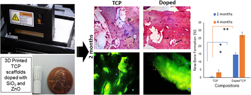
In this study, the effects of 3D-printed SiO2 and ZnO-doped tricalcium phosphate (TCP) scaffolds with interconnected pores were evaluated on the in vivo bone formation and healing properties of a rabbit tibial defect model. Pure and doped TCP scaffolds were fabricated by a ceramic powder-based 3D printing technique and implanted into critical sized rabbit tibial defects for up to 4 months. In vivo bone regeneration was evaluated using chronological radiological examination, histological evaluations, SEM micrographs, and fluorochrome labeling studies. Radiograph results showed that Si/Zn-doped samples had slower degradation kinetics than the pure TCP samples. 3D printing of TCP scaffolds improved bone formation. The addition of dopants in the TCP scaffolds improved osteogenic capabilities when compared to the pure scaffolds. In summary, our findings indicate that the addition of dopants to the TCP scaffolds enhanced bone formation and in turn leading to accelerated healing.

Type
Invited Article
Copyright
Copyright © Materials Research Society 2018 

Access options

Get access to the full version of this content by using one of the access options below. (Log in options will check for institutional or personal access. Content may require purchase if you do not have access.)

Footnotes

b)

This author was an editor of this journal during the review and decision stage. For the JMR policy on review and publication of manuscripts authored by editors, please refer to http://www.mrs.org/editor-manuscripts/.

References

REFERENCES

Bandyopadhyay, A., Bernard, S., Xue, W., and Bose, S.: Calcium phosphate-based resorbable ceramics: Influence of MgO, ZnO, and SiO2 dopants. J. Am. Ceram. Soc. 89, 26752688 (2006).CrossRefGoogle Scholar
Bose, S., Tarafder, S., Banerjee, S.S., Davies, N.M., and Bandyopadhyay, A.: Understanding in vivo response and mechanical property variation in MgO, SrO, and SiO2 doped β-TCP. Bone 48, 12821290 (2011).CrossRefGoogle Scholar
Liu, Q., Cen, L., Yin, S., Chen, L., Liu, G., Chang, J., and Cui, L.: A comparative study of proliferation and osteogenic differentiation of adipose-derived stem cells on akermanite and β-TCP ceramics. Biomaterials 29, 47924799 (2008).CrossRefGoogle ScholarPubMed
Huang, Y., Jin, X., Zhang, X., Sun, H., Tu, J., Tang, T., Chang, J., and Dai, K.: In vitro and in vivo evaluation of akermanite bioceramics for bone regeneration. Biomaterials 30, 50415048 (2009).CrossRefGoogle ScholarPubMed
Hutmacher, D.W.: Scaffolds in tissue engineering bone and cartilage. In The Biomaterials: Silver Jubilee Compendium (Elsevier, New York, New York, 2006); pp. 175189. https://doi.org/10.1016/B978-008045154-1.50021-6.CrossRefGoogle Scholar
Tarafder, S., Balla, V.K., Davies, N.M., Bandyopadhyay, A., and Bose, S.: Microwave-sintered 3D printed tricalcium phosphate scaffolds for bone tissue engineering. J. Tissue Eng. Regener. Med. 7, 631641 (2013).CrossRefGoogle ScholarPubMed
Karageorgiou, V. and Kaplan, D.: Porosity of 3D biomaterial scaffolds and osteogenesis. Biomaterials, 26, 54745491 (2005).CrossRefGoogle ScholarPubMed
Otsuki, B., Takemoto, M., Fujibayashi, S., Neo, M., Kokubo, T., and Nakamura, T.: Pore throat size and connectivity determine bone and tissue ingrowth into porous implants: Three-dimensional micro-CT based structural analyses of porous bioactive titanium implants. Biomaterials 27, 58925900 (2006).CrossRefGoogle ScholarPubMed
Bohner, M., Loosli, Y., Baroud, G., and Lacroix, D.: Commentary: Deciphering the link between architecture and biological response of a bone graft substitute. Acta Biomater. 7, 478484 (2011).CrossRefGoogle ScholarPubMed
Bohner, M. and Baumgart, F.: Theoretical model to determine the effects of geometrical factors on the resorption of calcium phosphate bone substitutes. Biomaterials 25, 35693582 (2004).CrossRefGoogle ScholarPubMed
Bose, S., Darsell, J., Kintner, M., Hosick, H., and Bandyopadhyay, A.: Pore size and pore volume effects on alumina and TCP ceramic scaffolds. Mater. Sci. Eng., C 23, 479486 (2003).CrossRefGoogle Scholar
Reffitt, D.M., Ogston, N., Jugdaohsingh, R., Cheung, H.F.J., Evans, B.A.J., Thompson, R.P.H., Powell, J.J., and Hampson, G.N.: Orthosilicic acid stimulates collagen type 1 synthesis and osteoblastic differentiation in human osteoblast-like cells in vitro. Bone 32, 127135 (2003).CrossRefGoogle ScholarPubMed
Gomes, P.S., Botelho, C., Lopes, M.A., Santos, J.D., and Fernandes, M.H.: Effect of silicon-containing hydroxyapatite coatings on the human in vitro osteoblastic response. Bone 44, S267 (2009).CrossRefGoogle Scholar
Fielding, G.A., Bandyopadhyay, A., and Bose, S.: Effects of silica and zinc oxide doping on mechanical and biological properties of 3D printed tricalcium phosphate tissue engineering scaffolds. Dent. Mater. 28, 113122 (2012).CrossRefGoogle ScholarPubMed
Roy, M., Fielding, G.A., Bandyopadhyay, A., and Bose, S.: Effects of zinc and strontium substitution in tricalcium phosphate on osteoclast differentiation and resorption. Biomater. Sci. 1, 7482 (2013).CrossRefGoogle ScholarPubMed
Kawamura, H., Ito, A., Miyakawa, S., Layrolle, P., Ojima, K., Ichinose, N., and Tateishi, T.: Stimulatory effect of zinc-releasing calcium phosphate implant on bone formation in rabbit femora. J. Biomed. Mater. Res. 50, 184190 (2000).3.0.CO;2-3>CrossRefGoogle Scholar
Yuan, H., De Bruijn, J.D., Li, Y., Feng, J., Yang, Z., De Groot, K., and Zhang, X.: Bone formation induced by calcium phosphate ceramics in soft tissue of dogs: A comparative study between porous α-TCP and β-TCP. J. Mater. Sci.: Mater. Med. 12, 713 (2001).Google ScholarPubMed
LeGeros, R.Z.: Properties of osteoconductive biomaterials: Calcium phosphates. Clin. Orthop. Relat. Res. 395, 8198 (2002).CrossRefGoogle Scholar
Gaasbeek, R.D., Toonen, H.G., van Heerwaarden, R.J., and Buma, P.: Mechanism of bone incorporation of β-TCP bone substitute in open wedge tibial osteotomy in patients. Biomaterials 26, 67136719 (2005).CrossRefGoogle ScholarPubMed
Bose, S., Banerjee, D., Robertson, S., and Vahabzadeh, S.: Enhanced in vivo bone and blood vessel formation by iron oxide and silica doped 3D printed tricalcium phosphate scaffolds. Ann. Biomed. Eng., 113 (2018). https://doi.org/10.1007/s10439-018-2040-8.Google ScholarPubMed
Bose, S. and Tarafder, S.: Calcium phosphate ceramic systems in growth factor and drug delivery for bone tissue engineering: A review. Acta Biomater. 8, 14011421 (2012).CrossRefGoogle ScholarPubMed
Patel, Z.S., Yamamoto, M., Ueda, H., Tabata, Y., and Mikos, A.G.: Biodegradable gelatin microparticles as delivery systems for the controlled release of bone morphogenetic protein-2. Acta Biomater. 4, 11261138 (2008).CrossRefGoogle ScholarPubMed
Suchanek, W., Yashima, M., Kakihana, M., and Yoshimura, M.: Hydroxyapatite ceramics with selected sintering additives. Biomaterials 18, 923933 (1997).CrossRefGoogle ScholarPubMed
Kanazawa, T., Umegaki, T., Yamashita, K., Monma, H., and Hiramatsu, T.: Effects of additives on sintering and some properties of calcium phosphates with various Ca/P ratios. J. Mater. Sci. 26, 417422 (1991).CrossRefGoogle Scholar
Pichavant, M.: Effects of B and H2O on liquidus phase relations in the haplogranite system at l kbar. Am. Mineral. 72, 10561070 (1987).Google Scholar
Yamaguchi, M.: Role of nutritional zinc in the prevention of osteoporosis. Mol. Cell. Biochem. 338, 241254 (2010).CrossRefGoogle Scholar
Anavi, Y., Avishai, G., Calderon, S., and Allon, D.M.: Bone remodeling in onlay beta-tricalcium phosphate and coral grafts to rat calvaria: Microcomputerized tomography analysis. J. Oral Implantol. 37, 379386 (2011).CrossRefGoogle ScholarPubMed
Hashizume, M. and Yamaguchi, M.: Effect of β-alanyl-L-histidinato zinc on differentiation of osteoblastic MC3T3-El cells: Increases in alkaline phosphatase activity and protein concentration. Mol. Cell. Biochem. 131, 1924 (1994).CrossRefGoogle Scholar
Li, X., Sogo, Y., Ito, A., Mutsuzaki, H., Ochiai, N., Kobayashi, T., Nakamura, S., Yamashita, K., and LeGeros, R.Z.: The optimum zinc content in set calcium phosphate cement for promoting bone formation in vivo. Mater. Sci. Eng., C 29, 969975 (2009).CrossRefGoogle ScholarPubMed
Patel, N., Best, S.M., Bonfield, W., Gibson, I.R., Hing, K.A., Damien, E., and Revell, P.A.: A comparative study on the in vivo behavior of hydroxyapatite and silicon substituted hydroxyapatite granules. J. Mater. Sci.: Mater. Med. 13, 11991206 (2002).Google ScholarPubMed
Camiré, C.L., Jegou Saint-Jean, S., Mochales, C., Nevsten, P., Wang, J.S., Lidgren, L., McCarthy, I., and Ginebra, M.P.: Material characterization and in vivo behavior of silicon substituted α-tricalcium phosphate cement. J. Biomed. Mater. Res., Part B 76, 424431 (2006).CrossRefGoogle ScholarPubMed
Bandyopadhyay, A., Shivaram, A., Tarafder, S., Sahasrabudhe, H., Banerjee, D., and Bose, S.: In vivo response of laser processed porous titanium implants for load-bearing implants. Ann. Biomed. Eng. 45, 249260 (2017).CrossRefGoogle ScholarPubMed
Arinzeh, T.L., Peter, S.J., Archambault, M.P., Van Den Bos, C., Gordon, S., Kraus, K., Smith, A., and Kadiyala, S.: Allogeneic mesenchymal stem cells regenerate bone in a critical-sized canine segmental defect. J. Bone Jt. Surg., Am. Vol. 85, 19271935 (2003).CrossRefGoogle Scholar
Jiang, T., Nukavarapu, S.P., Deng, M., Jabbarzadeh, E., Kofron, M.D., Doty, S.B., Abdel-Fattah, W.I., and Laurencin, C.T.: Chitosan–poly(lactide-co-glycolide) microsphere-based scaffolds for bone tissue engineering: In vitro degradation and in vivo bone regeneration studies. Acta Biomater. 6, 34573470 (2010).CrossRefGoogle ScholarPubMed
Nandi, S.K., Ghosh, S.K., Kundu, B., De, D.K., and Basu, D.: Evaluation of new porous β-tri-calcium phosphate ceramic as bone substitute in goat model. Small Rumin. Res. 75, 144153 (2008).CrossRefGoogle Scholar
Gibson, C.J., Thornton, V.F., and Brown, W.A.B.: Incorporation of tetracycline into impeded and unimpeded mandibular incisors of the mouse. Calcif. Tissue Res. 26, 2931 (1978).CrossRefGoogle ScholarPubMed
Dahners, L.E. and Bos, G.D.: Fluorescent tetracycline labeling as an aid to debridement of necrotic bone in the treatment of chronic osteomyelitis. J. Orthop. Trauma 16, 345346 (2002).CrossRefGoogle ScholarPubMed