INTRODUCTION
China contributes one of every seven new tuberculosis cases globally with an estimated 1·3 million new tuberculosis cases in China per annum [1]. Tuberculosis epidemics in China are also complicated by the emergence of drug-resistant Mycobacterium tuberculosis strains [Reference Zhao2]. Airborne transmission is the most common route by which M. tuberculosis is spread, and outbreaks often occur among people living in crowded, closed spaces over an extended period of time. Direct inoculation of M. tuberculosis into the skin has been reported after intralesional injection of steroids [Reference Kim3]. Acupuncture is a very popular form of alternative pain therapy in China, where in some clinics needles used for acupuncture or injections are usually reused, which may carry the risk of direct inoculation of M. tuberculosis if these needles are inadequately sterilized. Here, we report an outbreak of direct inoculation tuberculosis in 13 cases who developed tuberculous abscesses in the paraspinal muscles following intramuscular injection at a local acupuncture clinic in southeastern China.
METHODS
Index patient
The index patient was a 70-year-old man who complained of a painful swollen lump on the left nuchal region for 1 month. The patient received acupuncture and paraspinal muscular injection with lidocaine and triamcinolone in the left nuchal region at a local acupuncture clinic in the city of Wenzhou, Zhejiang, China, in April 2011. Local swelling gradually developed at the injection site, which failed to resolve despite 2 weeks of cephradine. The patient was referred to our hospital. Physical examination revealed a lump in the left nuchal region, which was 6 × 5 cm in size with no erythema and local heat. Magnetic resonance imaging (MRI) and computed tomography (CT) showed that the soft tissue mass was poorly confined in the left nuchal region of the paraspinal muscle of the cervical spine without involving the lamina or the facet joint (Fig. 1). The tuberculin skin test (TST) was positive (5–9 mm in size), but chest radiograph was normal. Sputum, urine and stool cultures were negative for M. tuberculosis. Debridement was performed 1 week later, which showed the presence of viscous pus that contained numerous acid-fast bacilli. Pus cultures were positive for M. tuberculosis.

Fig. 1. MRI axial (T1W image) and coronal (STIR image) cut section of the cervical spine of a 70-year-old man who complained of a painful swollen lump in the left nuchal region for 1 month show a poorly confined oval intramuscular lesion (arrows) in the left paraspinal muscle with diffuse muscle oedema (a) and (b). Sagittal plane MRI (contrast image) indicates paraspinal muscle infection (arrow) without any evidence of tumour (c). CT scan of the cervical spine revealed a soft tissue mass (arrow) without involvement of the lamina or the facet joint (d).
Secondary cases
From May 2011 to August 2011, 12 more patients with a swollen lump on the nuchal region or in the lower back or the buttocks region were referred to our hospital. These patients had attended the same local acupuncture clinic as the index patient and all received paraspinal muscle injections. We performed TST of these patients using standard procedures. A trained nurse injected intradermally 0·1 ml (2 IU) of TST produced from bacille Calmette-Guérin (Chengdu Institute of Biological Products, China) into the inner surface of the left forearm using the Mantoux method. An experienced physician measured the transverse induration (in mm) at the TST site 72 h after injection [Reference Fraser4]. T-SPOT.TB assays were performed as instructed by the manufacturer (T-SPOT.TB, Oxford Immunotec, UK). Acid-fast stain was performed using the Ziehl–Neelsen method. M. tuberculosis culture was performed using BACTEC™ 460TB System Mycobacterial Culture Media. Chest radiograph and lump MRI were additionally performed. Furthermore, lesions and biopsies were subjected to polymerase chain reaction (PCR) and gene sequencing by the Disease Control Center of Zhejiang Province, China. The gene sequencing products were compared with the genomic sequences of M. tuberculosis in GenBank.
RESULTS
Outbreak description
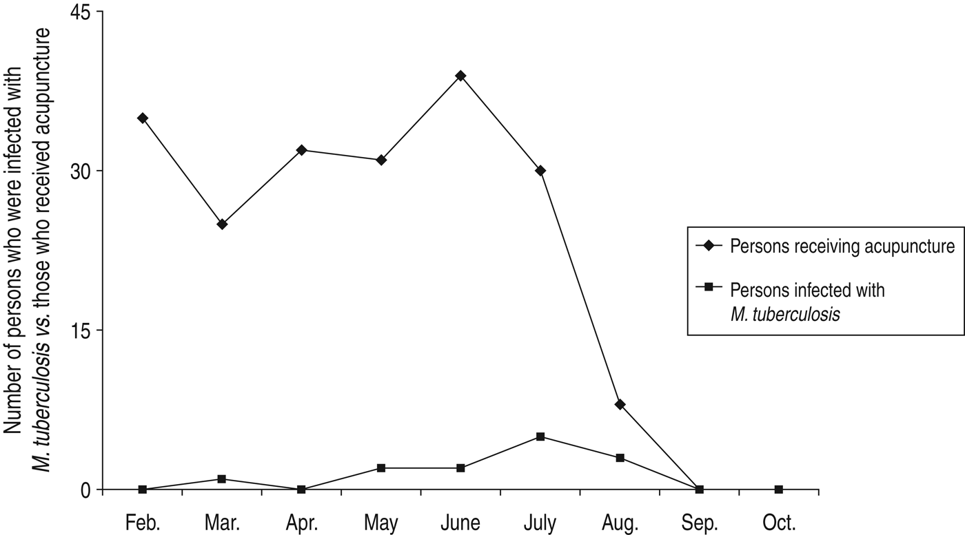
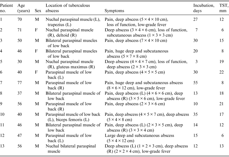
Over a 7-month period from February 2011 to August 2011, ~5000 persons received acupuncture from two acupuncturists at a local clinic. Roughly, 200 patients received intramuscular injection at the paraspinal muscle with 2% lidocaine and triamcinolone, of which 13 (6%) reported a swollen lump at the injection site (Fig. 2) Onsite investigation revealed that needles and syringes were reused after autoclave sterilization. The patients' demographic and baseline data are shown in Table 1. The patients included 10 men and three women with a median age of 47 (range 30–77) years. None of these patients had a history of positive TST or evidence of prior tuberculosis. No cervical or lumbar lumps were present in any patient at the time of acupuncture. The TST was positive for all patients. T-SPOT.TB tests were also positive for all patients (10/10) who underwent the test. PCR assays were positive for patients (100%, 5/5) who received the assays and the causative agent was identified as M. tuberculosis, Beijing type. Twelve (92·3%, 12/13) patients had positive acid-fast stains of pus specimens from debrided lump. Culture specimens were obtained from 13 patients, one by needle aspiration and 12 by surgical debridement or drainage biopsy. Routine bacterial cultures were negative in all patients while mycobacterial cultures of abscess specimens were positive in all 13 (100%, 13/13) patients. The mycobacteria were sensitive to isoniazid, rifampicin, pyrazinamide, ethambutol and streptomycin. The median incubation period was 15 (range 3–55) days and the median time from onset of symptoms to hospital visit was 35 (range 23–57) days. All cases were reported to the local Department of Health.

Fig. 2. Over a 7-month period from February 2011 to August 2011, ~5000 persons received acupuncture from two acupuncturists at a local clinic. About 200 patients received intramuscular injection at the paraspinal muscle with 2% lidocaine and triamcinolone, of which 13 (6%) reported a swollen lump at the injection site.
Table 1. Demographic and baseline characteristics of the patients

TST, Tuberculin skin test; M, male; F, female; L, left; R, right.
Disease characteristics
All patients developed abscesses in the paraspinal muscles or adjacent muscles such as the gluteus maximus, deltoid or trapezius that had received intramuscular injection (Table 1). Four (30·8%, 4/13) patients had abscesses in the paraspinal muscle of the cervical spine (one case also had abscesses on the right gluteal region and one case also had abscesses on the right deltoid muscle), and nine (69·2%, 9/13) patients had abscess on the paraspinal muscle of the lumbar spine (six cases also had abscesses in the gluteal region, one case also had abscesses on the left thigh). The median diameter of the masses was 7 (range 4–15) cm and the mean size of the masses was 2 × 3 × 4 (range 2 × 3 × 4 to 2 × 3 × 4) cm3. Eleven (84·6%, 11/13) patients had abscesses that drained thick yellow liquid. Histopathological examination of 20 biopsies from 13 patients showed three major distinct patterns: (a) prevailing abscesses with mild granulomatous reaction (36·7%, 11/30); (b) granulomatous nodular or diffuse inflammation with mixed granulomas (20%, 6/30); (c) massive necrosis with few granulomas, (10%, 3/30).
Nine (69·2%, 9/13) patients complained of pain and 11 (84·6%, 11/13) reported vague discomfort in the affected area. Four (30·8%, 4/13) patients with nuchal abscesses complained of limitation of movement of the cervical spine. Three (23·1%, 3/13) patients developed low-grade fever. Chest radiograph was normal in all patients. MRI was performed in all patients and revealed the presence of poorly confined soft tissue masses in the paraspinal muscle without involving the lamina or the facet joint. Contrast MRI was performed in nine patients, showing no evidence of tumour.
Management
The acupuncture clinic was asked to stop using reusable needles and syringes. All patients were treated with the combination regimen consisting of isoniazid (300 mg once daily), rifampin (450 mg once daily), ethambutol (750 mg once daily) and pyrazinamide (500 mg thrice daily) for 18 months. Abscesses were treated with repeat needle aspiration (one patient), surgical debridement (five patients) and surgical drainage (vacuum sealing drainage; seven patients). Dressing changes were continued for the next 4–8 weeks until the abscesses healed completely. The paraspinal tuberculosis abscesses were resolved completely. None of the patients reported recurrence of lesions after more than 6 months of follow up.
DISCUSSION
Primary inoculation tuberculosis is an exogenous infection which occurs when the integrity of the skin is breached. We report 13 cases of large paraspinal muscle lumps as a result of intramuscular injection using inadequately sterilized needles and syringes. Generally, inoculation tuberculous cutaneous lesions appear 2–4 weeks after inoculation and could present as an erythematous non-painful papule or nodule. The median incubation period for our series was ~2 weeks. The tuberculous lesions are typically confined to the injection site, as seen in all our patients. In our series, multiple abscesses in several patients were due to injections at multiple sites and abscess developed at each of the injected sites. Patients may complain of pain and limitation of movement and could develop low-grade fever. Our histopathological examination revealed the presence of acute non-specific inflammatory reaction, tuberculoid appearance and caseation necrosis, alone or in combination, in the lesions, reflecting different stages of disease development in these patients. In addition, the mycobacteria were readily detected by acid-fast bacilli stain.
The epidemiological link to the cluster was not recognized by our conventional outbreak investigation. The reused needles and syringes at the local clinic remain the likely cause as no new cases of inoculation tuberculosis occurred after reuse of autoclaved needles and syringes was prohibited. No patient had a history of positive TST or tuberculosis. Furthermore, a thorough examination revealed no open pulmonary tuberculosis or active tuberculous disease elsewhere, and an endogenous source from an underlying vertebral or facet joint infection was also ruled out by MRI. The nodular lesion was diagnosed as a tuberculous paraspinal abscess based on the data from acid-fast stain, mycobacterial culture and DNA sequencing. This finding is further supported by the fact that it took a median of ~2 weeks for the lesions to start to appear. By contrast, pyogenic bacterial infections typically have a shorter incubation period [Reference Fraser4–Reference Galil6].
The number of non-tuberculous mycobacterial infections has significantly increased in recent years [Reference Fraser4–Reference Villanueva7]. Mycobacterial abscesses are frequently associated with the use of contaminated medical devices [Reference Fraser4, Reference Ara8] or improper intake of certain medications [Reference Fraser4]. The exact source of mycobacterial abscesses in this report was not determined, and the origin of these bacteria is, at present, speculative. They probably were not present in the injected medicine, although it should be noted that the composition of these medicines is not publically available. If the medicine had been contaminated with M. tuberculosis, the number of infected patients would have been far greater. The use of reusable syringes in multiple patients also increased the risk of patient-to-patient transmission.
Anti-tuberculosis medicines were chosen as the primary therapeutic agents, as they are known to have consistent efficacy against mycobacterial abscesses [Reference Mushatt and Witzig9]. Moreover, surgical debridement is recommended, especially for those with necrosis, cellulitis or abscesses. In non-tuberculous mycobacterial skin and soft-tissue infections, the combination of surgical therapy and drug treatment reduce healing time by 50%, compared to surgical therapy or drug treatment alone [Reference Fraser4]. This study reinforces the importance of this combinatorial therapeutic approach.
Given the rising popularity of acupuncture both in and outside of China, it is important to ensure the safety of syringes and needles used in acupuncture by educating the acupuncturists and by implementing proper sterilization measures. Moreover, physicians should consider the possibility of mycobacterial infections, apart from other bacterial infections, in patients with a swollen paraspinal lump following intramuscular injection. Furthermore, education on infection control, including hygienic practice, should be emphasized to doctors of traditional Chinese medicine practising acupuncture and local injection.
ACKNOWLEDGEMENTS
The authors thank Yunxiang Zeng, Tieli Zhou, and Kate Huang for their skilful technical assistance.
DECLARATION OF INTEREST
None.


