Skip to main content Accessibility help
×
Hostname: page-component-745bb68f8f-mzp66 Total loading time: 0 Render date: 2025-01-25T01:14:06.425Z Has data issue: false hasContentIssue false

Section 3 - Noninvasive Investigation of Insular Epilepsy

Published online by Cambridge University Press:  09 June 2022

Dang Nguyen
Affiliation:
Université de Montréal
Jean Isnard
Affiliation:
Claude Bernard University Lyon
Philippe Kahane
Affiliation:
Grenoble-Alpes University Hospital
Get access

Summary

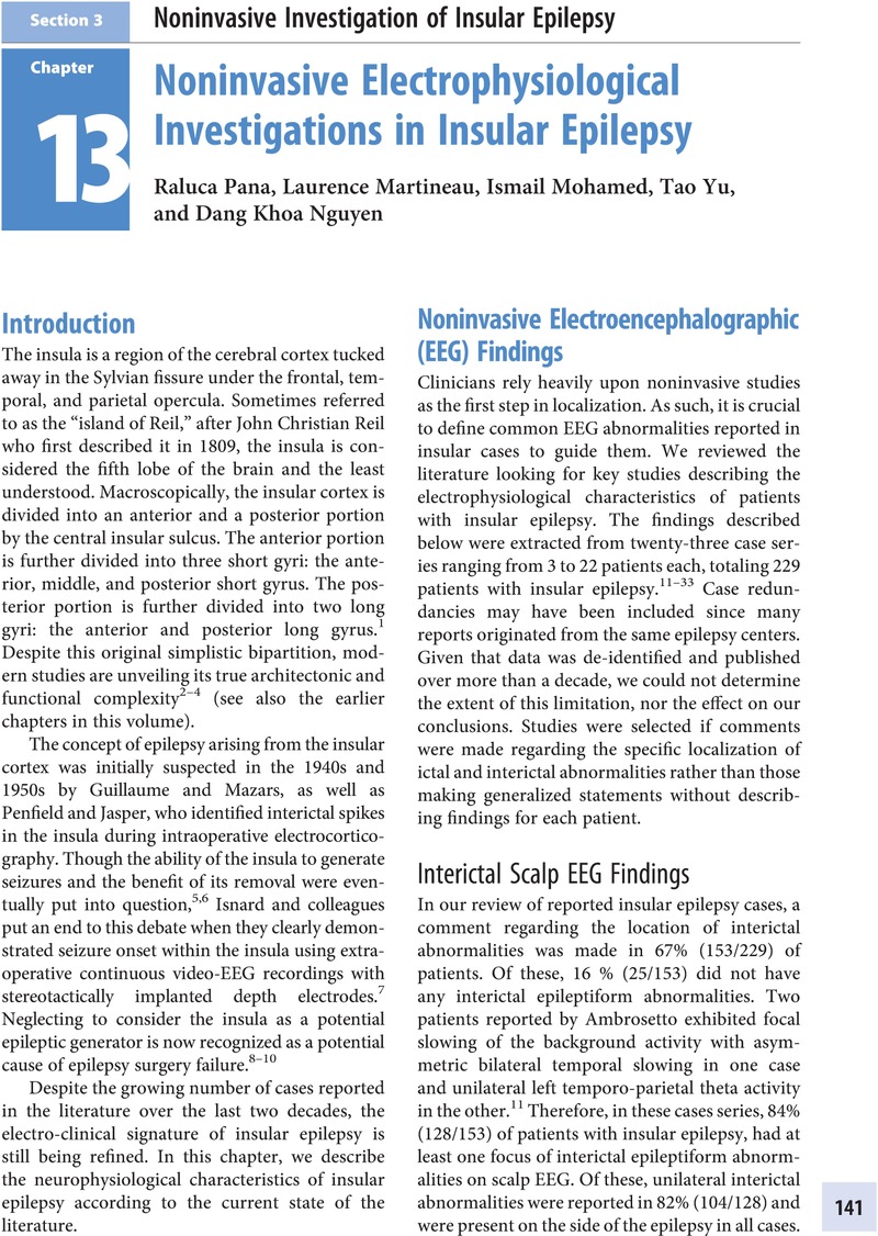
Image of the first page of this content. For PDF version, please use the ‘Save PDF’ preceeding this image.'
Type
Chapter
Information
Insular Epilepsies , pp. 141 - 202
Publisher: Cambridge University Press
Print publication year: 2022

Access options

Get access to the full version of this content by using one of the access options below. (Log in options will check for institutional or personal access. Content may require purchase if you do not have access.)

References

References

Stephani, C, Fernandez-Baca Vaca, G, Maciunas, R, Koubeissi, M, Luders, HO (2011). Functional neuroanatomy of the insular lobe. Brain Struct Funct. 216: 137149.Google Scholar
Morel, A, Gallay M, Baechler N, Wyss, M, Gallay, DS (2013). The human insula: Architectonic organization and postmortem MRI registration. Neuroscience. 236: 117135.Google Scholar
Mufson, EJ, Mesulam, M (1982). Insula of the old world monkey. II: Afferent cortical input and comments on the claustrum. J Comp Neurol. 212: 2337.Google Scholar
Jobst, BC, Gonzalez-Martinez, J, Isnard, J, Kahane, P, Lacuey, N, Lathoo, S, Nguyen, DK, Wu, C, Lado, F (2019). The insula and its epilepsies. Epileptic Disorders Epilepsy Currents. 19(1): 1121.Google Scholar
Guillaume, MMJ, Mazars, G (1949). Cinq cas de foyers épileptogènes insulaires opérés. Soc Française de Neurol (a). 766769.Google Scholar
Penfield, W, Faulk, ME (1955). The insula: Further observations on its function. Brain. 78: 445470.Google Scholar
Isnard, J, Guenot, M, Ostrowsky, K, et al (2000). The role of the insular cortex in temporal lobe epilepsy. Ann Neurol. 48: 614623.Google Scholar
Harroud, A, Bouthillier, A, Weil, A, Nguyen, DK (2012). Temporal lobe epilepsy surgery failures: A review. Epilepsy Res Treat. 201651.Google Scholar
Isnard, J, Ryvlin, P, Mauguière, F (2008). Insular epilepsy. In Luders, H (ed.), Textbook of Epilepsy Surgery. London: Informa Healthcare; 2008: 320333.Google Scholar
Laoprasert, P, Ojemann, JG, Handler, MH (2017). Insular epilepsy surgery. Epilepsia. 58(suppl 1): 3545.Google Scholar
Ambrosetto, G (1993). Treatable epilepsy and unilateral opercular neuronal migration disorder. Epilepsia. 34 (4): 604608.Google Scholar
Malak, R, Bouthillier, A, Carmant, L, et al (2009). Microsurgery of epileptic foci in the insular region. J Neurosurg. 110: 11531163.Google Scholar
Bouthillier, A, Nguyen, DK (2017). Epilepsy surgeries requiring an operculoinsular cortectomy: Operative technique and results. Neurosurgery. 81(4): 602612.Google Scholar
Von Lehe, M, Wellmer, J, Urbach, H, Schramm, J, Elger, CE, Clusmann, H (2009). Insular lesionectomy for refractory epilepsy: Management and outcome. Brain. 132(pt 4): 10481056.Google Scholar
Gras-Combe, G, Minotti, L, Hoffmann, D, Krainik, A, Kahane, P, Chabardes, S (2016). Surgery for nontumoral insular epilepsy explored by stereoelectroencephalography. Neurosurgery. 79(4): 578588.Google Scholar
Dylgjeri, S, Taussig, D, Chipaux, M (2014). Insular and insulo-opercular epilepsy in childhood: An SEEG study. Seizure. 23(4): 300308.Google Scholar
Proserpio, P, Cossu, M, Francione, S, Tassi, L, Mai, R, Didato, G, Castana, L, Cardinale, F, Sartori, I, Gozzo, F, Citterio, A, Schiariti, M, Lo Russo, G, Nobili, L (2011). Insular-opercular seizures manifesting with sleep related paroxysmal motor behaviors: A stereo-EEG study. Epilepsia. 52: 17811791.Google Scholar
Ryvlin, P, Minotti, L, Demarquay, G, Hirsch, E, Arzimanoglou, A, Hoffman, D, Guenot, M, Picard, F, Rheims, S, Kahane, P (2006). Nocturnal hypermotor seizures, suggesting frontal lobe epilepsy, can originate in the insula. Epilepsia. 47: 755765.CrossRefGoogle ScholarPubMed
Freri, E, Matricardi, S, Gozzo, F, Cossu, M, Granata, T, Tassi, L (2017). Peri-Sylvian, including insular, childhood epilepsy: Presurgical workup and surgical outcome. Epilepsia. (8): 13601369.Google Scholar
Gonzalez-Martinez, J, Mullin, J, Bulacio, J, Gupta, A, Enatsu, R, Najm, I, Bingaman, W, Wyllie, E, Lachwany, D (2014). Stereoelectroencephalography in children and adolescents with difficult-to-localize refractory focal epilepsy. Neursosurgery. 75(3): 258268.Google Scholar
Montavont, A, Mauguiere, F, Mazzola, L, et al. (2015). On the origin of painful somatosensory seizures. Neurology 84: 594601.Google Scholar
Tran, TP, Truong, VT, Wilk, M, et al. (2014). Different localizations underlying cortical gelastic epilepsy: case series and review of literature. Epilepsy Behav. 35: 3441.Google Scholar
Kakisaka, Y, Iwasaki, M, Alexopoulos, AV, Enatsu, R, Jin, K, Wang, ZI, Mosher, JC, Dubarry, AS, Nair, DR, Burgess, RC (2012). Magnetoencephalography in fronto-parietal opercular epilepsy. Epilepsy Res. 102: 7177.Google Scholar
Weil, AG, Le, NM, Jayakar, P (2016). Medically resistant pediatric insular-opercular/peri-Sylvian epilepsy. Part 2: Outcome following resective surgery. J Neurosurg Pediatr. 18(5): 523535.Google Scholar
Hagiwara, K, Jung, J, Bouet, R (2017). How can we explain the frontal presentation of insular lobe epilepsy? The impact of non-linear analysis of insular seizures. Clin Neurophysiol. 128(5): 780791.Google Scholar
Mohamed, IS, Gibbs, SA, Robert, M, Bouthillier, A, Leroux, JM, Khoa Nguyen, D (2013). The utility of magnetoencephalography in the presurgical evaluation of refractory insular epilepsy. Epilepsia. 54(11): 19501959.CrossRefGoogle ScholarPubMed
Xiao, H, Tran, TP, Boucher, Petrin M O, Mohamed, I, Bouthillier, A, DK, Nguyen (2016). Reflex operculoinsular seizures. Epileptic Disord. 2016; 18(1): 1925.Google Scholar
Levy, A, Tran, YPT, Boucher, O, Bouthillier, A, Nguyen, DK (2017). Operculo-Insular epilepsy. J Clin Neurophysiol. 34(5): 438447.Google Scholar
Wang, S, Tang, Y, Aung, T, Chen, C, Katagiri, M, Jones, SE, Prayson, RA, Krishnan, B, Gonzalez-Martinez, J, Burgess, RC, Najm, I, et al. (2019). Multimodal noninvasive evaluation in MRI-negative operculo-insular epilepsy. J Neurosurgery. Apr 12: 111.Google Scholar
Yin, C, Zhang, X, Chen, Z, Li, X, Wu, S, Lv P, Wang Y (2019). Detection and localization of interictal ripples with magnetoencephalography in the presurgical evaluation of drug-resistant insular epilepsy. Brain Research (1706): 147156.Google Scholar
Park, HM, Nakasato, N, Tominaga, T (2012). Localization of abnormal discharges causing insular epilepsy by magnetoencephalography. Tohoku J Exp Med 226: 207211.Google Scholar
Heers, M, Rampp, S, Stefan, H, Urbach, H, Elger, CE, von Lehe, M, Wellmer, J (2012). MEG-based identification of the epileptogenic zone in occult peri-insular epilepsy. Seizure 21: 128–113.Google Scholar
Perry, MS, Donahue, DJ, Malik, SI, Keator, CG, Hernandez, A, Reddy, RK, Perkins, Jr. FF, Lee, MR, Clarke, DF (2017). Magnetic resonance imaging–guided laser interstitial thermal therapy as treatment for intractable insular epilepsy in children. J Neurosurg Pediatr. 20: 575582.Google Scholar
Nguyen, DK, Nguyen, DB, Malak, R, Bouthillier, A (2009a). Insular cortex epilepsy: An overview. Can J Neurol Sci. 36(suppl. 2): S58S62.Google Scholar
Ghaziri, J, Tucholka, A, Girard, G (2017). The corticocortical structural connectivity of the human insula. Cereb Cortex. 27(2): 12161228.CrossRefGoogle ScholarPubMed
Dennis, EL, Jahanshad, N, McMahon, KL, Zubicaray, GI, Martin, NG, Hickie, IB, Toga, AW, Wrigth, MJ, Thompson, PM (2014). Development of insula connectivity between the ages 12 and 30 revealed by high angular resolution diffusion imaging. Hum Brain Mapp, April. 35(4): 17901800.Google Scholar
Kharkar, S, Knolton, R (2015). Magnetoencephalography in the presurgical evaluation of epilepsy. Epilepsy& Behavior. 46: 1926.Google Scholar
Zerouali, Y, Pouliot, P, Robert, M, Mohamed, I, Bouthillier, A, Lesage, F, Nguyen, DK (2016). Magnetoencephalographic signatures of insular epileptic spikes based on functional connectivity. Hum Brain Map. 37: 32503261.Google Scholar
Kakisaka, Y, Kakisaka, Y, Alkawadri, R, ZI, Wang, Enatsu, R, JC, Mosher, A-S, Dubarry, AV, Alexopoulos, RC, Burgess (2012). Sensitivity of scalp 10–20 EEG and magnetoencephalography Epileptic Disord. 2013; 15(1): 2731.CrossRefGoogle Scholar
Spencer, S, Huh, L (2008). Outcomes of epilepsy surgery in adults and children. Lancet Neurology. 7: 525537.Google Scholar
Yu, T, Ni, D, Zhang, X, Wang, X, Qiao, L, Zhou, X, Wang, Y, Li, Y, Zhang, G (2018). The role of magnetoencephalography in the presurgical evaluation of patients with MRI-negative operculo-insular epilepsy. Seizure. 61; 104110.Google Scholar
Jung, J, Bouet, R, Delpuech, C, Ryvlin, P, Isnard, J, Guenot, M, Bertrand, O, Hammers, A, Mauguière, F (2013). The value of magnetoencephalography for seizure-onset zone localization in magnetic resonance imaging-negative partial epilepsy. Brain 136; 31763186.Google Scholar
Taniguchi, M, Yoshimine, T, Kato, A, Maruno, M, Hirabuki, N, Nakamura, H, Imai, K, Okada, S, Hyakawa, T (1998). Dysembryoplastic neuroepithelial tumor in the insular cortex. Three-dimensional magnetoencephalographic localization of epileptic discharges. Neurological Research. (20) 433438.Google Scholar
Brodbeck, V, Spinelli, L, AM, Lascano, Wissmeier, M, MI, Vargas, Vulliemoz, S, Pollo, C, Schaller, K, CM, Michel, Seeck, M (2011). Electroencephalographic source imaging: A prospective study of 152 operated epileptic patients. Brain. 134; 28872897.Google Scholar
BE, Mouthaan, Rados, M, Boon, P, Carrette, E et al. (2019). Diagnostic accuracy of interictal source imaging in presurgical epilepsy evaluation: A systematic review from the E-PILEPSY consortium. Clinical Neurophysiology. 130; 845855.Google Scholar
Abdallah, C, Maillard, LG, Rikir, E, Jonas, J, Thiriaux, A, Gavaret, M, Bartolomei, F, Colnar-Coulbois, S, Vignal, JP, Koessler, L (2017). Localizing value of electrical source imaging: Frontal lobe, malformations of cortical development and negative MRI related epilepsies are the best candidates. NeuroImage: Clinical. 16; 310329.Google ScholarPubMed

References

Von Bonin, G, Bailey, P. The Neocortex of Macaca mulatta. J Am Med Assoc. 1948;137(12):1093. doi:10.1001/jama.1948.02890460089032.Google Scholar
Chen, BY, Boucher, O, Dugas, C, Nguyen, DK, Gioia, L. Insular Ischemic Stroke. In: Turgut, M, Yurttaş, C, Tubbs, RS, eds. Island of Reil (Insula) in the Human Brain: Anatomical, Functional, Clinical and Surgical Aspects.Springer; 2018: 203.Google Scholar
Nieuwenhuys, R. The insular cortex. A review. In: Progress in Brain Research. Vol. 195. Elsevier B.V.; 2012: 123163. doi:10.1016/B978-0-444-53860-4.00007-6.Google Scholar
Türe, U, DCH, Yaşargil, Al-Mefty, O, Yaşargil, MG. Topographic anatomy of the insular region. J Neurosurg. 1999;90(4):720733. doi:10.3171/jns.1999.90.4.0720.Google Scholar
Guenot, M, Isnard, J, Sindou, M. Surgical anatomy of the insula. Adv Tech Stand Neurosurg. 2004. doi:10.1007/978-3-7091-0558-0_7.Google Scholar
Jobst, BC, Gonzalez-Martinez, J, Isnard, J, et al. The insula and its epilepsies. Epilepsy Curr. 2019;19(1):1121. doi:10.1177/1535759718822847.CrossRefGoogle ScholarPubMed
TP, Naidich, Kang, E, GM, Fatterpekar, et al. The insula: Anatomic study and MR imaging display at 1.5 T. AJNR Am J Neuroradiol 2004;25(2):222232.Google Scholar
Gaillard, WD, Cross, JH, Duncan, JS, Stefan, H, Theodore, WH. Epilepsy imaging study guideline criteria: Commentary on diagnostic testing study guidelines and practice parameters. Epilepsia. 2011;52(9):17501756. doi:10.1111/j.1528-1167.2011.03155.x.Google Scholar
Bernasconi, A, Cendes, F, Theodore, WH, et al. Recommendations for the use of structural magnetic resonance imaging in the care of patients with epilepsy: A consensus report from the International League Against Epilepsy Neuroimaging Task Force. Epilepsia. 2019;60(6):10541068. doi:10.1111/epi.15612.Google Scholar
Nguyen, DK, Nguyen, DB, Malak, R, et al. Revisiting the role of the insula in refractory partial epilepsy. Epilepsia. 2009;50(3):510520. doi:10.1111/j.1528-1167.2008.01758.x.Google Scholar
Laoprasert, P, Ojemann, JG, Handler, MH. Insular epilepsy surgery. Epilepsia. 2017;58:3545. doi:10.1111/epi.13682.Google Scholar
Chevrier, MC, Bard, C, Guilbert, F, Nguyen, DK. Structural abnormalities in patients with insular/peri-insular epilepsy: spectrum, frequency, and pharmacoresistance. Am J Neuroradiol. 2013;34(11):21522156. doi:10.3174/ajnr.A3636.Google Scholar
Mohamed, AR, Bailey, CA, Freeman, JL, Maixner, W, Jackson, GD, Harvey, FAS. Intrinsic Epileptogenicity of Cortical Tubers Revealed by Intracranial EEG Monitoring Neurology. 2012; 79(23):2249–2257. doi:10.1212/wnl.0b013e3182768923.Google Scholar
Kuzniecky, R, Andermann, F. The Congenital Bilateral Peri-Sylvian Syndrome: Imaging Findings in a Multicenter Study. Am J Neuroradiol. 1994;15(1):139–144.Google Scholar
Potts, MB, Young, WL, Lawton, MT. Deep arteriovenous malformations in the basal ganglia, thalamus, and insula: Microsurgical management, techniques, and results. Neurosurgery. 2013;73(3):417429. doi:10.1227/NEU.0000000000000004.CrossRefGoogle ScholarPubMed
Jaiswal, M. Insular Cortex Epilepsy. In: Turgut, M, Yurttaş, C, RS, Tubbs, eds., Island of Reil (Insula) in the Human Brain: Anatomical, Functional, Clinical and Surgical Aspects. Springer; 2018: 199200.Google Scholar
Varadkar, S, Bien, CG, Kruse, CA, et al. Rasmussen’s encephalitis: Clinical features, pathobiology, and treatment advances. Lancet Neurol. 2014;13(2):195205. doi:10.1016/S1474-4422(13)70260-6.Google Scholar
Ryvlin, P, Minotti, L, Demarquay, G, et al. Nocturnal hypermotor seizures, suggesting frontal lobe epilepsy, can originate in the insula. Epilepsia. 2006;47(4):755-765.doi:10.1111/j.1528-1167.2006.00510.x.Google Scholar
Isnard, J, Guénot, M, Sindou, M, Mauguière, F. Clinical manifestations of insular lobe seizures: A stereo- electroencephalographic study. Epilepsia. 2004;45(9):10791090. doi:10.1111/j.0013-9580.2004.68903.x.Google Scholar
Bouthillier, A, Nguyen, DK. Epilepsy surgeries requiring an operculoinsular cortectomy: Operative technique and results. Clin Neurosurg. 2017;81(4):602611. doi:10.1093/neuros/nyx080.Google Scholar
Dylgjeri, S, Taussig, D, Chipaux, M, et al. Insular and insulo-opercular epilepsy in childhood: An SEEG study. Seizure. 2014;23(4):300308. doi:10.1016/j.seizure.2014.01.008.Google Scholar
Weil, AG, Le, NMD, Jayakar, P, et al. Medically resistant pediatric insular-opercular/peri-Sylvian epilepsy. Part 2: Outcome following resective surgery.J Neurosurg Pediatr. 2016;18(5):523535. doi:10.3171/2016.4.PEDS15618.Google Scholar
Bernasconi, A, Bernasconi, N, Bernhardt, BC, Schrader, D. Advances in MRI for “cryptogenic” epilepsies. Nat Rev Neurol. 2011;7(2):99108. doi:10.1038/nrneurol.2010.199.Google Scholar
Bastos, AC, Comeau, RM, Andermann, F, et al. Diagnosis of subtle focal dysplastic lesions: Curvilinear reformatting from three-dimensional magnetic resonance imaging. Ann Neurol. 1999;46(1):8894. doi:10.1002/1531-8249(199907)46:1<88::AID-ANA13>3.0.CO;2-4.Google Scholar
Bernasconi, A. Structural analysis applied to epilepsy. In: RI, Kuzniecky, GD, Jackson, eds., Magnetic Resonance in Epilepsy. 2nd ed. Academic Press; 2005: 249269.Google Scholar
Cendes, F, Theodore, WH, Brinkmann, BH, Sulc, V, Cascino, GD. Neuroimaging of Epilepsy. Vol. 136. 1st ed. Elsevier B.V.; 2016. doi:10.1016/B978-0-444-53486-6.00051-X.Google Scholar
Wang, ZI, Alexopoulos, A V., Jones, SE, Jaisani, Z, Najm, IM, Prayson, RA. The pathology of magnetic-resonance-imaging-negative epilepsy. Mod Pathol. 2013;26(8):10511058. doi:10.1038/modpathol.2013.52.Google Scholar
Wang, ZI, Alexopoulos, AV, Jones, SE, et al. Linking MRI postprocessing with magnetic source imaging in MRI-negative epilepsy. Ann Neurol. 2014;75(5). doi:10.1002/ana.24169.Google Scholar
Wang, ZI, Jones, SE, Jaisani, Z, et al. Voxel-based morphometric magnetic resonance imaging (MRI) postprocessing in MRI-negative epilepsies. Ann Neurol. 2015;77(6):1060–1075. doi:10.1002/ana.24407.Google Scholar
Bernasconi, A, Antel, SB, Collins, DL, et al. Texture analysis and morphological processing of magnetic resonance imaging assist detection of focal cortical dysplasia in extra-temporal partial epilepsy. Ann Neurol. 2001;49(6):770–775. doi:10.1002/ana.1013.Google Scholar
Kassubek, J, Huppertz, HJ, Spreer, J, Schulze-Bonhage, A. Detection and localization of focal cortical dysplasia by voxel-based 3-D MRI analysis. Epilepsia. 2002;43(6):596602. doi:10.1046/j.1528-1157.2002.41401.x.Google Scholar
Huppertz, HJ, Grimm, C, Fauser, S, et al. Enhanced visualization of blurred gray-white matter junctions in focal cortical dysplasia by voxel-based 3D MRI analysis. Epilepsy Res. 2005;67(1–2):35–50. doi:10.1016/j.eplepsyres.2005.07.009.Google Scholar
Wang, IZ, Ristic, AJ, Wong, CH, et al. Neuroimaging characteristics of MRI-negative orbitofrontal epilepsy with focus on voxel-based morphometric MRI postprocessing. Epilepsia. 2013;54(12):2195–2203. doi:10.1111/epi.12390.Google Scholar
Wang, S, Jin, B, Aung, T, et al. Application of MRI Post-processing in presurgical evaluation of non-lesional cingulate epilepsy. Front Neurol. 2018;9 (Nov.):1–7. doi:10.3389/fneur.2018.01013.Google Scholar
Wang, S, Tang, Y, Aung, T, et al. Multimodal noninvasive evaluation in MRI-negative operculoinsular epilepsy. J Neurosurg. 2019:111. doi:10.3171/2018.12.JNS182746.Google Scholar
Blümcke, I, Thom, M, Aronica, E, et al. The clinicopathologic spectrum of focal cortical dysplasias: A consensus classification proposed by an ad hoc Task Force of the ILAE Diagnostic Methods Commission. Epilepsia. 2011;52(1):158–174. doi:10.1111/j.1528-1167.2010.02777.x.Google Scholar
Engel, JJ, Van Ness, PC, Rasmussen, TB, Ojemann, LM. Outcome with respect to epileptic seizures. In: Engel J., Jr (ed). Surgical Treatment of the Epilepsies, ed 2.: Raven Press, 1993:609–621.Google Scholar
Besson, P, Andermann, F, Dubeau, F, Bernasconi, A. Small focal cortical dysplasia lesions are located at the bottom of a deep sulcus. Brain. 2008;131(12):32463255. doi:10.1093/brain/awn224.Google Scholar
Hofman, PAM, Fitt, GJ, Harvey, AS, Kuzniecky, RI, Jackson, G. Bottom-of-sulcus dysplasia: Imaging features. Am J Roentgenol. 2011;196(4):881885. doi:10.2214/AJR.10.4423.Google Scholar
Ying, Z, Wang, I, Blümcke, I, et al. A comprehensive clinico-pathological and genetic evaluation of bottom-of-sulcus focal cortical dysplasia in patients with difficult-to-localize focal epilepsy. Epileptic Disord. 2019;21(1):6577. doi:10.1684/epd.2019.1028.Google Scholar
Harvey, AS, Mandelstam, SA, Maixner, WJ, et al. The surgically remediable syndrome of epilepsy associated with bottom-of-sulcus dysplasia. Neurology. 2015;84(20):20212028. doi:10.1212/WNL.0000000000001591.Google Scholar
Antel, SB, Bernasconi, A, Bernasconi, N, et al. Computational models of MRI characteristics of focal cortical dysplasia improve lesion detection. Neuroimage. 2002;17(4):1755–1760. doi:10.1006/nimg.2002.1312.Google Scholar
Antel, SB, Collins, DL, Bernasconi, N, et al. Automated detection of focal cortical dysplasia lesions using computational models of their MRI characteristics and texture analysis. Neuroimage. 2003;19(4): 1748–1759. doi:10.1016/S1053-8119(03)00226-X.Google Scholar
Hong, SJ, Kim, H, Schrader, D, Bernasconi, N, Bernhardt, BC, Bernasconi, A. Automated detection of cortical dysplasia type II in MRI-negative epilepsy. Neurology. 2014;83(1):48–55. doi:10.1212/WNL.0000000000000543.Google Scholar
Jin, B, Krishnan, B, Adler, S, et al. Automated detection of focal cortical dysplasia type II with surface-based magnetic resonance imaging postprocessing and machine learning. Epilepsia. 2018;59(5):982–992. doi:10.1111/epi.14064.Google Scholar
Mo, JJ, Zhang, JG, Li, WL, et al. Clinical value of machine learning in the automated detection of focal cortical dysplasia using quantitative multimodal surface-based features. Front Neurosci. 2019;13(Jan.):111. doi:10.3389/fnins.2018.01008.Google Scholar
Wu, ST, Voltoline, R, Loos, W, et al. Toward a multimodal diagnostic exploratory visualization of focal cortical dysplasia. IEEE Comput Graph Appl. 2018;38(3):7389. doi:10.1109/MCG.2018.032421655.Google Scholar
Heers, M, Rampp, S, Stefan, H, et al. MEG-based identification of the epileptogenic zone in occult peri-insular epilepsy. Seizure. 2012;21(2):128133. doi:10.1016/j.seizure.2011.10.005.Google Scholar
Ma, D, Gulani, V, Seiberlich, N, et al. Magnetic resonance fingerprinting. Nature. 2013;495(7440):187–192. doi:10.1038/nature11971.Google Scholar
Ma, D, Jones, SE, Deshmane, A, et al. Development of high-resolution 3D MR fingerprinting for detection and characterization of epileptic lesions. J Magn Reson Imaging. 2019;49(5):13331346. doi:10.1002/jmri.26319.Google Scholar
De Ciantis, A, Barba, C, Tassi, L, et al. 7T MRI in focal epilepsy with unrevealing conventional field strength imaging. Epilepsia. 2016;57(3):445454. doi:10.1111/epi.13313.Google Scholar
Feldman, RE, Delman, BN, Pawha, PS, et al. 7T MRI in epilepsy patients with previously normal clinical MRI exams compared against healthy controls. PLoS One. 2019;14(3):124. doi:10.1371/journal.pone.0213642.Google Scholar

Section 1 References

Nguyen, DK, Nguyen, DB, Malak, R, et al. Revisiting the role of the insula in refractory partial epilepsy.Epilepsia. 2009;50:510520.Google Scholar
Laoprasert, P, Ojemann, JG, Handler, MH. Insular epilepsy surgery. Epilepsia. 2017;58(suppl. 1):3545.Google Scholar
Jobst, BC, Gonzalez-Martinez, J, Isnard, J, et al. The insula and its epilepsies. Epilepsy Curr. 2019;19:1121.Google Scholar
Obaid, S, Zerouali, Y, Nguyen, DK. Insular epilepsy: Semiology and noninvasive investigations. J Clin Neurophysiol. 2017;34:315323.Google Scholar
Fei, P, Soucy, JP, Obaid, S, Boucher, O, Bouthillier, A, Nguyen, DK. The value of regional cerebral blood flow SPECT and FDG PET in operculoinsular epilepsy. Clin Nucl Med. 2018; 43:e67e73.Google Scholar
Ryvlin, P, Picard, F. Invasive investigation of insular cortex epilepsy. J Clin Neurophysiol. 2017;34:328332.Google Scholar
Mullatti, N, Landre, E, Mellerio, C, et al. Stereotactic thermocoagulation for insular epilepsy: Lessons from successes and failures. Epilepsia. 2019;60:15651579.Google Scholar
Wang, X, Hu, W, McGonigal, A, et al. Electroclinical features of insulo-opercular epilepsy: an SEEG and PET study. Ann Clin Transl Neurol. 2019;6:11651177.Google Scholar
Chassoux, F, Semah, F, Bouilleret, V, et al. Metabolic changes and electro-clinical patterns in mesio-temporal lobe epilepsy: a correlative study. Brain, 2004;127:164174.Google Scholar
Chassoux, F, Artiges, E, Semah, F, et al. Determinants of brain metabolism changes in mesial temporal lobe epilepsy. Epilepsia. 2016;57:907919.Google Scholar
Mohamed, IS, Gibbs, SA, Robert, M, Bouthillier, A, Leroux, JM, Khoa Nguyen, D. The utility of magnetoencephalography in the presurgical evaluation of refractory insular epilepsy. Epilepsia. 2013;54:19501959.Google Scholar
Weil, AG, Le, NM, Jayakar, P, et al. Medically resistant pediatric insular-opercular / peri-Sylvian epilepsy. Part 2: Outcome following resective surgery. J Neurosurg Pediatr. 2016;18: 523535.Google Scholar
Gras-Combe, G, Minotti, L, Hoffmann, D, Krainik, A, Kahane, P, Chabardes, S. Surgery for nontumoral insular epilepsy explored by Stereoelectroencephalography. Neurosurgery. 2016;79:578588.Google Scholar
Dylgjeri, S, Taussig, D, Chipaux, M, et al. Insular and insulo-opercular epilepsy in childhood: An SEEG study. Seizure. 2014;23:300308.Google Scholar
Perry, MS, Donahue, DJ, Malik, SI, et al. Magnetic resonance imaging-guided laser interstitial thermal therapy as treatment for intractable insular epilepsy in children. Neurosurg Pediatr. 2017;20:575582.Google Scholar
Ding, H, Zhou, J, Guan, Y, et al. Bipolar electro-coagulation with cortextomy in the treatment of insular and insulo-opercular epilepsy explored by stereoelectro-encephalography. Epilepsy Res. 2018;145:1826.Google Scholar
Ahmed, R, Otsubo, H, Snead C III, et al. Diagnostic evaluation and surgical management of pediatric insular epilepsy utilizing magnetoencephalography and invasive EEG monitoring. Epilepsy Res. 2018;140:7281.Google Scholar
Desarnaud, S, Mellerio, C, Semah, F, et al. 18F-FDG-PET in drug-resistant epilepsy duto focal cortical dysplasia type 2: additional value of electroclinical data and coregistration with MRI. Eur J Nucl Med Mol Imaging. 2018;45:14491460.Google Scholar
von Lehe, M, Wellmer, J, Urbach, H, Schramm, J, Elger, CE, Clusmann, H. Insular lesionectomy for refractory epilepsy: management and outcome. Brain. 2009;132:10481056.Google Scholar
Chevrier, MC, Bard, C, Guilbert, F, Nguyen, DK. Structural abnormalities in patients with insular/peri-insular epilepsy: spectrum, frequency, and pharmacoresistance. AJNR Am J Neuroradiol. 2013;34:21522156.CrossRefGoogle ScholarPubMed
Semah, F, Chassoux, F, Landre, E, et al. FDG-PET in patients with insular lobe seizures and normal MRI. Neurology. 2008: 70(suppl 1):A402–A402.Google Scholar
Chassoux, F, Rodrigo, S, Semah, F, et al. FDG-PET improves surgical outcome in negative-MRI Taylor type focal cortical dysplasias. Neurology. 2010;75:21682175.Google Scholar
23.Chassoux, F, Landre, E, Mellerio, C, et al. Type II focal cortical dysplasia: Electro- clinical phenotypes and surgical outcome related to imaging. Epilepsia. 2012;53:349358.Google Scholar
Isnard, J, Guénot, M, Ostrowsky, K, Sindou, M, Mauguière, F. The role of the insular cortex in temporal lobe epilepsy. Ann Neurol. 2000;48:614623.Google Scholar
Isnard, J, Guénot, M, Sindou, M, Mauguière, F. Clinical manifestations of insular lobe seizures: a stereo-electroencephalographic study. Epilepsia. 2004;45:10791090.Google Scholar
Ryvlin, P, Minotti, L, Demarquay, G, et al. Nocturnal hypermotor seizures, suggesting frontal lobe epilepsy, can originate in the insula. Epilepsia. 2006;47:755765.Google Scholar
Chassoux, F, Navarro, V, Catenoix, H, Valton, L, Vignal, JP. Planning and management of SEEG. Neurophysiol Clin. 2018;48:2537.Google Scholar
Chassoux, F, Artiges, E, Semah, F, et al. 18 F-FDG-PET patterns of surgical success and failure in mesial temporal lobe epilepsy. Neurology. 2017;88:10451053.Google Scholar

Section 2 References

Friedland, RP et al. A centennial reexamination of “On the regulation of the blood-supply of the brain.Neurology. 1991;41:1014.Google Scholar
Atti, R. Sulla circolazione del cervello dell’uomo. Accad Lincei. 1880;5:237358.Google Scholar
Roy, CS, Sherrington, CS. On the regulation of the blood supply of the brain. J Physiol. 1890;11:85108.Google Scholar
Phillips, AA, Chan, FHN, Zheng, MMZ. Neurovascular coupling in humans: Physiology, methodological advances and clinical implications. J Cereb Blood Flow Metab. 2016;36:647664.Google Scholar
Iadecola, C. The neurovascular unit coming of age: A journey through neurovascular coupling in health and disease. Neuron. 2017;96:1742.Google Scholar
Gibbs, FA. Cerebral blood flow preceding and accompanying experimental convulsions. Arch Neurol Psychiat. 1933;30:10031010.Google Scholar
Penfield, W, von Santha, K, Cipriani, A. Cerebral blood flow during induced epileptiform seizures in animals and man. J Neurophys. 1939;2:257267.Google Scholar
Harris, S, Bruyns-Haylett, M, Kennerley, A, et al. The effects of focal epileptic activity on regional sensory-evoked neurovascular coupling and postictal modulation of bilateral sensory processing. J Cereb Blood Flow Metab. 2013;33:15951604.Google Scholar
Harris, S, Ma, H, Zhao, M. Coupling between gamma-band power and cerebral blood volume during recurrent acute neocortical seizures. NeuroImage. 2014;97:6270.Google Scholar
Broderson, P, Paulson, OB, Bolwig, TG, et al. Cerebral hyperemia in electrically induced epileptic seizures. Arch Neurol. 1973;28:334338.Google Scholar
Kuhl, DE, Barrio, JR, Huang, SC, et al. Quantifying local cerebral blood flow by N-isopropyl-p-[1231]lodoamphetamine (IMP) tomography. J Nucl Med. 1982;23:196203.Google Scholar
Magistretti, P, Uren, R, Blume, H. Delineation of epileptic focus by single photon emission tomography. Eur J Nucl Med. 1982;7:484485.Google Scholar
Fantini, S, Sassaroli, A, Tgavalekos, KT, et al. Cerebral blood flow and autoregulation: current measurement techniques and prospects for noninvasive optical methods. Neurophoton. 2016;3(3):031411.CrossRefGoogle ScholarPubMed
Kung, HF. New technetium 99 m-labeled brain perfusion imaging agents. Seminars in Nuclear Medicine. 1990;20:150158.Google Scholar
Farid, K, Sibon, I, Fernandez, P, et al. Tc-99 m HMPAO-SPECT with CT attenuation correction improves detection of epileptogenic areas. Clin Nuc Med. 2009;34:290291.Google Scholar
Rowe, CC, Berkovic, SF, Austin, MC, et al. Visual and quantitative analysis of interictal SPECT with technetium-99 m-HMPAO in temporal lobe epilepsy. J NucI Med. 1991;32:16881694.Google Scholar
Aungaroon, G, Trout, A, Radhakrishnan, R, et al. Impact of radiotracer injection latency and seizure duration on subtraction ictal SPECT co-registered to MRI (SISCOM) performance in children. Clinical Neurophysiology 2018;129:18421848.Google Scholar
Lee, SK, Lee, SY, Yun, CH, et al. Ictal SPECT in neocortical epilepsies: Clinical usefulness and factors affecting the pattern of hyperperfusion. Neuroradiology 2006;48:678684.Google Scholar
Stamoulis, C, Verma, N, Kaulas, H, et al. The promise of subtraction ictal SPECT co-registered to MRI for improved seizure localization in pediatric epilepsies: Affecting factors and relationship to the surgical outcome. Epilepsy Res. 2017;129: 5966.Google Scholar
Ramchuankiat, S., Jarumaneeroj, P, Limota, C, et al. Impact of injection time on migration of SPECT seizure onset in temporal lobe epilepsy. Conf Proc IEEE Eng Med Biol Soc. 2017:1465–1468.Google Scholar
Kim, DW, Lee, SK, Yun, CH, et al. Parietal lobe epilepsy: The semiology, yield of diagnostic workup, and surgical outcome. Epilepsia. 2004;45:641649.Google Scholar
Kun Lee, S, Young Lee, S, Kim, DW, et al. Occipital lobe epilepsy: Clinical characteristics, surgical outcome, and role of diagnostic modalities. Epilepsia 2005;46:688695.Google Scholar
Lee, SY, Lee, SK, CH, Yunet et al. Clinico-electrical characteristics of lateral temporal lobe epilepsy; anterior and posterior lateral temporal lobe epilepsy. J Clin Neurol. 2006;2:118125.Google Scholar
Lee, JJ, Lee, SK, Lee, SY, et al. Frontal lobe epilepsy: Clinical characteristics, surgical outcomes and diagnostic modalities. Seizure. 2008;17:514523.Google Scholar
Lee, JJ, Lee, SK, Choiet JW et al.Ictal SPECT using an attachable automated injector: Clinical usefulness in the prediction of ictal onset zone. Acta Radiol. 2009;50:11601168.Google Scholar
Kim, S, Holder, DL, Laymon, CM, et al. Clinical value of the first dedicated, commercially available automatic injector for ictal brain SPECT in presurgical evaluation of pediatric epilepsy: Comparison with manual injection. J Nucl Med. 2013;54:732738.Google Scholar
Heers, M, Rampp, S, Stefan, H, et al. MEG-based identification of the epileptogenic zone in occult peri-insular epilepsy. Seizure 2012;21:128133.Google Scholar
Burtscher, J, Schwarzer, C. The opioid system in temporal lobe epilepsy: Functional role and therapeutic potential. Front Mol Neurosci. 2017;10:425.Google Scholar
Eid, T, Behar, K, Dhaher, R, et al, Roles of glutamine synthetase inhibition in epilepsy. Neurochem Res. 2012;37:23392350.Google Scholar
Bjørnsen, LP, Eid, T, Holmseth, S, et al. Changes in glial glutamate transporters in human epileptogenic hippocampus: Inadequate explanation for high extracellular glutamate during seizures. Neurobiol Dis. 2007;25:319330.Google Scholar
Devous, MD Sr, Thisted, RA, Morgan, GF, et al. SPECT brain imaging in epilepsy: A meta-analysis. J Nuc Med. 1998;39:285293.Google Scholar
Oommen, KJ, Saba, S, Oommen, JA, et al. The relative localizing value of interictal and immediate postictal SPECT in seizures of temporal lobe origin. J Nucl Med. 2004;45:20212025.Google Scholar
O’Brien, TJ, O’Connor, MK, Mullan, BP, et al. Subtraction ictal SPET co-registered to MRI in partial epilepsy: Description and technical validation of the method with phantom and patient studies. Nuc Med Comm. 1998;19:3149.Google Scholar
Tan, KM, Britton, JW, Buchhalter JR et al. Influence of subtraction ictal SPECT on surgical management in focal epilepsy of indeterminate localization: A prospective study. Epilepsy Research 2008;82:190193.Google Scholar
Sulc, V, Stykel, S, Hanson, DP, et al. Statistical SPECT processing in MRI-negative epilepsy surgery. Neurology 2014;82:932939.Google Scholar
Long, Z, Hanson, DP, Mullan, BP, et al. Analysis of brain SPECT images coregistered with MRI in patients with epilepsy: Comparison of three methods. J Neuroimaging. 2018;28:307312.Google Scholar
Perissinotti, A, Niñerola-Baizán, A, Rubí, S, et al. PISCOM: A new procedure for epilepsy combining ictal SPECT and interictal PET. EJNMMI. 2018;45:23582367.Google Scholar
Sala-Padro, J, Fong, M, Rahman, Z, et al. A study of perfusion changes with Insula Epilepsy using SPECT. Seizure: Euro J Epilepsy. 2019;69:4450.Google Scholar
Fei, P, Soucy, JP, Sami Obaid, S, et al. The value of SPECT and PET in operculo-insular epilepsy. Clin Nucl Med. 2018;43:e67e73.Google Scholar
Mesulam, MM, Mufson, EJ. Insula of the old world monkey. III: Efferent Cortical Output and Comments on Function. Journal of Comparative Neurology 1982;212:3852.Google Scholar
Deen, B, Pitskel, NB, Pelphrey, KA. Three systems of insular functional connectivity identified with cluster analysis. Cerebral Cortex. 2011;21:14981506.Google Scholar
Cauda, F, D’Agata, F, Sacco, K, Duca, S, Geminiani, G, Vercelli, A. Functional connectivity of the insula in the resting brain. NeuroImage. 2011;55:823.Google Scholar
Jakab, A, Molnar, PP, Bogner, P, Beres, M, Berenyi, EL. Connectivity-based parcellation reveals interhemispheric differences in the insula. Brain Topogr. 2012;25:264271.Google Scholar
Barba, C, Minotti, L, Job, AS, Kahane, P. The insula in temporal plus epilepsy. Clin Neurophysiol. 2017;34:324327.Google Scholar

References

Cereda, C, Ghika, J, Maeder, P, Bogousslavsky, J. Strokes restricted to the insular cortex. Neurology. 2002;59:19501955.Google Scholar
Penfield, W, Faulk, ME. The insula: Further observations on its function. Brain. 1955;78:445470.CrossRefGoogle ScholarPubMed
Kurth, F, Zilles, K, Fox, PT, Laird, AR, Eickhoff, SB. A link between the systems: Functional differentiation and integration within the human insula revealed by meta-analysis. Brain Struct Funct. 2010;214:519537.Google Scholar
Mazzola, L, Mauguière, F, Isnard, J. Electrical stimulations of the human insula: Their contribution to the ictal semiology of insular seizures. J Clin Neurophysiol. 2017;307314.Google Scholar
Bamiou, DE, Musiek, FE, Luxon, LM. The insula (island of Reil) and its role in auditory processing. Literature review. Brain Res Brain Res Rev. 2003;42:143154.Google Scholar
Bamiou, DE, Musiek, FE, Stow, I, Stevens, J, Cipolotti, L, Brown, MM, et al. Auditory temporal processing deficits in patients with insular stroke. Neurology. 2006;67:614619.Google Scholar
Spreen, O, Benton, AL, Fincham, RW. Auditory agnosia without aphasia. Arch Neurol. 1965;13:8492.Google Scholar
Habib, M, Daguin, G, Milandre, L, Royere, ML, Rey, M, Lanteri, A, et al. Mutism and auditory agnosia due to bilateral insular damage – role of the insula in human communication. Neuropsychologia. 1995;33:327339.Google Scholar
Griffiths, TD, Warren, JD, Dean, JL, Howard, D. “When the feeling’s gone”: a selective loss of musical emotion. J Neurol Neurosurg Psychiatry. 2004;75:344345.Google Scholar
Boucher, O, Turgeon, C, Chapoux, S, Ménard, L, Rouleau, I, Lassonde, M, et al. Hyperacusis following unilateral damage to the insular cortex: a three-case report. Brain Res. 2015;1606:102112.Google Scholar
Boucher, O, Citherlet, D, Hébert-Seropian, Nguyen DK. Neuropsychological deficits due to insular damage. In Turgut, M, Yurttaş, C, Tubbs, RS (eds.), Island of Reil (Insula) in the Human Brain. Springer International Publising AG, 2018:223238.Google Scholar
Borovsky, A, Saygin, AP, Bates, E, Dronkers, N. Lesion, correlates of conversational speech production deficits. Neuropsychologia. 2007;45:25252533.Google Scholar
Baier, B, Conrad, J, Zu Eulenberg, P, Best, C, Müller-Forell, W, Birklein, F, et al. Insular strokes cause no vestibular deficits. Stroke. 2013;44:26042606.Google Scholar
Menon, V, Uddin, LQ. Saliency, switching, attention, and control: A network model of insula function. Brain Struct Funct. 2010;214:655667.Google Scholar
Wu, AS, Witgert, ME, Lang, FF, Xiao, L, Bekele, BN, Meyers, CA, et al. Neurocognitive function before and after surgery for insular gliomas. J Neurosurg. 2011;115:11151125.Google Scholar
Sanai, N, Polley, MY, Berger, MS. Insular glioma resection: assessment of patient morbidity, survival, and tumor progression. J Neurosurg. 2010;112:19.Google Scholar
Baxendale, S, Wilson, SJ, Baker, GA, Barr, W, Helmstaedter, C, Hermann, BP, et al. Indications and expectations for neuropsychological assessment in epilepsy surgery in children and adults. Epileptic Disord. 2019;21:221234.Google Scholar
Borg, C, Bedoin, N, Peyron, R, Bogey, S, Laurent, B, Thomas-Antérion, C. Impaired emotional processing in a patient with a left posterior insula-SII lesion. Neurocase. 2013;19:592603.Google Scholar
Cho, HJ, Kim, SJ, Hwang, SJ, Jo, MK, Kim, HJ, Seeley, WW, et al. Social-emotional dysfunction after isolated right anterior insular infarction. J Neurol. 2012;259:764767.Google Scholar
Thomas-Antérion, C, Creac’h, C, Dionet, E, Borg, C, Extier, C, Faillenot, I, et al. De novo artistic activity following insular-SII ischemia. Pain. 2010;150:121127.Google Scholar
Phillips, ML, Young, AW, Senior, C, Brammer, M, Andrew, C, Calder, AJ, et al. A specific neural substrate for perceiving facial expressions of disgust. Nature. 1997;389:495498.Google Scholar
Wicker, B, Keysers, C, Plailly, J, Royet, JP, Gallese, V, Rizzolatti, G. Both of us disgusted in my insula: The common neural basis of seeing and feeling disgust. Neuron. 2003;40:655664.Google Scholar
Bernhardt, BC, Singer, T. The neural basis of empathy. Ann Rev Neurosci. 2012;35:123.Google Scholar
Driscoll, DM, Dal Monte, O, Solomon, J, Krueger, F, Grafman, J. Empathic deficits in combat veterans with traumatic brain injury: a voxel-based lesion symptom mapping study. Cogn Behav Neurol. 2012;25:160166.Google Scholar
Chen, P, Wang, G, Ma, R, Jing, F, Zhang, Y, Wang, Y, et al. Multidimensional assessment of empathic abilities in patients with insular glioma. Cogn Affect Behav Neurosci. 2016;16:962975.Google Scholar
Clark, L, Bechara, A, Damasio, H, Aitken, MR, Sahakian, BJ, Robbins, TW. Differential effects of insular and ventromedial prefrontal cortex lesions on risky decision-making. Brain. 2008;131:13111322.Google Scholar
Weller, JA, Levin, IP, Shiv, B, Bechara, A. The effects of insula damage on decision-making for risky gains and losses. Soc Neurosci. 2009;4:347358.Google Scholar
Clark, L, Studer, B, Bruss, J, Tranel, D, Bechara, A. Damage to insula abolishes cognitive distortions during simulated gambling. Proc Natl Acad Sci U S A. 2014;111:60986103.Google Scholar
Boucher, O, Rouleau, I, Escudier, F, Malenfant, A, Denault, C, Charbonneau, S, et al. Neuropsychological performance before and after partial or complete insulectomy in patients with epilepsy. Epilepsy Behav. 2015;43:5360.Google Scholar
Denis, DJ, Marouf, R, Rainville, P, Bouthillier, A, Nguyen, DK. Effects of insular stimulation on thermal nociception. Eur J Pain. 2016;20:800810.Google Scholar
Hébert-Seropian, B, Boucher, O, Sénéchal, C, Rouleau, I, Bouthillier, A, Lepore, F, et al. Does unilateral resection disturb personality? A study with epileptic patients. J Clin Neurosci. 2017;43:121125.Google Scholar
Boucher, O, Rouleau, I, Lassonde, M, Lepore, F, Bouthillier, A, Nguyen, DK. Social information processing following resection of the insular cortex. Neuropsychologia. 2015; 71:110.Google Scholar
Von Siebenthal, Z, Boucher, O, Rouleau, I, Lassonde, M, Lepore, F, Nguyen, DK. Decision-making impairments following insular and medial temporal lobe resection for drug-resistant epilepsy. Soc Cogn Affect Neurosci. 2017;12:128137.Google Scholar

Save book to Kindle

To save this book to your Kindle, first ensure [email protected] is added to your Approved Personal Document E-mail List under your Personal Document Settings on the Manage Your Content and Devices page of your Amazon account. Then enter the ‘name’ part of your Kindle email address below. Find out more about saving to your Kindle.

Note you can select to save to either the @free.kindle.com or @kindle.com variations. ‘@free.kindle.com’ emails are free but can only be saved to your device when it is connected to wi-fi. ‘@kindle.com’ emails can be delivered even when you are not connected to wi-fi, but note that service fees apply.

Find out more about the Kindle Personal Document Service.

Available formats
×

Save book to Dropbox

To save content items to your account, please confirm that you agree to abide by our usage policies. If this is the first time you use this feature, you will be asked to authorise Cambridge Core to connect with your account. Find out more about saving content to Dropbox.

Available formats
×

Save book to Google Drive

To save content items to your account, please confirm that you agree to abide by our usage policies. If this is the first time you use this feature, you will be asked to authorise Cambridge Core to connect with your account. Find out more about saving content to Google Drive.

Available formats
×