Skip to main content Accessibility help
×
Hostname: page-component-78c5997874-s2hrs Total loading time: 0 Render date: 2024-11-03T09:31:21.027Z Has data issue: false hasContentIssue false

14 - Nanoscale Electromagnetic Measurements for Life Science Applications

Published online by Cambridge University Press:  21 September 2017

T. Mitch Wallis
Affiliation:
National Institute of Standards and Technology, Boulder
Pavel Kabos
Affiliation:
National Institute of Standards and Technology, Boulder
Get access

Summary

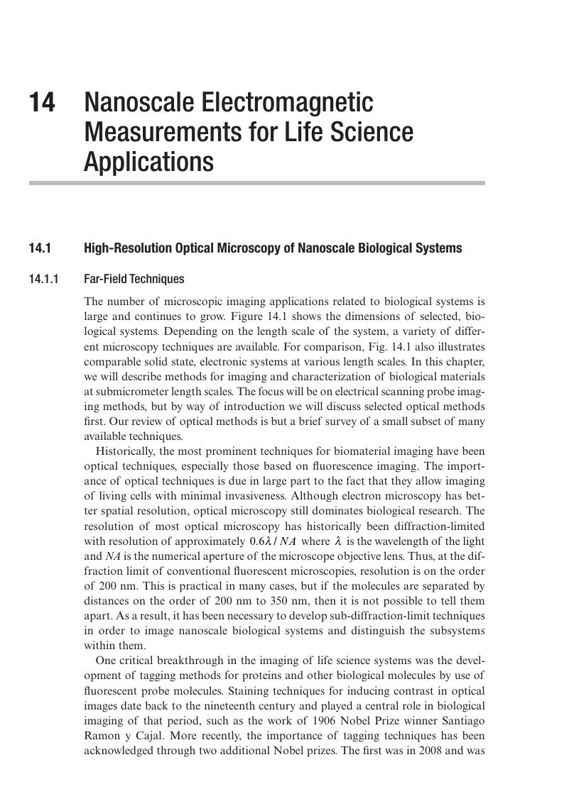
Image of the first page of this content. For PDF version, please use the ‘Save PDF’ preceeding this image.'
Type
Chapter
Information
Publisher: Cambridge University Press
Print publication year: 2017

Access options

Get access to the full version of this content by using one of the access options below. (Log in options will check for institutional or personal access. Content may require purchase if you do not have access.)

References

Hell, S. W., “Nobel Lecture: Nanoscopy with Freely Propagating Light,” Reviews of Modern Physics 87 (2015) pp. 11691181.CrossRefGoogle Scholar
Moerner, W. E., “Nobel Lecture: Single-Molecule Spectroscopy, Imaging, and Photocontrol: Foundations for Super-resolution Microscopy,” Reviews of Modern Physics 87 (2015) pp. 11831212.Google Scholar
Habuchi, S., “Super-resolution Molecular and Functional Imaging of Nanoscale Architectures in Life and Materials Science,” Frontiers in Bioengineering and Biotechnology 2 (2014) art. no. 20.Google Scholar
Axelrod, D., “Cell Substrate Contacts Illuminated by Total Internal Reflection Fluorescence,” Journal of Cell Biology, 89 (1981) pp. 141145.Google Scholar
Betzig, E., “Nobel Lecture: Single Molecules, Cells, and Super-resolution Optics,” Reviews of Modern Physics 87 (2015) pp. 11531167.CrossRefGoogle Scholar
Ash, E. A. and Nicholls, G., “Super Resolution Aperture Scanning Microscope,” Nature 237 (1972) pp. 510512.Google Scholar
Tetard, L., Passian, A., Venmar, K. T., Lynch, R. M., Voy, B. H., Shekhawat, G., Dravid, V. P. and Thundat, T., “Imaging Nanoparticles in Cells by Nanomechanical Holography,” Nature Nanotechnology 3 (2008) pp. 501505.Google Scholar
Guckenberger, R., Heim, M., Cevc, G., Knapp, H. F., Wiegräbe, W., and Hillebrand, A., “Scanning Tunneling Microscopy of Insulators and Biological Specimens Based on Lateral Conductivity of Ultrathin Water Films,” Science 266 (1994) pp. 15381540.Google Scholar
Hoh, J. J. and Hansma, P. K., “Atomic Force Microscopy for High Resolution Imaging in Cell Biology,” Trends in Cell Biology 2 (1992) pp. 208213.Google Scholar
Setia, Aman and Kishore Reddy, S., “Advancements in Microwave Breast Imaging Techniques,” IJREAS 2 (2012) pp. 16791690.Google Scholar
Burdette, E. C., Cain, F. L., and Seals, J., “In vivo Probe Measurement Technique for Determining Dielectric Properties at VHF Through Microwave Frequencies,” IEEE Transactions on Microwave Theory and Techniques MTT-28 (1980) pp. 414427.Google Scholar
Athe, T. W., Stuchly, M. A., and Stuchly, S. S., “Measurement of Radio Frequency Permittivity of Biological Tissues with an Open-Ended Coaxial Line: Part I,” IEEE Transactions on Microwave Theory and Techniques MTT-30 (1982) pp. 8286.CrossRefGoogle Scholar
Stuchly, M. A., Athey, T. W., Samaras, G. M., and Taylor, G. E., “Measurement of Radio Frequency Permittivity of Biological Tissues with an Open-Ended Coaxial Line: Part II – Experimental Results,” IEEE Transactions on Microwave Theory and Techniques MTT-30 (1982) pp. 8792.Google Scholar
Reznik, A. N. and Yurasova, N. V., “Electrodynamics of Near Field Probing: Application to Medical Diagnostics,” Journal of Applied Physics 98 (2005) art. no. 114701.Google Scholar
Alanenyz, E., Lahtinenyx, T. and Nuutineny, J., “Measurement of Dielectric Properties of Subcutaneous Fat with Open-Ended Coaxial Sensors,” Physics in Medicine Biology 43 (1998) pp. 475485.CrossRefGoogle Scholar
Wu, X. and Romahi, O. M., “Near-Field Scanning Microwave Microscopy for Detection of Subsurface Biological Anomalies,” Antennas and Propagation Society International Symposium 3 (2004) pp. 24442447.Google Scholar
Sihvola, A., Electromagnetic Mixing Formulas and Applications (Institution of Engineering and Technology, 1999).CrossRefGoogle Scholar
Reznik, A. N. and Yurasova, N. V., “Detection of Contrast Objects Inside Biological Media by Near-Field Microwave Diagnostics,” Technical Physics 51 (2006) pp. 8699.Google Scholar
Klein, L. A. and Swift, C. T., “An Improved Model for the Dielectric Constant of Sea Water at Microwave Frequencies,” IEEE Transactions on Antennas and Propagation AP-25 (1977) pp. 104111.Google Scholar
Yang, L., Weerasinghe, S., Smith, P. E., and Pettitt, B. M.Dielectric Response of Triplex DNA in Ionic Solution from Simulations,” Biophysical Journal 69 (1995) pp. 15191527.Google Scholar
Cuervo, A., Dans, P. D., Carrascosa, J. L., Orozco, M., Gomila, G., and Fumagalli, L., “Direct Measurement of the Dielectric Polarization Properties of DNA,” PNAS (2014) pp. E3624E3630.Google ScholarPubMed
Biagi, M. Ch., Fabregas, R., Gramse, G., Van Der Hofstadt, M., Juárez, A., Kienberger, F., Fumagalli, L., and Gomila, G., “Nanoscale Electric Permittivity of Single Bacterial Cells at Gigahertz Frequencies by Scanning Microwave Microscopy,” ACS Nano 10 (2016) pp. 280288.Google Scholar
Gramse, G., Edwards, M. A., Fumagalli, L., and Gomila, G., “Theory of Amplitude Modulated Electrostatic Force Microscopy for Dielectric Measurements in Liquids at MHz Frequencies,” Nanotechnology 24 (2013) art. no. 415709.CrossRefGoogle ScholarPubMed
Gramse, G., Edwards, M. A., Fumagalli, L., and Lluch, G. Gomila, “Dynamic Electrostatic Force Microscopy in Liquid Media,” Applied Physics Letters 101 (2012) art. no. 213108.Google Scholar
Gramse, G., Dols-Perez, A., Edwards, M. A., Fumagalli, L., and Gomila, G., “Nanoscale Measurement of the Dielectric Constant of Supported Lipid Bilayers in Aqueous Solutions with Electrostatic Force Microscopy,” Biophysical Journal 104 (2013) pp. 12571262.Google Scholar
Wei, T. X., Xiang, X. -D., Wallace-Freedman, W. G., and Schultz, P. G., “Scanning Tip Microwave Near-Field Microscope,” Applied Physics Letters 68 (1996) pp. 35063508.Google Scholar
Tabib-Azar, M., Katz, J. L., and LeClair, S. R., “Evanescent Microwaves: A Novel Super-Resolution Noncontact Nondestructive Imaging Technique for Biological Applications,” IEEE Transactions on Instrumentation and Measurement 48 (1999) pp. 11111116.Google Scholar
Farina, M., Lucesoli, A., di Donato, A., Mencarelli, D., Maccari, L., Venanzoni, G., Morini, A., and Rozzi, T., “Algorithm for Reduction of Noise in Ultramicroscopy and Application to Near-Field Microwave Microscopy,” Electronics Letters 46 (2010) pp. 5052.CrossRefGoogle Scholar
Coakley, K. J., Imtiaz, A., Wallis, T. M., Weber, J. C., Berweger, S., and Kabos, P., “Adaptive and Robust Statistical Methods for Processing Near-Field Scanning Microwave Microscopy Images,” Ultramicroscopy 150 (2015) pp. 19.CrossRefGoogle ScholarPubMed
LOCFIT package can be obtained via www.locfit.info/Google Scholar
Gramse, G., Kasper, M., Fumagalli, L., Gomila, G., Hinterdorfer, P. and Kienberger, F., “Calibrated Complex Impedance and Permittivity Measurements with Scanning Microwave Microscopy,” Nanotechnology 25 (2014) art. no. 145703.Google Scholar
Fabiani, S., Lucesoli, A., di Donato, A., Mencarelli, D., Venanzoni, G., Morini, A., Rozzi, T., and Farina, M., “Dual-Channel Microwave Scanning Probe Microscopy for Nanotechnology and Molecular Biology,” Proceedings of the 40th European Microwave Conference (2010) pp. 767770.Google Scholar
Farina, M., Mencarelli, D., Di Donato, A., Venanzoni, G., and Morini, A., “Calibration Protocol for Broadband Near-Field Microwave Microscopy,” IEEE Transactions on Microwave Theory and Techniques 59 (2011) pp. 27692776.Google Scholar
Kochanski, G. P., “Nonlinear Alternating-Current Tunneling Microscopy,” Physical Review Letters 62 (1989) pp. 22852288.CrossRefGoogle ScholarPubMed
Farina, M., Di Donato, A., Mencarelli, D., Venanzoni, G., Morini, A. and Pietrangelo, T., “Imaging of Biological Structures by Near-Field Microwave Microscopy,” Proceedings of the 45th European Microwave Conference (2015) pp. 666669.Google Scholar
Tselev, A., Velmurugan, J., Ievlev, A. V., Kalinin, S. V., and Kolmakov, A., “Seeing through Walls at the Nanoscale: Microwave Microscopy of Enclosed Objects and Processes in Liquids,” ACS Nano 10 (2016) pp. 35623570.Google Scholar

Save book to Kindle

To save this book to your Kindle, first ensure [email protected] is added to your Approved Personal Document E-mail List under your Personal Document Settings on the Manage Your Content and Devices page of your Amazon account. Then enter the ‘name’ part of your Kindle email address below. Find out more about saving to your Kindle.

Note you can select to save to either the @free.kindle.com or @kindle.com variations. ‘@free.kindle.com’ emails are free but can only be saved to your device when it is connected to wi-fi. ‘@kindle.com’ emails can be delivered even when you are not connected to wi-fi, but note that service fees apply.

Find out more about the Kindle Personal Document Service.

Available formats
×

Save book to Dropbox

To save content items to your account, please confirm that you agree to abide by our usage policies. If this is the first time you use this feature, you will be asked to authorise Cambridge Core to connect with your account. Find out more about saving content to Dropbox.

Available formats
×

Save book to Google Drive

To save content items to your account, please confirm that you agree to abide by our usage policies. If this is the first time you use this feature, you will be asked to authorise Cambridge Core to connect with your account. Find out more about saving content to Google Drive.

Available formats
×