


Introduction
Nematodes of the genus Serpentirhabdias (Tkach et al., Reference Tkach, Kuzmin and Snyder2019), within the family Rhabdiasidae (Railliet, Reference Railliet1899), exhibit alternating free-living and parasitic life cycles, infesting the lungs of various snake species. Currently, 22 valid species are described (Kuzmin and Tkach, Reference Kuzmin and Tkach2024; Velázquez-Brito et al., Reference Velázquez-Brito, García-Prieto, Garduño-montes de Oca, Sosa-Jiménez, Vera-Chávez and León-Règagnon2024), characterized by 2–8 mm in length, with rounded anterior and pointed posterior ends. Their mouths feature 6 equally sized lips arranged in pairs, with the presence/absence of onchia in the oesophagostome. This character is useful for dividing Serpentirhabdias species into 2 groups: 5 species with onchia and 17 without (Velázquez-Brito et al., Reference Velázquez-Brito, García-Prieto, Garduño-montes de Oca, Sosa-Jiménez, Vera-Chávez and León-Règagnon2024). Moreover, the 5 species with onchia lack a buccal capsule, while species without onchia usually have a small and thin-walled buccal cavity (Velázquez-Brito et al., Reference Velázquez-Brito, García-Prieto, Garduño-montes de Oca, Sosa-Jiménez, Vera-Chávez and León-Règagnon2024). The oesophagus is club-shaped with a bulbous posterior. They possess distinct excretory glands and an amphidelphic reproductive system, with seminal receptacles formed by thick-walled parts of the oviduct. The vulva is located at the equatorial or pre-equatorial position, and their tails are conical with pointed tips (Tkach et al., Reference Tkach, Kuzmin and Snyder2019).
Species of Serpentirhabdias have been documented in various global regions: 10 species in the Neotropics, 7 in the northern part of the Palaearctic, 3 in the Nearctic, 1 in South Africa and 1 in Australia (see Table S1 for species distribution). Notably, Serpentirhabdias fuscovenosa (Railliet, Reference Railliet1899; Goodey, Reference Goodey1924) has reportedly been found in both the Nearctic and Palaearctic regions (Baker, Reference Baker1978, Reference Baker1987), though they may represent distinct species (Machado et al., Reference Machado, Kuzmin, Tkach, Dos Santos, Gonçalves and de V2018; Kuzmin et al., Reference Kuzmin, Tkach and Melo2020). Species found in the Palaearctic region, corresponding to reported host distributions (refer to the reptile-database.org – Uetz et al., Reference Uetz, Freed, Aguilar, Reyes, Kudera and Hošek2024), are typically restricted to the temperate zones of Asia and Europe (Table S1). Importantly, no species have been reported infecting snakes in the Oriental (Southeast Asia) region. Additionally, all Serpentirhabdias spp. have been reported infecting snakes primarily in the families Viperidae and Colubridae, with a few species in Dipsadidae and Boidae, but none in Elapidae. This highlights a research gap and lack of attention to the lungworms of snakes within this wildlife, particularly snakes, within this biogeographic region.
The monocled cobra, Naja kaouthia Lesson, 1831, is common and widely distributed across the Oriental regions and Thailand (Stuart and Wogan, Reference Stuart and Wogan2012; Ratnarathorn et al., Reference Ratnarathorn, Harnyuttanakorn, Chanhome, Evans and Day2019, Reference Ratnarathorn, Nadolski, Sumontha, Hauser, Suntrarachun, Khunsap, Laoungbua, Radcliffe, Vasaruchapong, Tawan and Chanhome2023). Belonging to the family Elapidae, all species of this genus are deadly venomous and of medical importance (Mattison, Reference Mattison2007). This cobra species primarily preys on amphibians, reptiles, small mammals, and chicks (Cox et al., Reference Cox, Hoover, Chanhome and Thirakhupt2012), which facilitates its role as a host for parasitic helminths, given its behaviour as an active predator (Jintakune, Reference Jintakune2004). Being highly adaptable to anthropogenic environments, this cobra species plays a crucial ecological role within human-altered habitats (Ratnarathorn et al., Reference Ratnarathorn, Sanunsilp, Laoungbua, Tawan, Sumontha, Kongrit and Thaweepworadej2024), potentially serving as a reservoir for pathogens and, thereby, presenting an attractive subject for zoonotic research. The study of parasitic infections in snakes highlights their relevance to wildlife health and management, particularly in the context of exotic pet ownership and interactions with wild animals (e.g. Charoennitiwat et al., Reference Charoennitiwat, Thaenkham, Tongpon, Chaisiri, Laoungbua, Tawan, Kanjanapruthipong, Ampawong, Chan and Ratnarathorn2024b).
During our survey of parasitic helminths in snakes, a high prevalence of helminths was found inside the lung, trachea and heart of the monocled cobras captured from central Thailand. These helminths were classified as Serpentirhabdias due to their general morphology, parasitism in snakes and organ infected (Tkach et al., Reference Tkach, Kuzmin and Snyder2019). Only parasitic female nematodes were found in the cobras, as males are reportedly free-living (Tkach et al., Reference Tkach, Kuzmin and Snyder2019; Kuzmin et al., Reference Kuzmin, Tkach and Melo2019). Supported by preliminary genetic results, the biological data addressed the species distinction of the specimens in the genus, leading to the suspicion of new species. Consequently, the present study was initiated.
A substantial number of lungworm specimens were analysed using both molecular phylogenetic techniques and detailed morphological examinations. The primary objective of the morphological analysis was to identify distinguishing features of the new nematode species within its taxonomic context, complementing insights from genetic data. Phylogenetic relationships were explored through the reconstruction of genetic sequences, including cytochrome c oxidase subunit 1 (COI) and large subunit nuclear ribosomal RNA (28S rRNA). The study aims to provide strong molecular and morphological evidence supporting the description of the newly identified Serpentirhabdias orientalis sp. nov., the first report of this species in Southeast Asia and in elapid snakes.
Materials and methods
Host and parasite specimen preparation
Five carcasses of wild monocled cobras (Naja kaouthia) were obtained from the Snake Farm at Queen Saovabha Memorial Institute, Thai Red Cross Society in Bangkok, Thailand. These cobras, captured by locals and rescuers from the central region, Thailand near Bangkok, died during quarantine. Before dissection, the snakes were examined using criteria from Cox et al. (Reference Cox, Hoover, Chanhome and Thirakhupt2012), assessing morphological measurements to confirm species and collect host data. Dissections followed protocols by Ratnarathorn and Kongrit (Reference Ratnarathorn and Kongrit2024) to investigate lung, heart and trachea parasites. Organs were placed in Petri dishes with 0.85% saline and examined under stereomicroscopes (Olympus, SZ30 and SZ51, Japan). Micro-dissecting needles were used to isolate parasites, which were recorded and preserved in 70% ethanol at – 20 °C in the Department of Helminthology, Faculty of Tropical Medicine. Moreover, 10 specimens were preserved in glutaraldehyde for scanning electron microscopy (SEM).
Morphological study
For morphological studies, 10 complete female helminth specimens were selected from the preserved 70% ethanol stock to create permanent slides. Three specimens in excellent condition were chosen as the holotype and paratypes. Each specimen was stained with acetocarmine and dehydrated by sequential immersion in ethanol (70%, 80%, 90%, 95% and 100%) for 45 min each. For clearing and preparation, specimens were placed in a 1:1 ethanol: xylene solution for 45 min, then briefly in xylene, before being mounted in Permount™ on glass slides, covered with coverslips, and incubated at 60 °C for several days. The remaining specimens (n = 47) were mounted using Lactophenol.
A comprehensive examination was conducted using an inverted microscope (Zeiss, Primovert, Germany) equipped with a Zeiss Axiocam and ZEN2 blue edition software. All measurements were recorded in micrometers (µm). Taxonomic keys for identifying Serpentirhabdias species and relevant morphological features were derived from Tkach et al. (Reference Tkach, Kuzmin and Snyder2019), Kuzmin et al. (Reference Kuzmin, Tkach and Melo2019, Reference Kuzmin, Tkach and Melo2020, Reference Kuzmin, du Preez and Svitin2021) and Velázquez-Brito et al. (Reference Velázquez-Brito, García-Prieto, Garduño-montes de Oca, Sosa-Jiménez, Vera-Chávez and León-Règagnon2024). Illustrations (see Figure 1) were generated using a light microscope with a camera lucida (Leitz, Wetzlar, Germany).

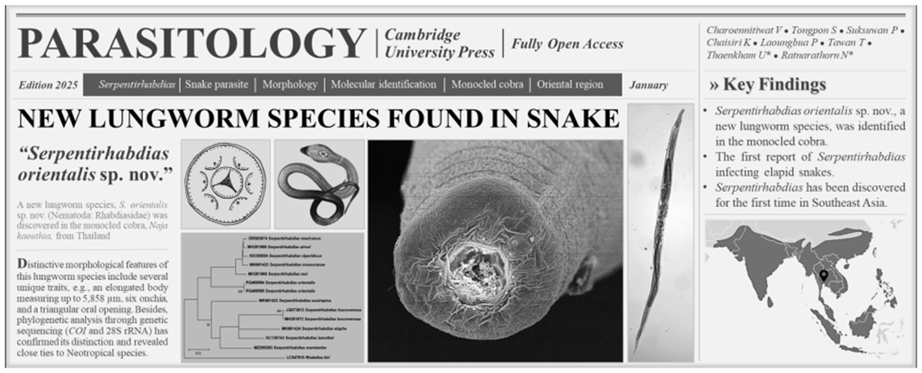
Figure 1. Serpentirhabdias orientalis sp. nov. (A) Entire body; (B) anterior end, ventral view, with dashed lines indicating optical cross-sectional views at oesophagostome levels for C and D; (C) anterior end, apical view; (D) optical cross-section at the intermediate-depth level of the oesophagostome, apical view; (E) anterior part of the body, lateral view; (F) anterior part of the body, ventral view; (G) posterior end and tail, lateral view; (H) vulva region at mid-body, lateral view; and (I) egg in the vulva. AN, anus; CP, cephalic papilla; IN, intestine; LEG, large excretory gland; NR, nerve ring; ON, onchia; OS, oesophagus; PH, phasmid; RA, radial muscle of the oesophagus; SEG, small excretory gland; SY, syngonium; and VU, vulva.
For the SEM analysis, 10 specimens were initially immersed in a solution of 2.5% glutaraldehyde in 0.1 M sucrose phosphate buffer (SPB) for primary fixation, followed by secondary fixation in 1% osmium tetroxide in 0.1 M SPB. The specimens were then dehydrated using ethanol and dried with a critical point dryer (CPD300 auto, Leica, Wetzlar, Germany). They were coated with gold using a sputter coater (Q150R PLUS, Quorum, East Sussex, England). Specimen preparation was carried out at the Department of Tropical Pathology, Faculty of Tropical Medicine, Mahidol University, while SEM imaging (SU8010, Hitachi High-Tech, Japan) was conducted at the Central Instrument Facility, Faculty of Science (Phaya Thai), Mahidol University (see Figure 2 for illustrations).

Figure 2. Scanning electron micrographs of Serpentirhabdias orientalis sp. nov. (A) anterior region with lips (black arrows), apical view; (B) mouth opening with cephalic papillae (white arrows), apical view, enlarged from A; (C) tail with anus [AN], ventral view; (D) cuticle at mid-body; (E) magnified image of the cephalic papilla; (F) magnified image of the anus, lateral view; and (G) vulva opening, lateral view.
To assess the morphological variation among the 52 helminth specimens (with 5 incomplete individuals excluded), 19 distinct morphological characteristics were measured, all expressed in the same measurement unit (μm). The measurements are presented as the mean, followed by the standard deviation, with the range in parentheses. These included body length, body width, body width at the oesophago–intestinal junction, buccal cavity width, buccal cavity depth, oesophagus length, oesophageal width at the anterior end, minimum oesophageal width, oesophageal bulb width, the distance from the anterior end to the nerve-ring, the distance from the anterior end to the excretory pore, the distance from the flexure of the posterior syngonium to the tail end, the distance from the anterior end to the vulva, the distance from the anterior end to the flexure of the anterior syngonium, the lengths of the large and small excretory glands, tail length, egg length and egg width (see Tables 1 and S1). A variance–covariance matrix was employed to analyse these 19 characteristics. Additionally, 4 characteristics were included to further examine variations among specimens from different hosts: oesophagus length as a percentage of body length, distance from the anterior end to the vulva as a percentage of body length, tail length, tail length as a percentage of body length and the number of eggs in the uteri (see Table S2). These 4 were analysed in conjunction with the original 19 using a correlation model. The resulting multivariable data matrices were analysed with principal component analysis (PCA) via PAST version 4.06b software (Hammer et al., Reference Hammer, Harper and Ryan2001). Both variance–covariance and correlation matrices generated two-dimensional scatter plots that illustrated the morphological variation among specimens and across hosts, along with the corresponding percentage variances. Additionally, to identify the morphological traits causing divergence among host groups, the Discriminant Analysis function in the programme was used.
Table 1. Information and measurement characters for Serpentirhabdias orientalis sp. nov., S. viperidicus, S. atroxi, S. mexicanus, S. moi and S. mussuranae. Diagnostic characters for the new species are indicated in bold type. All measurements, in micrometres (μm), are presented as the mean, followed by the standard deviation, with the range in parentheses

Molecular and phylogenetic study
For DNA extraction, 2 specimens were homogenized and processed using the DNeasy Blood & Tissue Kit (Qiagen, Germany) according to the manufacturer’s protocol. The extracted genomic DNA was eluted with 30 µL of nuclease-free water.
The amplification focused on partial sequences of 2 genes: the mitochondrial cytochrome c oxidase subunit I (COI) and the nuclear 28S ribosomal RNA (28S rRNA), chosen for their utility in molecular identification and genetic diversity studies in nematodes (Chan et al., Reference Chan, Chaisiri, Dusitsittipon, Jakkul, Charoennitiwat, Komalamisra and Thaenkham2020; Thaenkham et al., Reference Thaenkham, Chaisiri and Chan2022). The primers used were LCO 5′-GGTCAACAAATCATAAAGATATTGGT-3′ and HCO 5′-TAAACTTCAGGGTGACCAAAAAATCA-3′ for COI, and #500 5′-ACTTTGAAGAGAGAGTTCAAGAG-3′ and #501 5′-TCGGAAGGAACCAGCTACTA-3′ for 28S rRNA. The expected amplicon lengths were 721 bp for COI and 681 bp for 28S rRNA.
PCRs were performed using a T100™ thermocycler (Bio-Rad) in a 30 µL mixture containing 15 µL of 2X i-Taq master mix (Biotechnology, Gyeonggi, South Korea), 10 µM of each primer, and 1 ng/µL of DNA. For the COI primers, the thermocycling profile included an initial denaturation at 94 °C for 5 min, followed by 35 cycles of 94 °C for 1 min, 50 °C for 1 min and 72 °C for 1 min, with a final extension at 72 °C for 10 min (Folmer et al., Reference Folmer, Black, Hoeh, Lutz and Vrijenhoek1994). For the 28S rRNA primers, the profile began with an initial denaturation at 94 °C for 3 min, followed by 55 cycles of 94 °C for 30 sec, 54 °C for 30 sec and 72 °C for 1 min, with a final extension at 72 °C for 7 min (Dare et al., Reference Dare, Nadler and Forbes2008). PCR amplicons were visualized on a 1% agarose gel stained with SYBR Safe (Thermo Fisher Scientific, USA). Selected products were sequenced using Fast Next-generation sequencing (Tsingke, Beijing, China). Nucleotide sequences were submitted to the NCBI database with accession numbers PQ469594–95 for COI and PQ471682–83 for 28S rRNA.
Partial sequences of the genes were manually inspected and edited using BioEdit version 7.2.5. Phylogenetic analysis was conducted using maximum likelihood in MEGA-X, with sequence alignment performed by ClustalX 2.1. Best-fit nucleotide substitution models were selected for each gene: Tamura-Nei (TN93) with a discrete gamma distribution (+G) and by assuming that a certain fraction of sites is evolutionarily invariable (+I) for COI and Kimura 2-parameter (K2) with a discrete gamma distribution (+G) for 28S rRNA. Phylogenetic trees were constructed with 1000 bootstrap replicates to ensure robust analysis (Hall, Reference Hall1999; Thompson et al., Reference Thompson, Gibson and Higgins2002; Tamura et al., Reference Tamura, Stecher, Peterson, Filipski and Kumar2013). The COI and 28S rRNA analyses incorporated the sequences of all available species within Serpentirhabdias and other species of the family Rhabditidae. For the COI analysis, Rhabdias kiri was used as the outgroup, while various Rhabditid species served as outgroups for the 28S rRNA analysis. All sequences were retrieved from GenBank (see Figure 5).
Results
Taxonomy
Phylum: Nematoda Diesing, 1861
Class: Chromadorea Inglis, 1983
Order: Rhabditida Chitwood, 1933
Family: Rhabdiasidae (Railliet, Reference Railliet1899)
Genus: Serpentirhabdias (Tkach et al., Reference Tkach, Kuzmin and Snyder2019)
Species: Serpentirhabdias orientalis sp. nov. (Charoennitiwat et al., 2025; Table 1, Figures 1–3).

Figure 3. Permanent slides (acetocarmine dye, A–E) and semi-permanent slides (lactophenol, F and G) of Serpentirhabdias orientalis sp. nov.: (A) entire body; (B) anterior region, ventral view; (C) vulva region at mid-body, lateral view; (D) posterior end and tail, lateral view; (E) anterior end showing onchia (black arrows), ventral view, with dashed lines indicating optical cross-sectional views at the oral part for F, and at the oesophagus for G; (F) oral part showing lips and triangular opening, apical view; and (G) optical cross-section at the intermediate level of the oesophagus, apical view. A, anus; DR ,deirid; IN, intestine; LEG, large excretory gland; OS, oesophagus; Oss, oesophagostome; PH, phasmid, RA, radial muscle of oesophagus, SEG, small excretory gland; SY, syngonium, and VU, vulva.
Type-host: Naja kaouthia Lesson, 1831
Type-locality: Bangkok’s suburbs (e.g. Don Mueang district, Prawet district, etc.) in Thailand. Coordinates were not recorded.
Collection date: November 10th, 2020 to August 26th, 2023.
Site of infection: Lung, trachea and heart
Parasite intensity: 6–216 worms, mean approximately 58 (based on 5 host specimens)
ZooBank LSID:
Etymology: The specific epithet ‘orientalis’ indicates that the nematode species is discovered in oriental region. We propose the colloquial English name for this nematode as the ‘Oriental snake lungworm’.
General description
Based on 57 parasitic adult female specimens (Table 1): body elongated, with anterior end rounded, and posterior end tapering (Figures 1A and 3A). Body length 5144 ± 431 (3360–5858). Width 207 ± 23 (137–249), and width 120 ± 15 (74–148) at oesophago–intestinal junction. Cuticle thin and smooth (Figure 2D). Oral opening triangular and wide in apical view (Figures 1C, 2A, and 3F, G); buccal cavity width 23 ± 3 (14–30) and depth 17 ± 2 (11–24). Oesophagus 351 ± 28 (251–402) in length, representing 7 ± 0.4 (6–8) % of body length, with 38 ± 4 (24–51) width at anterior end and 38 ± 4 (24–49) minimum width; oesophageal bulb 71 ± 11 (36–99) width. Six lips similar in shape and size, arranged in 2 lateral groups of 3 (Figures 1C and 2A). Each lip with small, cone-like papillae (Figure 2B and E). Six onchia project outward from top of radial muscle in oesophagostome (Figures 1B, D, and 3E). Distance from anterior end to nerve-ring 182 ± 23 (111–234); anterior end to excretory pore 279 ± 26 (191–325). Excretory gland length 521 ± 44 (340–593) and 555 ± 46 (362–621) in smaller and larger glands, respectively (Figures 1E, F, and 3B). Distance from anterior end to anterior syngonium flexure 892 ± 183 (365–1485) and from posterior syngonium flexure to tail end 986 ± 169 (605–1478). Distance from anterior end to vulva 2387 ± 237 (1497–2950), representing 46 ± 3 (42–59) % of body length. Uteri containing 34 ± 16 (12–76) eggs (Figures 1H and 3C). Egg size (width × length) 51 ± 6 (30–62) × 84 ± 7 (60–99) (Figure 1I). Tail narrow, elongated, gradually tapering posteriorly (Figures 1G, 2C, and 3D). Tail length 264 ± 24 (209–317), representing 5 ± 0.5 (4–7) % of body length.
Type materials
A parasitic gravid female specimen, designated as the holotype (Voucher no.: MUMNH-NEM0033; specimen code: SN063_Holotype), and 2 parasitic female paratypes (Voucher nos.: MUMNH-NEM0034–0035; specimen codes: SN127_Paratype1–2) were deposited at the Mahidol University Museum of Natural History. All specimens were collected from the lungs of the monocled cobras (Naja kaouthia), with the holotype (project ID: SN063; AASL ID: AAS107 [EL-Nk-029]) collected on August 3rd, 2023, and the paratypes (project ID: SN127; AASL ID: AAS091 [EL-Nk-027]) on November 9th, 2023, by Vachirapong Charoennitiwat and his team at the Department of Helminthology, Faculty of Tropical Medicine, Mahidol University. Measurements for both the holotype and paratypes are provided in Table S2, with their morphological characteristics consistent with the general description.
Remarks
Serpentirhabdias orientalis sp. nov. is placed within the genus Serpentirhabdias based on key features, including 6 laterally arranged lips, a thin body cuticle without noticeable cuticular inflations, a vulva positioned near the mid-body with surrounding papillae, and the absence of a buccal capsule (Figures 2A–G and 3A–G). This species is also characterized by its lung parasitism in snakes, aligning it with other members of the genus (Tkach et al., Reference Tkach, Kuzmin and Snyder2019; Kuzmin et al., Reference Kuzmin, Giese, Melo, da Costa, Maschio and Dos Santos2016; Velázquez-Brito et al., Reference Velázquez-Brito, García-Prieto, Garduño-montes de Oca, Sosa-Jiménez, Vera-Chávez and León-Règagnon2024).
The genus Serpentirhabdias consists of 22 globally distributed species (Table S1), divided into 2 morphological groups based on the presence or absence of onchia in the oesophagostome. Six species, including Serpentirhabdias orientalis sp. nov., possess onchia (Figures 1B, D, and 3E), grouping it with Serpentirhabdias atroxi (Kuzmin et al., Reference Kuzmin, Giese, Melo, da Costa, Maschio and Dos Santos2016), Serpentirhabdias viperidicus (Morais et al., Reference Morais, Aguiar, Müller, Narciso, da Silva and da Silva2017), Serpentirhabdias moi (Machado et al., Reference Machado, Kuzmin, Tkach, Dos Santos, Gonçalves and de V2018), Serpentirhabdias mussuranae (Kuzmin et al., Reference Kuzmin, Tkach and Melo2019) and Serpentirhabdias mexicanus (Velázquez-Brito et al., Reference Velázquez-Brito, García-Prieto, Garduño-montes de Oca, Sosa-Jiménez, Vera-Chávez and León-Règagnon2024). All these species share the absence of a buccal capsule. Moreover, these 6 species are categorized into 2 subgroups based on the shape of their oral openings (Velázquez-Brito et al., Reference Velázquez-Brito, García-Prieto, Garduño-montes de Oca, Sosa-Jiménez, Vera-Chávez and León-Règagnon2024): the rounded-mouth species – S. atroxi, S. viperidicus and S. mexicanus – and the triangular-mouth species – S. moi, S. mussuranae and S. orientalis sp. nov (Figures 1C, 2A, and 3F).
Serpentirhabdias orientalis sp. nov. exhibits significant morphological distinctions from S. moi in several key traits. In particular, S. orientalis sp. nov. is much larger than S. moi in body length (ave. 5,144 [3360–5858] vs 2500 [2200–2600]), body width (ave. 207 [137–249] vs 109 [103–119]) and tail length (ave. 264 [209–317] vs 121 [114–128]). Further notable differences are observed in the dimensions of the buccal cavity, with S. orientalis sp. nov. having both a wider (ave. 23 [14–30]) and deeper (ave. 17 [11–24]) buccal cavity compared to S. moi (ave. 10 [10–11] and 7 [6–7], respectively). The length of the oesophagus also varies significantly, as S. orientalis sp. nov. has an average length of 351 (251–402), while S. moi averages 243 (233–256). Reproductive traits also differ significantly between the 2 species, with S. orientalis sp. nov. having a higher average number of eggs in its uteri (ave. 34 [12–76]) compared to S. moi (ave. 10 [6–13]). In terms of geographical distribution and host specificity, Serpentirhabdias orientalis sp. nov infects the lung, bronchi and heart of the monocled cobra, Naja kaouthia (Elapidae) in Southeast Asia, primarily Thailand. In contrast, S. moi parasitizes only the lung of the Linnaeus’ Sipo, Chironius exoletus (Colubridae), in the Neotropical region, specifically Brazil (Machado et al., Reference Machado, Kuzmin, Tkach, Dos Santos, Gonçalves and de V2018).
Serpentirhabdias orientalis sp. nov. can be differentiated from S. mussuranae by its longer tail length, with an average of 131 (120–150) for S. mussuranae. Additionally, the distance from the anterior end to the excretory pore is greater in the new species (ave. 279 [191–325]) compared to S. mussuranae (ave. 168 [160–185]). Notable differences also exist in buccal cavity dimensions, as S. orientalis sp. nov. has a greater width (ave. 23 [14–30] vs 14 [13–15]) and depth (ave. 17 [11–24] vs 11 [8–12]). Furthermore, S. orientalis sp. nov. infects multiple organs, contrasting with S. mussuranae, which infects only the lung. In terms of geographical distribution, S. mussuranae is found in the Neotropical region. Finally, regarding host specificity, S. mussuranae infects the mussurana, Clelia clelia within the Colubridae family (Kuzmin et al., Reference Kuzmin, Tkach and Melo2019).
Variation
To assess the homogeneity of S. orientalis sp. nov. without population or strain diversification, morphological measurements of female specimens were analysed. The results indicated no significant morphological clustering among individuals, as demonstrated by a two-dimensional plot of the PCA of PC1 and PC2, which revealed no clear separation (Figure 4A). This absence of differentiation is further supported by PCA accounting for 87.975% of the total variance, with a marked decline in eigenvalues from PC1 (77.379%) to PC2 (10.596%), and from PC2 to PC3 (3.253%).

Figure 4. Principal component analysis of Serpentirhabdias orientalis sp. nov. specimens: (A) Principal component analysis of all female samples based on 19 examined morphological characters (87.975% of total variance). Black dots and lines represent individual specimens and their group; (B) Principal component analysis of all female specimens based on 23 examined morphological characters (81.405% of total variance). Coloured dots represent individual female S. orientalis sp. nov. specimens hosted in different snake specimens, and corresponding-coloured lines reveal their clustering.
The intensity of S. orientalis sp. nov. infections ranged from 6 to 216 parasites, with a mean of 58. PCA analysis of all female specimens revealed 5 overlapping clusters, accounting for 81.405% of the total variance, indicating some degree of morphological variation among Serpentirhabdias specimens from different snake hosts (Figure 4B). Further analysis identified body length as the primary measurement contributing to the observed divergence, particularly among hosts represented by the green and purple clusters (Figure 4B). These findings suggest that body length may be a key morphological feature influenced by host factors, underscoring the need for further investigation into the host-related mechanisms driving this variation.
Genetic characterization and phylogenetic position
Both phylogenetic analyses (COI and 28S rRNA genes) confirmed the genus Serpentirhabdias as a monophyletic group (Figures 4A–B), with only the COI analysis recognizing S. orientalis sp. nov. as a distinct species within the genus (Figure 5A). In the COI phylogeny, the species of Serpentirhabdias, whether characterized by the presence or absence of onchia, clustered in accordance with their distribution and other morphological traits. Within the group possessing onchia, S. orientalis sp. nov. (in the Oriental region) exhibited a close phylogenetic relationship with S. mussuranae and S. moi (in the Neotropical region), both of which share a triangular oral opening. In contrast, S. mexicanus was closely related to S. viperidicus and S. atroxi (also in Neotropics), which possess a round oral opening. The 28S rRNA phylogeny displayed low genetic variation, making it difficult to clearly distinguish S. orientalis sp. nov. However, it confirmed that the genus Serpentirhabdias is distinct from other genera of reptilian/amphibian lungworms. All species within the genus Serpentirhabdias are associated with snake hosts, while the genera Rhabdias, Entomelas and Pneumonema infect frogs and toads, legless lizards, as well as skinks and salamanders. This finding also indicates that Neotropical species are separated from those in other regions (Figure 5B).

Figure 5. Phylogenetic analysis of available lungworm species sequences from the genus Serpentirhabdias and the Rhabdiasidae outgroup, using COI (A) and 28S rRNA (B) genes. These analyses were performed with the maximum likelihood method in MEGAX, with branch length scale bars representing the number of substitutions per site. (A): coloured characteristics represent variations in rhabditid helminths sourced from GenBank. Serpentirhabdias orientalis sp. nov., found in the oriental region in this study, is highlighted in red (font/line), while other coloured fonts/lines represent species from various biogeographic regions. Dashed lines indicate species with or without onchia. Symbols represent the shape of the oral opening: round (⚫), triangular (▲), and no data/suspected triangular (▲?). (B): coloured fonts/lines represent different genera of helminths and the specificity of the infected host groups.
The genetic distance matrix revealed a clear distinction between S. orientalis sp. nov. and its congeners, with genetic divergence for the COI gene ranging from 12% to 14% and for the 28S rRNA gene from 0.2% to 4%. Additionally, the genetic sequence variation in the COI and 28S rRNA genes between the 2 specimens of S. orientalis sp. nov. was 0%, indicating complete genetic similarity.
Natural history
Serpentirhabdias orientalis sp. nov. exhibits a high prevalence in its host, the monocled cobra (Naja kaouthia), with all 5 examined specimens testing positive for infection. This species is predominantly located in the lung, but it is occasionally found in the trachea and heart of the snakes, which were captured in Bangkok and surrounding areas. This finding is particularly significant as it represents the first identification of Serpentirhabdias sp. in the Oriental region and within the Elapid family.
Currently, only adult female parasites have been documented in the host, with no investigation into lesions or other life cycle aspects at this stage. However, the presence of numerous eggs in the intestinal contents of the host suggests that larvae may hatch externally.
Discussion
A new species of snake lungworm, Serpentirhabdias orientalis sp. nov., was discovered in the respiratory organs of the monocled cobra (Naja kaouthia) in Thailand, marking the first identification of a lungworm in an Elapid snake. This worm species belongs to the genus Serpentirhabdias, within the family Rhabdiasidae, known for infecting carnivorous reptiles (Tkach et al., Reference Tkach, Kuzmin and Snyder2019). Detailed taxonomic analyses revealed that the specimens exhibit distinct geographical, host-specific, morphological, and genetic characteristics that differentiate them from all previously described species within the genus Serpentirhabdias.
Parasite species in the genus Serpentirhabdias are known for their strong host specificity (Table S1), presumably co-evolving with their reptilian hosts over time. Phylogenetic analyses in this study supports this notion, revealing that S. orientalis sp. nov. in Southeast Asia has a deep evolutionary relationship with its host. This long-standing evolutionary history likely reflects the specialized nature of the host–parasite relationship, further indicating a co-evolutionary dynamic shaped by these interactions (e.g. Kuzmin et al., Reference Kuzmin, Tkach and Brooks2007; Buckingham and Ashby, Reference Buckingham and Ashby2022). Comparisons have shown that different Serpentirhabdias species tend to infect specific snake families (Table S1), highlighting the close link between parasite evolution and host diversification. However, 2 exceptions exist: Serpentirhabdias fuscovenosa, reported to infect species from 3 snake families – Colubridae, Boidae and Viperidae (Martínez-Salazar and León-Règagnon, Reference Martínez-Salazar and León-Règagnon2006; Tkach et al., Reference Tkach, Kuzmin and Snyder2019; Morais et al., Reference Morais, Aguiar, Müller, Narciso, da Silva and da Silva2017; Kuzmin and Tkach, Reference Kuzmin and Tkach2024) – and Serpentirhabdias eustreptos, which parasitises both Viperidae and Colubridae (MacCallum, Reference MacCallum1916; Kuzmin and Tkach, Reference Kuzmin and Tkach2024). The discovery of S. orientalis sp. nov. suggests that Southeast Asia may harbour additional, yet undiscovered, parasite species with similarly specialized host associations.
In terms of geographical distribution, the genus Serpentirhabdias comprises 22 species found globally (Table S1). In Asia, 7 species have been reported, including Serpentirhabdias fuscovenosa (Railliet, Reference Railliet1899; Goodey, Reference Goodey1924), in the Holarctic region; in the Western Palaearctic region, Serpentirhabdias elaphe (Sharpilo, Reference Sharpilo1976) and Serpentirhabdias martinoi (Kurochkin and Gus’kov, Reference Kurochkin and Guskov1963); in the Eastern Palaearctic region, Serpentirhabdias horigutii (Yamaguti, Reference Yamaguti1943), Serpentirhabdias agkistrodonis (Sharpilo, Reference Sharpilo1976) and Serpentirhabdias vibakari (Kuzmin, Reference Kuzmin1996); and in the Eastern Palaearctic islands, Serpentirhabdias kurilensis (Sharpilo, Reference Sharpilo1976). Prior to this study, no species from this genus had been reported in the Oriental region or Southeast Asia, highlighting the lack of research on lungworms in these areas. Given that Southeast Asia, and Thailand in particular, is home to approximately 250 species of snakes that inhabit a variety of ecosystems (Cox et al., Reference Cox, Hoover, Chanhome and Thirakhupt2012; Ratnarathorn et al., Reference Ratnarathorn, Sanunsilp, Laoungbua, Tawan, Sumontha, Kongrit and Thaweepworadej2024), it is plausible that other, as yet undiscovered, species of lungworms may exist in this region. Further investigations into other snake species in the same and neighbouring habitats may reveal additional infections by S. orientalis sp. nov. or other related helminth species. Continued exploration is warranted.
Serpentirhabdias orientalis sp. nov. represents the eighth species of this genus reported in Asia, and the first discovery in the Oriental region. The morphological characteristics of this new species, particularly the structure of the onchia, along with phylogenetic analysis, suggest an evolutionary relationship that is more closely aligned with species found in the Neotropical region (Kuzmin et al., Reference Kuzmin, Giese, Melo, da Costa, Maschio and Dos Santos2016, Reference Kuzmin, du Preez and Svitin2021; Morais et al., Reference Morais, Aguiar, Müller, Narciso, da Silva and da Silva2017; Machado et al., Reference Machado, Kuzmin, Tkach, Dos Santos, Gonçalves and de V2018; Velázquez-Brito et al., Reference Velázquez-Brito, García-Prieto, Garduño-montes de Oca, Sosa-Jiménez, Vera-Chávez and León-Règagnon2024). The morphological and molecular (COI) evidence supporting a close relationship between S. orientalis sp. nov. and Neotropical species presents a puzzling biogeographical scenario regarding how the parasites dispersed between these 2 continents.
Given that these parasites are highly host-dependent (e.g. Barrett et al., Reference Barrett, Thrall, Burdon and Linde2008; Tkach et al., Reference Tkach, Kuzmin and Snyder2019), their evolutionary history differs from that of their snake hosts. Snakes from South America and Southeast Asia share a common ancestor that dispersed across these continents around 100–90 million years ago, during the breakup of Gondwana, particularly in the case of boas and pythons (Pyron et al., Reference Pyron, Burbrink and Wiens2013). Notably, there is no direct evidence of any snake species crossing between Southeast Asia and South America, or vice versa, following the continental isolation after the breakup of Gondwana (Scotese, Reference Scotese2004; Vidal and Hedges, Reference Vidal and Hedges2009; Pyron et al., Reference Pyron, Burbrink and Wiens2013). The presence of lungworms with close evolutionary ties across these 2 continents suggests that the parasites may have evolved after the diversification of snakes and/or potentially dispersed via other means such as host co-transport (Chaisiri et al., Reference Chaisiri, Dusitsittipon, Panitvong, Ketboonlue, Nuamtanong, Thaenkham, Morand and Dekumyoy2019; Cowie et al., Reference Cowie, Ansdell, Dunavan and Rollins2022). Besides, morphological and genetic similarities observed in these lungworms might be attributed to convergent evolution, a process where unrelated organisms independently evolve the similarities due to analogous selective pressures in similar environments (Losos, Reference Losos2011). These aspects remain unexplored and represents an intriguing area for future research. Investigating the evolutionary timeline of these parasites in relation to their hosts’ distribution could shed light on the broader biogeographical patterns at play.
Despite the monophyly of the genus Serpentirhabdias, the positioning of S. orientalis sp. nov. differs between the COI and 28S rRNA phylogenies The COI analysis indicated a close relationship with Neotropical lungworms, while the 28S rRNA analysis did not provide a clear distinction. This discrepancy may be attributed to the significantly lower genetic divergence observed in the 28S rRNA compared to the COI, suggesting that the COI gene is more effective for species identification (e.g. Kuzmin et al., Reference Kuzmin, Tkach and Melo2019; Velázquez-Brito et al., Reference Velázquez-Brito, García-Prieto, Garduño-montes de Oca, Sosa-Jiménez, Vera-Chávez and León-Règagnon2024), whereas the 28S rRNA appears to be limited to genus-level differentiation. Future research focusing on genetics of other species within Serpentirhabdias, as well as the examination of additional genetic markers, may yield greater insights into the relationships among these parasites.
The male morphology of S. orientalis sp. nov. requires further investigation to enhance species identification. Future studies should explore sexual dimorphism and aspects of the nematode’s life cycle to provide a more comprehensive understanding of this species. The PCA of female specimens revealed distinct clusters, suggesting that individual host specimens may influence the morphological traits of the parasite. In particular, body length was identified as the primary parameter contributing to the observed divergence, likely reflecting differences in the maturity of the nematodes. However, the small sample size from each host highlights the necessity for larger studies to better assess variability related to host factors (Charoennitiwat et al., Reference Charoennitiwat, Chaisiri, Ampawong, Laoungbua, Chanhome, Vasaruchapong, Tawan, Thaenkham and Ratnarathorn2023, Reference Charoennitiwat, Chaisiri, Kanjanapruthipong, Ampawong, Chanhome, Vasaruchapong, Thaenkham and Ratnarathorn2024a, Reference Charoennitiwat, Thaenkham, Tongpon, Chaisiri, Laoungbua, Tawan, Kanjanapruthipong, Ampawong, Chan and Ratnarathorn2024b).
Ecologically, the presence of S. orientalis sp. nov. in the cobras prompts questions about the potential impact of parasitic infections on snake populations. Although the observed worm burden of up to 216 individuals per host may not necessarily indicate a high-intensity infection, the effect on host fitness remains uncertain. Given that monocled cobras are commonly found in rural and urban settings (Ratnarathorn et al., Reference Ratnarathorn, Sanunsilp, Laoungbua, Tawan, Sumontha, Kongrit and Thaweepworadej2024), further studies are needed to assess the ecological and conservation implications of this parasitism, particularly in stressed habitats.
From a veterinary and ecological standpoint, the discovery of S. orientalis sp. nov. underscores the importance of monitoring parasitic infections in reptiles. Although there is no evidence suggesting zoonotic potential, increasing wildlife–human interactions call for vigilance in understanding parasite dynamics. Additionally, parasitic infections in captive reptiles present veterinary challenges, emphasizing the need for biosecurity measures in the reptile trade, exotic pet care and conservation efforts (e.g. Charoennitiwat et al., Reference Charoennitiwat, Thaenkham, Tongpon, Chaisiri, Laoungbua, Tawan, Kanjanapruthipong, Ampawong, Chan and Ratnarathorn2024b).
In conclusion, this study reports the discovery and description of S. orientalis sp. nov., a new species of lungworm found in the respiratory organs of the monocled cobra (Naja kaouthia) in Thailand. This marks the first record of a Serpentirhabdias species in Southeast Asia and in an elapid snake. Detailed morphological and molecular analyses confirmed the distinctiveness of S. orientalis based on traits such as body size, buccal cavity dimensions and reproductive characteristics, differentiating it from closely related species. The phylogenetic analysis showed a close relationship with species found in the Neotropical region, raising questions about the biogeographical patterns of parasite dispersal. The high prevalence of infection in the cobras and its potential ecological and conservation impacts were noted.
Supplementary materials
The supplementary material for this article can be found at https://doi.org/10.1017/S0031182025000174.
Data availability
The data that support the findings of this study are available from the first and corresponding authors upon reasonable request.
Acknowledgements
Sincerely thank Chanisara Kaenkaew, Pakteema Namjad and all supporting staffs from the Snake Farm, Queen Saovabha Memorial Institute, the Department of Helminthology, Faculty of Tropical Medicine, the Department of Tropical Pathology, Faculty of Tropical Medicine, and the Department of Biology, Faculty of Science, Mahidol University for facilitating working spaces and convivences.
Author contributions
VC conducted data investigation, performed formal data analyses, curated data, and made methodology (helminth and host specimen preparation, morphological examination, SEM, and genetics), visualization, and the writing of the original draft. NR played a role in research conceptualization, methodology (helminth and host specimen preparation and SEM), formal data analyses, visualization, validation, funding, and the writing and editing of the original and final drafts. UT contributed to conceptualization, funding and provided recommendations. ST participated in methodology (PCA and SEM), the writing of the original draft and visualization. PS was responsible for methodology (including helminth and host specimen preparation, SEM and morphological examination). KC contributed to visualization, validation and provided recommendations. PL and TT assisted with specimen preparation, processing and resource management.
Financial support
This project is funded by National Research Council of Thailand (NRCT) and Mahidol University (Contact ID: N42A660912).
Competing interests
None.
Ethical standards
All procedures performed by researchers, snake handlers and veterinarians in handling snakes were approved by the Safety Committee of Queen Saovabha Memorial Institute (Document No. SN001). The authors confirm that the field studies did not involve endangered or protected species. The study was also approved by the Ethics Committee of Queen Saovabha Memorial Institute (Approval Protocol Number: QSMI-ACUC-11-2021).


 Open access