Hostname: page-component-745bb68f8f-d8cs5 Total loading time: 0 Render date: 2025-01-08T16:00:25.389Z Has data issue: false hasContentIssue false

Impact of calcium, vitamin D, vitamin K, oestrogen, isoflavone and exercise on bone mineral density for osteoporosis prevention in postmenopausal women: a network meta-analysis

Published online by Cambridge University Press:  04 September 2019

Zijun Xu
Affiliation:
Department of Community Health and Behavioral Medicine, School of Public Health, Shanghai Jiao Tong University, Shanghai 200000, People’s Republic of China
Huwen Wang
Affiliation:
Department of Community Health and Behavioral Medicine, School of Public Health, Shanghai Jiao Tong University, Shanghai 200000, People’s Republic of China
Yue Shi
Affiliation:
Department of Community Health and Behavioral Medicine, School of Public Health, Shanghai Jiao Tong University, Shanghai 200000, People’s Republic of China
Qiuming Shen
Affiliation:
Department of Community Health and Behavioral Medicine, School of Public Health, Shanghai Jiao Tong University, Shanghai 200000, People’s Republic of China
Lhakpa Tsamlag
Affiliation:
Department of Community Health and Behavioral Medicine, School of Public Health, Shanghai Jiao Tong University, Shanghai 200000, People’s Republic of China
Zezhou Wang
Affiliation:
Department of Cancer Prevention, Shanghai Cancer Center, Fudan University, Shanghai 200000, People’s Republic of China Department of Oncology, Shanghai Medical College, Fudan University, Shanghai 200000, People’s Republic of China
Shoukai Yu
Affiliation:
Department of Community Health and Behavioral Medicine, School of Public Health, Shanghai Jiao Tong University, Shanghai 200000, People’s Republic of China
Tian Shen
Affiliation:
Department of Community Health and Behavioral Medicine, School of Public Health, Shanghai Jiao Tong University, Shanghai 200000, People’s Republic of China
Ying Wang
Affiliation:
Department of Community Health and Behavioral Medicine, School of Public Health, Shanghai Jiao Tong University, Shanghai 200000, People’s Republic of China
Yong Cai*
Affiliation:
Department of Community Health and Behavioral Medicine, School of Public Health, Shanghai Jiao Tong University, Shanghai 200000, People’s Republic of China
*
*Corresponding author: Yong Cai, fax +86 21 63850472, email [email protected]
Rights & Permissions [Opens in a new window]

Abstract

The aim of this network meta-analysis is to compare bone mineral density (BMD) changes among different osteoporosis prevention interventions in postmenopausal women. We searched MEDLINE, Embase and Cochrane Library from inception to 24 February 2019. Included studies were randomised controlled trials (RCT) comparing the effects of different treatments on BMD in postmenopausal women. Studies were independently screened by six authors in three pairs. Data were extracted independently by two authors and synthesised using Bayesian random-effects network meta-analysis. The results were summarised as mean difference in BMD and surface under the cumulative ranking (SUCRA) of different interventions. A total of ninety RCT (10 777 participants) were included. Ca, vitamin D, vitamin K, oestrogen, exercise, Ca + vitamin D, vitamin D + vitamin K and vitamin D + oestrogen were associated with significantly beneficial effects relative to no treatment or placebo for lumbar spine (LS). For femoral neck (FN), Ca, exercise and vitamin D + oestrogen were associated with significantly beneficial intervention effects relative to no treatment. Ranking probabilities indicated that oestrogen + vitamin D is the best strategy in LS, with a SUCRA of 97·29 % (mean difference: +0·072 g/cm2 compared with no treatment, 95 % credible interval (CrI) 0·045, 0·100 g/cm2), and Ca + exercise is the best strategy in FN, with a SUCRA of 79·71 % (mean difference: +0·029 g/cm2 compared with placebo, 95 % CrI –0·00093, 0·060 g/cm2). In conclusion, in postmenopausal women, many interventions are valuable for improving BMD in LS and FN. Different intervention combinations can affect BMD at different sites diversely.

Type
Full Papers
Copyright
© The Authors 2019 

Osteoporosis is a common bone metabolic disease characterised by low bone mass and high fracture risk(1). Bone mineral density (BMD) decline increases the risk of fragility fractures, mainly of the spinal vertebrae, hip and radius(Reference Diez2). Hip and radial fractures are usually caused by falling, while vertebral fractures usually occur without external force(Reference Karinkanta, Piirtola and Sievanen3). Vertebral fractures may result in back pain, decreased body height and deformity(Reference Rao and Singrakhia4). Hip fractures are common at the intracapsular where the femoral neck (FN) is broken. Severe fractures can lead to prolonged bed rest, which increases mortality risk(Reference Piscitelli, Brandi and Nuti5). Hormonal changes in postmenopausal women lead to accelerated bone loss and osteoporosis(Reference Camacho, Petak and Binkley6), making them more vulnerable to osteoporosis and fragility fractures.

Ca, vitamin D and exercise are considered to be effective intervention methods to prevent bone loss, as mentioned in worldwide osteoporosis guidelines(Reference Camacho, Petak and Binkley6Reference Lorenc, Gluszko and Franek11). Guidelines also suggest oestrogens(Reference Khan and Fortier12), ‘natural’ oestrogens (isoflavones)(Reference Camacho, Petak and Binkley6) and vitamin K(Reference Camacho, Petak and Binkley6) supplements for prevention of bone loss in postmenopausal women. Many therapeutic treatments for osteoporosis are provided by guidelines but cannot completely restore bone integrity. People of all ages should pay attention to osteoporosis prevention, especially postmenopausal women(Reference Curry, Krist and Owens13). The effects of Ca, vitamin D, vitamin K, oestrogen, isoflavone and exercise singly or in combination on BMD in postmenopausal women have not been investigated in a network so far. It is uncertain which preventive measures can better reduce bone loss and should be chosen under particular conditions, such as when having limited budget, resource, time or when one is not suitable for a specific intervention.

Network meta-analysis is a relatively new meta-analysis technique that compares the therapeutic effects of different interventions based on both direct and indirect comparisons(Reference Mills, Ioannidis and Thorlund14). A randomised controlled trial (RCT) design can evaluate the effects of an intervention(Reference Grossman and Mackenzie15). The aim of the present study is to conduct a network meta-analysis of the existing RCT to compare the BMD changes generated by different combinations of osteoporosis prevention interventions in postmenopausal women and to rank the interventions for practical applications.

Methods

Search strategy and study selection

The present study was conducted following the Preferred Reporting Items for Systematic Reviews and Meta-analyses statement extension for network meta-analysis(Reference Hutton, Salanti and Caldwell16). We systematically searched MEDLINE, Embase and Cochrane Library from inception of each database to 24 February 2019. The keywords and MeSH terms used in the search strategy included Ca, vitamin D, vitamin K, oestrogen, isoflavone, exercise, postmenopausal, BMD and random. The full search strategies used in MEDLINE, Embase and Cochrane Library are provided in eMethod1 in the Supplement. Searches for Ca, vitamin D, vitamin K, oestrogen, isoflavone and exercise were conducted separately.

To make the present study both rigorous and manageable, six authors followed the same standard to conduct the literature review process in three independent pairs: X. Z. J. and W. H. W., W. Z. Z. and L. T. and S. Q. M. and S. Y. These three pairs of authors independently selected different possible interventions based on titles and abstracts (X. Z. J. and W. H. W.: Ca, vitamin K and exercise; W. Z. Z. and L. T.: vitamin D; S. Q. M. and S. Y.: oestrogen and isoflavone). All relevant systematic reviews and meta-analyses were reviewed to extract extra eligible trials. After removing duplicated trials from the databases and from systematic reviews and meta-analyses, the full texts of potentially relevant trials were reviewed by two authors independently (X. Z. J. and W. H. W.). Any disagreement between the two authors was resolved by consensus after discussion with a third investigator (C. Y.).

Inclusion and exclusion criteria

The inclusion criteria were as follows:

  1. (1) Study design: RCT and quasi-RCT, which uses a quasi-random method (such as medical record number) for allocating participants to different interventions;

  2. (2) Participants: postmenopausal women with natural or surgical menopause;

  3. (3) Intervention: single or combined treatment with Ca, vitamin D, vitamin K, oestrogen, isoflavone and exercise;

  4. (4) Comparison: no treatment, placebo for supplements or any intervention mentioned in (3);

  5. (5) Outcome: absolute mean difference in BMD, measured by dual-energy X-ray absorptiometry(Reference Kanis and Gluer17);

  6. (6) Time: study duration longer than 2 months.

Trials were excluded if:

  1. (1) they were abstracts, letters, conference reports without full text, duplications or not published in English;

  2. (2) the investigated postmenopausal women had any disease affecting bone metabolism, including musculoskeletal disease, renal failure, liver disorders, hyperparathyroidism, hyperthyroidism, diabetes mellitus, arthritis or cancer;

  3. (3) the intervention included dietary restriction, health education or other drugs that may affect bone metabolism, including bisphosphonate, fluoride, tamoxifen, calcitonin, corticosteroids, progestin, androgen or placebos for these drugs.

Data extraction and risk-of-bias assessment

Two authors (X. Z. J. and W. H. W.) extracted data from all eligible publications independently. Information including trial name, first author, year of publication, country, population, number of participants, average age, years since menopause (YSM), BMI, study duration, blinding, interventions and mean difference in BMD was extracted.

Two authors (X. Z. J. and W. H. W.) independently assessed the risk of bias with the Cochrane risk of bias assessment tool described in the Cochrane Handbook(Reference Higgins and Altman18), including the following seven categories: random-sequence generation (selection bias), allocation concealment (selection bias), blinding of participants and personnel (performance bias), blinding of outcome assessment (detection bias), incomplete outcome data (attrition bias), selective reporting (reporting bias) and other bias. Each category was judged as low risk, unclear risk or high risk. Discrepancies in data extraction and risk-of-bias assessment were resolved through discussion.

Statistical analysis

To compare all interventions simultaneously, a Bayesian network meta-analysis using Markov chain Monte Carlo simulation was conducted(Reference Lu and Ades19) to incorporate both indirect and direct comparisons. Treatment effects were estimated by random-effects network meta-analysis(Reference DerSimonian and Laird20). The generalised linear models were conducted with a logit link function with four chains and 20 000 iterated simulations, and the initial 5000 iterations were discarded as burn-in.

Effect sizes were summarised as weighted mean differences and 95 % credible intervals (95 % CrI) presented in forest plots. Trials reporting mean difference in BMD without standard deviation or standard error were included in the analysis, with standard deviation or standard error imputed when feasible(Reference Higgins and Altman18,Reference Furukawa, Barbui and Cipriani21) . The correlation between BMD at baseline and the end of intervention was calculated for all studies with complete outcome reports. The mean correlation was used to estimate the standard deviation or standard error in studies without available standard deviation or standard error values(Reference Abrams, Gillies and Lambert22). If two or more groups received the same intervention with different dosages, these groups were combined into a single group.

The relative ranking of osteoporosis prevention interventions and BMD changes was presented as rank probabilities and surface under the cumulative ranking (SUCRA) probabilities. SUCRA, which ranges between 0 and 100 %, was calculated by cumulative ranking probability, which represents the likelihood of being the best intervention(Reference Salanti, Ades and Ioannidis23,Reference Rucker and Schwarzer24) . In the present study, a higher SUCRA score represented a better intervention and increased BMD.

Between-study heterogeneity was assessed using the I² statistic, which ranges from 0 to 100 %. Between-study heterogeneity was also assessed by τ, which is independent of the study size(Reference Turner, Davey and Clarke25). The assumption of transitivity across treatment comparisons was assessed by comparing the distribution of BMI, the potential effect modifier, across the different pairwise comparisons using box plots(Reference Jansen and Naci26). Another important prerequisite for effective results is the consistency of direct and indirect evidence from the same treatment comparison, so the node-splitting model was used to assess potential inconsistency(Reference Bucher, Guyatt and Griffith27,Reference Dias, Welton and Caldwell28) . Publication bias was assessed using funnel plots(Reference Chaimani, Higgins and Mavridis29). Sensitivity analyses were performed by repeating the meta-analysis using the minimum and maximum correlation values of mean differences in BMD, adjusting the mean differences in BMD according to intervention duration and excluding studies with single group sample size less than 15.

Network meta-analysis was conducted using R software (version 3.5.1) with the gemtc(Reference Neupane, Richer and Bonner30) and rjags packages, JAGS (Plummer M, version 4.3.0) and STATA (version 13)(Reference Shim, Yoon and Shin31).

Results

Study selection

A total of 15 041 studies were identified from the three electronic databases (Fig. 1), among which 346 systematic reviews or meta-analyses were considered to be relevant to the topic and received full-text review. Of all the extracted articles considered eligible, 266 were extracted from systematic reviews or meta-analyses and another 549 were identified after screening the titles and abstracts from the databases; 642 articles received full-text review after removing duplicates. Of these studies, a total of ninety RCT met the inclusion criteria.

Fig. 1. Flow diagram of literature search and study. RCT, randomised controlled trial; BMD, bone mineral density; DXA, dual-energy X-ray absorptiometry.

Study characteristics

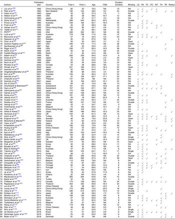
The characteristics of the RCT included are summarised in Tables 1 and 2. There were ninety RCT published between 1992 and 2018 that were included, and they had an average duration of 15·6 months. The present study included 10 777 participants with an average age of 62·7 years (range of average age, 42·7–82·4 years), an average YSM of 11·4 (range of average YSM, 0·9–32·5) and an average BMI of 25·4 kg/m2 (range of average BMI, 19·7–31·0 kg/m2). The population of three RCT were institutionalised women, and the remaining were non-institutionalised women.

Table 1. Description of included trials

YSM, years since menopause; LS, lumbar spine; FN, femoral neck; TC, trochanter; ITC, intertrochanter; WT, Ward’s triangle; TH, total hip; TB, total body; NA, not available; PEPI, Postmenopausal Estrogen/Progestin Interventions.

* Institutionalised women.

Only have thigh bone mineral density.

Table 2. Description of individual groups in included trials*

YSM, years since menopause; NA, not available; Ex, exercise; D, vitamin D; Est, oestrogen; PEPI, Postmenopausal Estrogen/Progestin Interventions; K, vitamin K; Iso, isoflavone; WBV, whole body vibration.

* Groups which did not meet the inclusion criteria are not shown.

Injection.

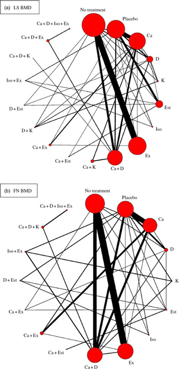
There were eighteen different intervention combination groups presented in the analysis: no treatment, placebo, Ca, vitamin D, vitamin K, oestrogen, isoflavone, exercise, Ca + vitamin D, Ca + vitamin K, Ca + oestrogen, Ca + exercise, vitamin D + vitamin K, vitamin D + oestrogen, isoflavone + exercise, Ca + vitamin D + vitamin K, Ca + vitamin D + exercise and Ca + vitamin D + isoflavone + exercise. The result of transitivity analysis conducted to assess the distribution of BMI across the different pairwise comparisons is shown in online Supplementary Fig. S1.

Among the ninety included RCT, seventy-four of them (n 8973, eighteen interventions) reported lumbar spine (LS) BMD, fifty-five (n 6707, sixteen interventions) reported FN BMD and 36, 11, 21, 25, 15 and 21 RCT reported trochanter, intertrochanter, Wald’s triangle, total hip, radius and total body BMD, respectively. Only the BMD values for LS and FN were included in the network meta-analysis because studies measuring the BMD of these two sites accounted for more than half the number of studies included and involved relatively complete intervention types (a total of eighteen different interventions were available in the present study).

Risk of bias

The risk of bias in the included RCT is shown in the Supplementary material (online Supplementary Table S1 and online Supplementary Fig. S2). Of the ninety RCT, the risk of bias was low for random-sequence generation in thirty-four RCT (37·8 %), allocation concealment in twenty RCT (22·2 %), blinding of participants and personnel in twenty-three RCT (25·6 %), blinding of outcome assessment in eighteen RCT (20·0 %), incomplete outcome data in thirty-six RCT (40·0 %) and other bias in eighty-eight RCT (97·8 %).

Publication bias

Funnel plots for publication bias in the network meta-analysis suggest no evidence of publication bias, but the fact that some studies were not in the 95 % CrI indicates the presence of heterogeneity (online Supplementary Fig. S3).

Lumbar spine

Network meta-analysis for the mean differences in LS BMD included seventy-four RCT (8973 participants) that used eighteen different types of interventions (Fig. 2(a)). The effects of each intervention are presented in Fig. 3(a). Ca (0·015 g/cm2, 95 % CrI 0·0024, 0·028 g/cm2), vitamin D (0·019 g/cm2, 95 % CrI 0·0078, 0·031 g/cm2), vitamin K (0·027 g/cm2, 95 % CrI 0·012, 0·42 g/cm2), oestrogen (0·050 g/cm2, 95 % CrI 0·033, 0·067 g/cm2), exercise (0·018 g/cm2, 95 % CrI 0·010, 0·025 g/cm2), Ca + vitamin D (0·024 g/cm2, 95 % CrI 0·011, 0·038 g/cm2), vitamin D + vitamin K (0·042 g/cm2, 95 % CrI 0·025, 0·059 g/cm2) and vitamin D + oestrogen (0·072 g/cm2, 95 % CrI 0·045, 0·100 g/cm2) were associated with significantly beneficial effects relative to no treatment. Ca (0·011 g/cm2, 95 % CrI 0·00052, 0·022 g/cm2), vitamin D (0·015 g/cm2, 95 % CrI 0·0028, 0·027 g/cm2), oestrogen (0·046 g/cm2, 95 % CrI 0·031, 0·060 g/cm2) Ca + vitamin D (0·020 g/cm2, 95 % CrI 0·0068, 0·033 g/cm2) were associated with beneficial effects compared with placebo. Vitamin D + vitamin K (0·027 g/cm2, 95 % CrI 0·0092, 0·044 g/cm2) was associated with positive effect with Ca. Oestrogen (0·031 g/cm2, 95 % CrI 0·014, 0·047 g/cm2), vitamin D + vitamin K (0·023 g/cm2, 95 % CrI 0·0071, 0·039 g/cm2) and vitamin D + oestrogen (0·053 g/cm2, 95 % CrI 0·026, 0·080 g/cm2) were associated with beneficial effect compared with vitamin D. Ca + vitamin D + exercise (0·028 g/cm2, 95 % CrI 0·0044, 0·053 g/cm2) had a beneficial effect compared with Ca + vitamin D. Ca + oestrogen (–0·030 g/cm2, 95 % CrI –0·058, –0·0022 g/cm2) and isoflavone + exercise (–0·048 g/cm2, 95 % CrI –0·072, –0·024 g/cm2) were related to negative effects relative to oestrogen.
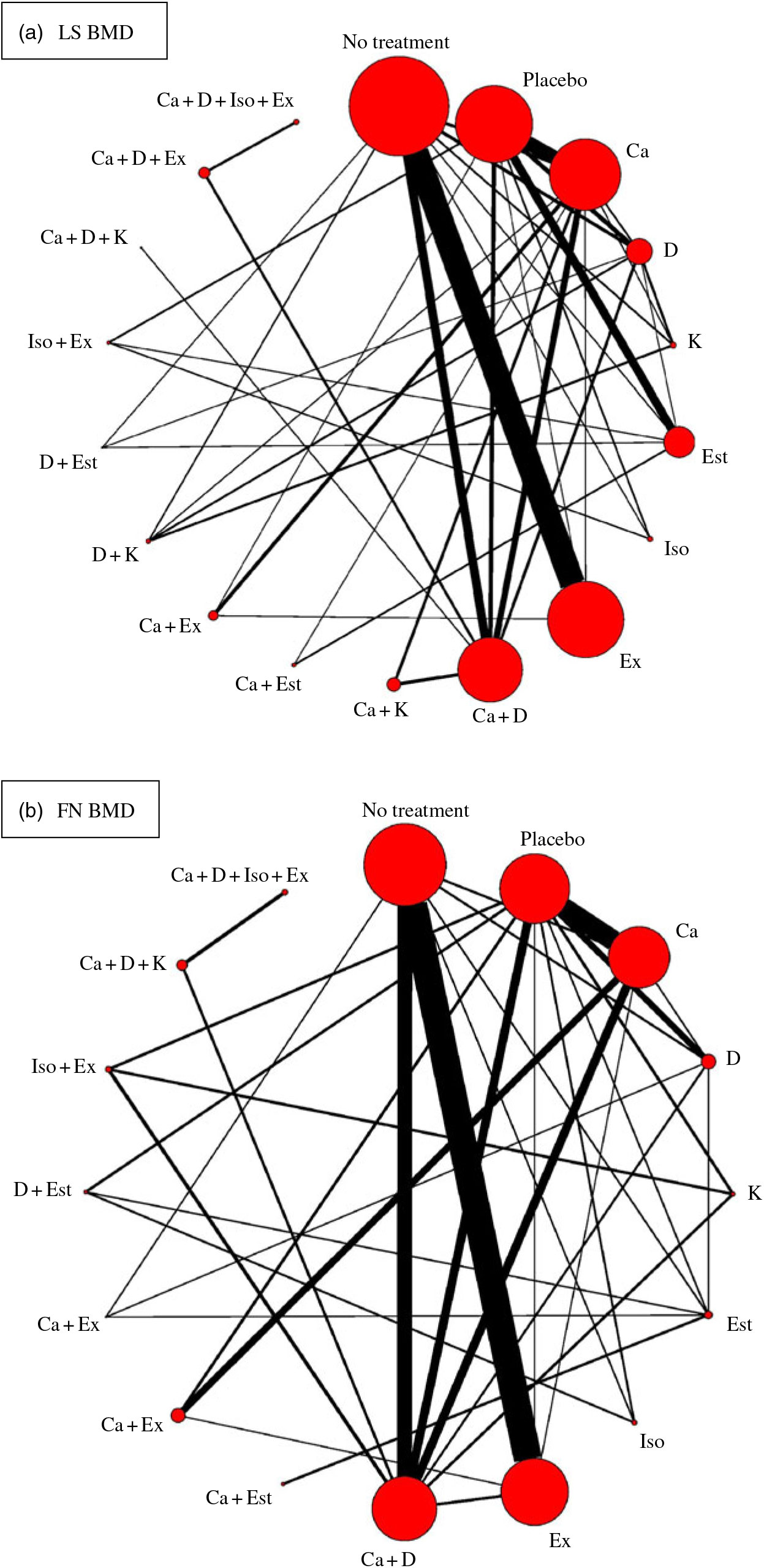
Fig. 2. Network plots for included studies with available direct comparisons for lumbar spine (LS) and femoral neck (FN) bone mineral density. Each node indicates an intervention and each line connecting two nodes indicates a direct comparison between two interventions. The size of the nodes and the thickness of the edges are weighted according to the number of participants evaluating each intervention and direct comparison, respectively. D, vitamin D; Est, oestrogen; Ex, exercise; K, vitamin K; Iso, isoflavone.

Fig. 3. Effect size for change in bone mineral density (BMD) using forest plots. LS, lumbar spine; D, vitamin D; Est, oestrogen; Ex, exercise; K, vitamin K; Iso, isoflavone; FN, femoral neck.

Within the network, there are thirty-seven intervention pairs for which both direct and indirect comparisons are available. Only the comparison between Ca and placebo (P = 0·037) and that between Ca + vitamin D and Ca (P = 0·031) showed significant evidence of inconsistency (online Supplementary Fig. S4).

The overall network heterogeneity τ was 0·021, and I² was 95·94. The heterogeneity of each comparison is shown in online Supplementary Table S2.

Femoral neck

Network meta-analysis for the mean differences in FN BMD included fifty-five RCT (n 6707) with sixteen different types of interventions (Fig. 2(b)). The effects of each intervention are presented in Fig. 3(b). Ca (0·031 g/cm2, 95 % CrI 0·0058, 0·058 g/cm2), exercise (0·028 g/cm2, 95 % CrI 0·014, 0·042 g/cm2) and vitamin D + oestrogen (0·050 g/cm2, 95 % CrI 0·0080, 0·092 g/cm2) were associated with significant beneficial intervention effects relative to no treatment.

Within the network, both direct and indirect comparisons are available for thirty-two intervention pairs. None of them showed significant evidence of inconsistency (online Supplementary Fig. S5).

The overall heterogeneity τ was 0·019 and I² was 96·59 in this network. The heterogeneity of each comparison is shown in online Supplementary Table S3.

Ranking probability

As shown in Table 3, the SUCRA values demonstrated that vitamin D + oestrogen had the highest SUCRA values for change of BMD in the LS (97·29 %), followed by Ca + vitamin D and exercise (86·86 %) and oestrogen (85·70 %). Ca + exercise had the highest SUCRA values for change of BMD in the FN (79·71 %), followed by Ca + oestrogen (79·38 %) and vitamin D + oestrogen (78·33 %). As for single interventions, oestrogen might be the best intervention to improve BMD in the LS (85·70 %) and Ca for FN (60·58 %). Most intervention combinations had higher SUCRA values than single interventions. The details of cumulative rank probabilities are supplied in the Supplementary material (online Supplementary Tables S4 and S5).

Table 3. Intervention rankings using surface under the cumulative ranking (SUCRA) values
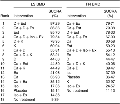
LS, lumbar spine; BMD, bone mineral density; FN, femoral neck; D, vitamin D; Est, oestrogen; Ex, exercise; Iso, isoflavone; K, vitamin K.

Sensitivity analysis

The minimum and maximum correlation values between BMD at baseline and the end of the intervention used to impute missing sd of BMD change were subject to a sensitivity analysis (online Supplementary Tables S6–S9). The findings were similar to those of the primary analysis. Another sensitivity analysis was conducted using the mean difference of BMD change after 15 months of intervention, which was the average duration of intervention in the included studies (online Supplementary Tables S6–S9). For LS BMD, the ranking of exercise was higher, from 12th to 5th, Ca + vitamin D and exercise appeared to be the highest rank and the rankings of higher ranked interventions remained stable. For FN BMD, the ranking of exercise was also higher, from 8th to 4th. Other findings were similar to those of the primary analysis. The last sensitivity analysis was conducted by excluding studies with group sample size less than 15. Higher ranked interventions remained ranking high.

Discussion

To our knowledge, this network meta-analysis is the first to compare the effects of various osteoporosis prevention methods on BMD in postmenopausal women, including Ca, vitamin D, vitamin K, oestrogen, isoflavone, exercise and their combinations. In this network meta-analysis, direct and indirect evidence from ninety RCT including 10 777 postmenopausal women was combined to compare the effect size of each intervention on BMD in both the LS and the FN. The results showed that compared with placebo or no treatment, many interventions can prevent bone loss. In addition, different single or combined interventions may have different impacts on different sites. However, some of the interventions had limited participants or involved limited studies, which may exaggerate or reduce the effect size of those interventions.

Ca and vitamin D supplements have long been considered as ways to prevent osteoporosis, and their effectiveness is consistent with our findings. Ca and vitamin D combined with exercise have beneficial effects on BMD in both the LS and the FN. It was found that the effect of Ca alone on FN BMD is greater than that of LS, which may be due to the different sensitivity of different sites to Ca supplementation, but the exact mechanism needs to be investigated further. Low bone density can not only cause fractures but also lead to bone pain and body metamorphosis(Reference Rao and Singrakhia4). This is the reason that BMD was chosen as the primary outcome in our study, although there has been some controversy about whether Ca and vitamin D effectively improve BMD and fracture rates(Reference Abrahamsen122) and Ca and vitamin D supplements may not prevent women from fracture(Reference Zhao, Zeng and Wang123). Fracture prevention requires all-round efforts, including improving BMD, maintaining muscle strength, maintaining a sense of balance and creating a safe home(Reference Johansson, Kanis and Oden124). Increasing BMD is important, but it is not the only component of fracture prevention.

Vitamin K plays an important role in the γ-carboxylation of osteocalcin, allowing osteocalcin to bind Ca and thus rendering it functional(Reference Hamidi, Gajic-Veljanoski and Cheung125). The effect size of vitamin K on BMD was different between the LS and the FN. Vitamin K ranked 6th among the eighteen interventions for LS but 14th among the sixteen interventions for FN, indicating that vitamin K supplementation can increase LS BMD but not FN BMD. This result is consistent with Fang’s meta-analysis that assessed the effects of vitamin K on BMD(Reference Fang, Hu and Tao126). Another meta-analysis showed that vitamin K2 can improve vertebral BMD in postmenopausal women with osteoporosis, while it did not have any effect in postmenopausal women without osteoporosis(Reference Huang, Wan and Lu127). The present study also showed that vitamin K2 might have a higher adverse reaction rate than control treatment. Considering the adverse reactions and different effects on postmenopausal women with and without osteoporosis, vitamin K should be carefully chosen for osteoporosis prevention.

Oestrogen is mainly generated by the ovaries in premenopausal women. Functional decline of the ovaries after menopause reduces oestrogen secretion. Oestrogen acts on osteoblasts and osteoclasts, thus affecting bone metabolism(Reference Zallone128). In this network meta-analysis, oestrogen + vitamin D was demonstrated to be the most effective way to improve LS BMD, and oestrogen + Ca was the most effective way to improve FN BMD. Oestrogen alone can be effective as well, a similar result to those of previous studies(Reference Fitzpatrick129). There were no interventions of oestrogen + Ca or vitamin D in our study, so the effects of these combinations remain unknown. If such studies are conducted in the future, this analysis can be updated. A previous meta-analysis showed that hormone replacement therapy (including oestrogen and progesterone) has a consistent, favourable, and large effect on bone density at all sites(Reference Wells, Tugwell and Shea130). However, considering the possible side effects of oestrogen and the limitations of access to oestrogen(Reference Lebech131), it should be taken under the guidance of a physician.

Isoflavone is a compound that has oestrogen-like activity in plants, and it exerts a weak oestrogenic effect by binding to the oestrogen receptor(Reference Setchell and Lydeking-Olsen132). It is still unknown whether its mechanism of action on bone turnover is the same as that of oestrogen(Reference Rickard, Monroe and Ruesink133). Isoflavone (not soya protein or foods containing isoflavone) was found to have a very limited effect on BMD in both the LS and the FN in the present study. Many studies, even meta-analyses, have shown inconsistent results about the role of isoflavone on BMD. In Taku’s meta-analysis, soya isoflavone extract supplements were found to have no effects on FN, total hip or trochanter BMD in menopausal women, and they concluded that it can only increase LS BMD(Reference Taku, Melby and Takebayashi134). Ricci’s meta-analysis reported that isoflavone mixtures cannot decrease bone loss in perimenopausal and postmenopausal western women(Reference Ricci, Cipriani and Chiaffarino135). Another two meta-analyses showed that lower doses were not effective at increasing BMD, while intake of more than 80–90 mg/d tended to have a beneficial effect(Reference Ma, Qin and Wang136,Reference Liu, Ho and Su137) . The effect of isoflavone on BMD is limited, but one study demonstrated that isoflavone may be safer than hormonal therapy for prevention of bone loss in postmenopausal women(Reference Xu, Qi and Deng138).

Exercise was shown to improve BMD to a certain extent in our study. The benefits of exercise lie not only in increasing BMD but also in improving muscle strength to prevent falling. Many meta-analyses have been conducted on different kinds of exercise. Kelley’s studies reported that aerobic exercise had a moderately positive effect on BMD in both the LS and the FN(Reference Kelley139,Reference Kelley140) , while resistance exercise did not maintain or improve BMD in either the LS or the FN(Reference Kelley and Kelley141). Most studies have suggested that combined exercise interventions effectively preserve postmenopausal women’s BMD(Reference Zhao, Zhang and Zhang142). Some meta-analyses have also suggested that exercise did not improve BMD in the FN(Reference Kelley and Kelley143). The studies may have had different results because of the different exercise protocols they used. In our study, exercise + Ca and vitamin D effectively prevented BMD loss. Exercise, as an intervention that can contribute to many other chronic non-communicable diseases in older people(Reference Penedo and Dahn144), is worthy of wide promotion.

Although there was high statistical heterogeneity indicated by I² in this network, it may be due to the large sample size in the study. The τ, which is independent of the study sample size, indicated low between-study heterogeneity. What is more, a node-splitting model was used to assess the potential inconsistency. Three other sensitivity analyses were conducted, which produced stable, consistent results. BMI, as a potential effect modifier, is generally thought to have a positive correlation with BMD(Reference Kumar, Sharma and Mittal145). However, study also indicated that BMI was not a determinant of BMD in postmenopausal women in an Asian population. What is more, mean differences in BMD were used to minimise the impact of baseline BMI in our study.

Limitations

The present study has several limitations. First, we did not conduct subgroup analyses of women with different YSM, BMI or osteoporosis status to define the best intervention methods for women with varying YSM, BMI and BMD. These information were not available from all included studies. Moreover, each type of intervention was combined into a single category, which makes it impossible to distinguish between high and low dosages or between slightly different forms of intervention (e.g. vitamin D2 v. D3, aerobic v. resistant exercise). The purpose of our research was to compare different kinds of interventions. Further studies should explore the effect sizes of different dosages and interventions in a network meta-analysis.

Second, we only included studies that employed oestrogen intervention and excluded studies that employed progesterone or androgens (such as hormone replacement therapy and tibolone), because it is unknown whether the effects of oestrogen on BMD will change if combined with progesterone or androgens. However, one study demonstrated that the effect size on BMD does not differ between tibolone and any oestrogen compound(Reference Doren, Nilsson and Johnell146). Progesterone can prevent endometrial hyperplasia during long-term oestradiol replacement(Reference Moyer, de Lignieres and Driguez147). If oestrogen is used to prevent postmenopausal osteoporosis, physicians’ guidance is necessary according to individual circumstances to decide the dosage and use of progesterone and androgens.

Third, the gemtc package is currently the most suitable package for analysing our study’s data. However, because of the limitations of the package, not all results of the comparisons between each pair of interventions were shown in the network forest plot, such as Ca + oestrogen compared with no treatment or placebo. Thus, mean differences were used to define if there was an effect or not in our study because some 95 % CrI of the effect sizes were not available.

Conclusion

The present study demonstrated that many interventions were valuable for improving BMD in the LS and FN of postmenopausal women. It confirmed the need for postmenopausal women to improve BMD through preventive measures such as nutrients or oestrogen. It also confirmed that different single or combined preventions can affect BMD at different sites in different orders. This reveals to medical and health workers and postmenopausal women which methods can be selected preferentially to prevent bone loss.

Acknowledgements

This work was supported by the grants from the National Natural Science Foundation of China (71603167, 71673187 and 71603166) and the Shanghai Key Discipline Construction Project in Public Health (15GWZK1002). The National Natural Science Foundation of China and the Shanghai Key Discipline Construction Project in Public Health had no role in the design, analysis or writing of this article.

Z. X., H. W., Y. S., Q. S., L. T., Z. W. and Y. C. designed and conducted the study. Z. X. and H. W. analysed the data. All authors participated in the interpretation of data. Z. X. drafted the manuscript. All authors helped to revise the manuscript and accept this version for publication. Y. C. is the supervisor.

There are no conflicts of interest.

Supplementary material

To view supplementary material for this article, please visit https://doi.org/10.1017/S0007114519002290

Footnotes

These authors contributed equally to this work and should be considered co-first authors.

References

NIH Consensus Development Panel on Osteoporosis Prevention, Diagnosis, and Therapy (2001) Osteoporosis prevention, diagnosis, and therapy. JAMA 285, 785795.CrossRefGoogle Scholar
Diez, F (2002) Guidelines for the diagnosis of osteoporosis by densitometric methods. J Manipulative Physiol Ther 25, 403415.CrossRefGoogle Scholar
Karinkanta, S, Piirtola, M, Sievanen, H, et al. (2010) Physical therapy approaches to reduce fall and fracture risk among older adults. Nat Rev Endocrinol 6, 396407.CrossRefGoogle ScholarPubMed
Rao, RD & Singrakhia, MD (2003) Painful osteoporotic vertebral fracture. Pathogenesis, evaluation, and roles of vertebroplasty and kyphoplasty in its management. J Bone Joint Surg Am 85, 20102022.CrossRefGoogle ScholarPubMed
Piscitelli, P, Brandi, ML, Nuti, R, et al. (2010) The TARGET project in Tuscany: the first disease management model of a regional project for the prevention of hip re-fractures in the elderly. Clin Cases Miner Bone Metab 7, 251254.Google ScholarPubMed
Camacho, PM, Petak, SM, Binkley, N, et al. (2016) American Association of Clinical Endocrinologists and American College of Endocrinology clinical practice guidelines for the diagnosis and treatment of postmenopausal osteoporosis – 2016. Endocr Pract 22, Suppl. 4, 142.CrossRefGoogle Scholar
Cosman, F, de Beur, SJ, LeBoff, MS, et al. (2014) Clinician’s guide to prevention and treatment of osteoporosis. Osteoporos Int 25, 23592381.CrossRefGoogle ScholarPubMed
Papaioannou, A, Morin, S, Cheung, AM, et al. (2010) 2010 clinical practice guidelines for the diagnosis and management of osteoporosis in Canada: summary. CMAJ 182, 18641873.CrossRefGoogle ScholarPubMed
Radominski, SC, Bernardo, W, Paula, AP, et al. (2017) Brazilian guidelines for the diagnosis and treatment of postmenopausal osteoporosis. Rev Bras Reumatol Engl Ed 57, Suppl. 2, 452466.CrossRefGoogle ScholarPubMed
Compston, J, Bowring, C, Cooper, A, et al. (2013) Diagnosis and management of osteoporosis in postmenopausal women and older men in the UK: National Osteoporosis Guideline Group (NOGG) update 2013. Maturitas 75, 392396.CrossRefGoogle Scholar
Lorenc, R, Gluszko, P, Franek, E, et al. (2017) Guidelines for the diagnosis and management of osteoporosis in Poland: update 2017. Endokrynol Pol 68, 604609.CrossRefGoogle ScholarPubMed
Khan, A & Fortier, M (2014) Osteoporosis in menopause. JOGC 36, 839840.Google ScholarPubMed
Curry, SJ, Krist, AH, Owens, DK, et al. (2018) Screening for osteoporosis to prevent fractures: US Preventive Services Task Force recommendation statement. JAMA 319, 25212531.Google ScholarPubMed
Mills, EJ, Ioannidis, JP, Thorlund, K, et al. (2012) How to use an article reporting a multiple treatment comparison meta-analysis. JAMA 308, 12461253.CrossRefGoogle ScholarPubMed
Grossman, J & Mackenzie, FJ (2005) The randomized controlled trial: gold standard, or merely standard? Perspect Biol Med 48, 516534.CrossRefGoogle ScholarPubMed
Hutton, B, Salanti, G, Caldwell, DM, et al. (2015) The PRISMA extension statement for reporting of systematic reviews incorporating network meta-analyses of health care interventions: checklist and explanations. Ann Intern Med 162, 777784.CrossRefGoogle ScholarPubMed
Kanis, JA & Gluer, CC (2000) An update on the diagnosis and assessment of osteoporosis with densitometry. Committee of Scientific Advisors, International Osteoporosis Foundation. Osteoporos Int 11, 192202.CrossRefGoogle ScholarPubMed
Higgins, JPT & Altman, DG (2008) Chapter 8: Assessing risk of bias in included studies. In Cochrane Handbook for Systematic Reviews of Interventions, version 5.0.0 (updated February 2008) [JPT Higgins and S Green, editors]. The Cochrane Collaboration. www.cochrane-handbook.org.CrossRefGoogle Scholar
Lu, G & Ades, AE (2004) Combination of direct and indirect evidence in mixed treatment comparisons. Stat Med 23, 31053124.CrossRefGoogle ScholarPubMed
DerSimonian, R & Laird, N (1986) Meta-analysis in clinical trials. Control Clin Trials 7, 177188.CrossRefGoogle ScholarPubMed
Furukawa, TA, Barbui, C, Cipriani, A, et al. (2006) Imputing missing standard deviations in meta-analyses can provide accurate results. J Clin Epidemiol 59, 710.CrossRefGoogle ScholarPubMed
Abrams, KR, Gillies, CL & Lambert, PC (2005) Meta-analysis of heterogeneously reported trials assessing change from baseline. Stat Med 24, 38233844.CrossRefGoogle ScholarPubMed
Salanti, G, Ades, AE & Ioannidis, JP (2011) Graphical methods and numerical summaries for presenting results from multiple-treatment meta-analysis: an overview and tutorial. J Clin Epidemiol 64, 163171.CrossRefGoogle ScholarPubMed
Rucker, G & Schwarzer, G (2015) Ranking treatments in frequentist network meta-analysis works without resampling methods. BMC Med Res Methodol 15, 58.CrossRefGoogle ScholarPubMed
Turner, RM, Davey, J, Clarke, MJ, et al. (2012) Predicting the extent of heterogeneity in meta-analysis, using empirical data from the Cochrane Database of Systematic Reviews. Int J Epidemiol 41, 818827.CrossRefGoogle ScholarPubMed
Jansen, JP & Naci, H (2013) Is network meta-analysis as valid as standard pairwise meta-analysis? It all depends on the distribution of effect modifiers. BMC Med 11, 159.CrossRefGoogle ScholarPubMed
Bucher, HC, Guyatt, GH, Griffith, LE, et al. (1997) The results of direct and indirect treatment comparisons in meta-analysis of randomized controlled trials. J Clin Epidemiol 50, 683691.CrossRefGoogle ScholarPubMed
Dias, S, Welton, NJ, Caldwell, DM, et al. (2010) Checking consistency in mixed treatment comparison meta-analysis. Stat Med 29, 932944.CrossRefGoogle ScholarPubMed
Chaimani, A, Higgins, JP, Mavridis, D, et al. (2013) Graphical tools for network meta-analysis in STATA. PLOS ONE 8, e76654.CrossRefGoogle ScholarPubMed
Neupane, B, Richer, D, Bonner, AJ, et al. (2014) Network meta-analysis using R: a review of currently available automated packages. PLOS ONE 9, e115065.CrossRefGoogle ScholarPubMed
Shim, S, Yoon, BH, Shin, IS, et al. (2017) Network meta-analysis: application and practice using STATA. Epidemiol Health 39, e2017047.CrossRefGoogle ScholarPubMed
Lau, EM, Woo, J, Leung, PC, et al. (1992) The effects of calcium supplementation and exercise on bone density in elderly Chinese women. Osteoporos Int 2, 168173.CrossRefGoogle Scholar
Reid, IR, Ames, RW, Evans, MC, et al. (1993) Effect of calcium supplementation on bone loss in postmenopausal women. NEJM 328, 460464.CrossRefGoogle Scholar
Hatori, M, Hasegawa, A, Adachi, H, et al. (1993) The effects of walking at the anaerobic threshold level on vertebral bone loss in postmenopausal women. Calcif Tissue Int 52, 411414.CrossRefGoogle Scholar
Nelson, ME, Fiatarone, MA, Morganti, CM, et al. (1994) Effects of high-intensity strength training on multiple risk factors for osteoporotic fractures. A randomized controlled trial. JAMA 272, 19091914.CrossRefGoogle ScholarPubMed
Ushiroyama, T, Okamura, S, Ikeda, A, et al. (1995) Efficacy of ipriflavone and 1 alpha vitamin D therapy for the cessation of vertebral bone loss. Int J Gynaecol Obstet 48, 283288.CrossRefGoogle ScholarPubMed
Ooms, ME, Roos, JC, Bezemer, PD, et al. (1995) Prevention of bone loss by vitamin D supplementation in elderly women: a randomized double-blind trial. J Clin Endocrinol Metab 80, 10521058.Google ScholarPubMed
Prince, R, Devine, A, Dick, I, et al. (1995) The effects of calcium supplementation (milk powder or tablets) and exercise on bone density in postmenopausal women. J Bone Miner Res 10, 10681075.CrossRefGoogle Scholar
Pruitt, LA, Taaffe, DR & Marcus, R (1995) Effects of a one-year high-intensity versus low-intensity resistance training program on bone mineral density in older women. J Bone Miner Res 10, 17881795.CrossRefGoogle Scholar
Haines, CJ, Chung, TK, Leung, PC, et al. (1995) Calcium supplementation and bone mineral density in postmenopausal women using estrogen replacement therapy. Bone 16, 529531.CrossRefGoogle ScholarPubMed
Taaffe, DR, Pruitt, L, Pyka, G, et al. (1996) Comparative effects of high- and low-intensity resistance training on thigh muscle strength, fiber area, and tissue composition in elderly women. Clin Physiol 16, 381392.CrossRefGoogle ScholarPubMed
Anonymous (1996) Effects of hormone therapy on bone mineral density: results from the Postmenopausal Estrogen/Progestin Interventions (PEPI) trial. The Writing Group for the PEPI. JAMA 276, 13891396.CrossRefGoogle Scholar
Lord, SR, Ward, JA, Williams, P, et al. (1996) The effects of a community exercise program on fracture risk factors in older women. Osteoporos Int 6, 361367.CrossRefGoogle ScholarPubMed
Mizunuma, H, Okano, H, Soda, M, et al. (1997) Prevention of postmenopausal bone loss with minimal uterine bleeding using low dose continuous estrogen/progestin therapy: a 2-year prospective study. Maturitas 27, 6976.CrossRefGoogle ScholarPubMed
Naessen, T, Berglund, L & Ulmsten, U (1997) Bone loss in elderly women prevented by ultralow doses of parenteral 17beta-estradiol. Am J Obstet Gynecol 177, 115119.CrossRefGoogle ScholarPubMed
Chen, JT, Shiraki, M, Hasumi, K, et al. (1997) 1-alpha-Hydroxyvitamin D3 treatment decreases bone turnover and modulates calcium-regulating hormones in early postmenopausal women. Bone 20, 557562.CrossRefGoogle ScholarPubMed
Dawson-Hughes, B, Harris, SS, Krall, EA, et al. (1997) Effect of calcium and vitamin D supplementation on bone density in men and women 65 years of age or older. NEJM 337, 670676.CrossRefGoogle ScholarPubMed
Gambacciani, M, Ciaponi, M, Cappagli, B, et al. (1997) Effects of combined low dose of the isoflavone derivative ipriflavone and estrogen replacement on bone mineral density and metabolism in postmenopausal women. Maturitas 28, 7581.CrossRefGoogle Scholar
Riggs, BL, O’Fallon, WM, Muhs, J, et al. (1998) Long-term effects of calcium supplementation on serum parathyroid hormone level, bone turnover, and bone loss in elderly women. J Bone Miner Res 13, 168174.CrossRefGoogle ScholarPubMed
Storm, D, Eslin, R, Porter, ES, et al. (1998) Calcium supplementation prevents seasonal bone loss and changes in biochemical markers of bone turnover in elderly New England women: a randomized placebo-controlled trial. J Clin Endocrinol Metab 83, 38173825.Google ScholarPubMed
Castelo-Branco, C, Pons, F, Vicente, JJ, et al. (1999) Preventing postmenopausal bone loss with ossein-hydroxyapatite compounds. Results of a two-year, prospective trial. J Reprod Med 44, 601605.Google ScholarPubMed
Adami, S, Gatti, D, Braga, V, et al. (1999) Site-specific effects of strength training on bone structure and geometry of ultradistal radius in postmenopausal women. J Bone Miner Res 14, 120124.CrossRefGoogle Scholar
Gorai, I, Chaki, O, Taguchi, Y, et al. (1999) Early postmenopausal bone loss is prevented by estrogen and partially by 1alpha-OH-vitamin D3: therapeutic effects of estrogen and/or 1alpha-OH-vitamin D3. Calcif Tissue Int 65, 1622.CrossRefGoogle ScholarPubMed
Iwamoto, I, Kosha, S, Noguchi, S, et al. (1999) A longitudinal study of the effect of vitamin K2 on bone mineral density in postmenopausal women a comparative study with vitamin D3 and estrogen-progestin therapy. Maturitas 31, 161164.CrossRefGoogle Scholar
Ruml, LA, Sakhaee, K, Peterson, R, et al. (1999) The effect of calcium citrate on bone density in the early and mid-postmenopausal period: a randomized placebo-controlled study. J Reprod Med 6, 303311.Google Scholar
Rhodes, EC, Martin, AD, Taunton, JE, et al. (2000) Effects of one year of resistance training on the relation between muscular strength and bone density in elderly women. Br J Sports Med 34, 1822.CrossRefGoogle ScholarPubMed
Shiraki, M, Shiraki, Y, Aoki, C, et al. (2000) Vitamin K2 (menatetrenone) effectively prevents fractures and sustains lumbar bone mineral density in osteoporosis. J Bone Miner Res 15, 515521.CrossRefGoogle ScholarPubMed
Iwamoto, J, Takeda, T & Ichimura, S (2000) Effect of combined administration of vitamin D3 and vitamin K2 on bone mineral density of the lumbar spine in postmenopausal women with osteoporosis. J Orthop Sci 5, 546551.CrossRefGoogle Scholar
Ongphiphadhanakul, B, Piaseu, N, Tung, SS, et al. (2000) Prevention of postmenopausal bone loss by low and conventional doses of calcitriol or conjugated equine estrogen. Maturitas 34, 179184.CrossRefGoogle ScholarPubMed
Kerr, D, Ackland, T, Maslen, B, et al. (2001) Resistance training over 2 years increases bone mass in calcium-replete postmenopausal women. J Bone Miner Res 16, 175181.CrossRefGoogle ScholarPubMed
Iwamoto, J, Takeda, T & Ichimura, S (2001) Effect of menatetrenone on bone mineral density and incidence of vertebral fractures in postmenopausal women with osteoporosis: a comparison with the effect of etidronate. J Orthop Sci 6, 487492.CrossRefGoogle Scholar
Chailurkit, LO, Ongphiphadhanakul, B, Piaseu, N, et al. (2001) Biochemical markers of bone turnover and response of bone mineral density to intervention in early postmenopausal women: an experience in a clinical laboratory. Clin Chem 47, 10831088.CrossRefGoogle Scholar
Iwamoto, J, Takeda, T & Ichimura, S (2001) Effect of exercise training and detraining on bone mineral density in postmenopausal women with osteoporosis. J Orthop Sci 6, 128132.CrossRefGoogle Scholar
Son, SM & Chun, YN (2001) Effect of oral therapy with alphacalcidol or calcium in Korean elderly women with osteopenia and low dietary calcium. Nutr Res 21, 13471355.CrossRefGoogle Scholar
Arrenbrecht, S & Boermans, AJ (2002) Effects of transdermal estradiol delivered by a matrix patch on bone density in hysterectomized, postmenopausal women: a 2-year placebo-controlled trial. Osteoporos Int 13, 176183.CrossRefGoogle Scholar
Hans, D, Genton, L, Drezner, MK, et al. (2002) Monitored impact loading of the hip: initial testing of a home-use device. Calcif Tissue Int 71, 112120.CrossRefGoogle ScholarPubMed
Ushiroyama, T, Ikeda, A & Ueki, M (2002) Effect of continuous combined therapy with vitamin K(2) and vitamin D(3) on bone mineral density and coagulofibrinolysis function in postmenopausal women. Maturitas 41, 211221.CrossRefGoogle Scholar
Haines, CJ, Yim, SF, Chung, TK, et al. (2003) A prospective, randomized, placebo-controlled study of the dose effect of oral estradiol on bone mineral density in postmenopausal Chinese women. Maturitas 45, 169173.CrossRefGoogle Scholar
Going, S, Lohman, T, Houtkooper, L, et al. (2003) Effects of exercise on bone mineral density in calcium-replete postmenopausal women with and without hormone replacement therapy. Osteoporos Int 14, 637643.CrossRefGoogle Scholar
Jessup, JV, Horne, C, Vishen, RK, et al. (2003) Effects of exercise on bone density, balance, and self-efficacy in older women. Biol Res Nurs 4, 171180.CrossRefGoogle Scholar
Cooper, L, Clifton-Bligh, PB, Nery, ML, et al. (2003) Vitamin D supplementation and bone mineral density in early postmenopausal women. Am J Clin Nutr 77, 13241329.CrossRefGoogle ScholarPubMed
Grados, F, Brazier, M, Kamel, S, et al. (2003) Prediction of bone mass density variation by bone remodeling markers in postmenopausal women with vitamin D insufficiency treated with calcium and vitamin D supplementation. J Clin Endocrinol Metab 88, 51755179.CrossRefGoogle ScholarPubMed
Uesugi, T, Toda, T, Okuhira, T, et al. (2003) Evidence of estrogenic effect by the three-month-intervention of isoflavone on vaginal maturation and bone metabolism in early postmenopausal women. Endocr J 50, 613619.CrossRefGoogle ScholarPubMed
Verschueren, SM, Roelants, M, Delecluse, C, et al. (2004) Effect of 6-month whole body vibration training on hip density, muscle strength, and postural control in postmenopausal women: a randomized controlled pilot study. J Bone Miner Res 19, 352359.CrossRefGoogle ScholarPubMed
Chan, K, Qin, L, Lau, M, et al. (2004) A randomized, prospective study of the effects of Tai Chi Chun exercise on bone mineral density in postmenopausal women. Arch Phys Med Rehabil 85, 717722.CrossRefGoogle Scholar
Ishida, Y & Kawai, S (2004) Comparative efficacy of hormone replacement therapy, etidronate, calcitonin, alfacalcidol, and vitamin K in postmenopausal women with osteoporosis: the Yamaguchi Osteoporosis Prevention study. Am J Med 117, 549555.CrossRefGoogle ScholarPubMed
Harwood, RH, Sahota, O, Gaynor, K, et al. (2004) A randomised, controlled comparison of different calcium and vitamin D supplementation regimens in elderly women after hip fracture: the Nottingham Neck of Femur (NONOF) study. Age Ageing 33, 4551.CrossRefGoogle ScholarPubMed
Inanir, A, Ozoran, K, Tutkak, H, et al. (2004) The effects of calcitriol therapy on serum interleukin-1, interleukin-6 and tumour necrosis factor-alpha concentrations in post-menopausal patients with osteoporosis. J Int Med Res 32, 570582.CrossRefGoogle ScholarPubMed
Englund, U, Littbrand, H, Sondell, A, et al. (2005) A 1-year combined weight-bearing training program is beneficial for bone mineral density and neuromuscular function in older women. Osteoporos Int 16, 11171123.CrossRefGoogle ScholarPubMed
Moschonis, G & Manios, Y (2006) Skeletal site-dependent response of bone mineral density and quantitative ultrasound parameters following a 12-month dietary intervention using dairy products fortified with calcium and vitamin D: the Postmenopausal Health Study. Br J Nutr 96, 11401148.CrossRefGoogle ScholarPubMed
Yasui, T, Miyatani, Y, Tomita, J, et al. (2006) Effect of vitamin K2 treatment on carboxylation of osteocalcin in early postmenopausal women. Gynecol Endocrinol 22, 455459.CrossRefGoogle ScholarPubMed
Korpelainen, R, Keinanen-Kiukaanniemi, S, Heikkinen, J, et al. (2006) Effect of impact exercise on bone mineral density in elderly women with low BMD: a population-based randomized controlled 30-month intervention. Osteoporos Int 17, 109118.CrossRefGoogle Scholar
Huang, HY, Yang, HP, Yang, HT, et al. (2006) One-year soy isoflavone supplementation prevents early postmenopausal bone loss but without a dose-dependent effect. J Nutr Biochem 17, 509517.CrossRefGoogle ScholarPubMed
Wu, J, Oka, J, Higuchi, M, et al. (2006) Cooperative effects of isoflavones and exercise on bone and lipid metabolism in postmenopausal Japanese women: a randomized placebo-controlled trial. Metabolism 55, 423433.CrossRefGoogle Scholar
Nuti, R, Bianchi, G, Brandi, ML, et al. (2006) Superiority of alfacalcidol compared to vitamin D plus calcium in lumbar bone mineral density in postmenopausal osteoporosis. Rheumatol Int 26, 445453.CrossRefGoogle ScholarPubMed
Maddalozzo, GF, Widrick, JJ, Cardinal, BJ, et al. (2007) The effects of hormone replacement therapy and resistance training on spine bone mineral density in early postmenopausal women. Bone 40, 12441251.CrossRefGoogle ScholarPubMed
Woo, J, Hong, A, Lau, E, et al. (2007) A randomised controlled trial of Tai Chi and resistance exercise on bone health, muscle strength and balance in community-living elderly people. Age Ageing 36, 262268.CrossRefGoogle Scholar
Bolton-Smith, C, McMurdo, ME, Paterson, CR, et al. (2007) Two-year randomized controlled trial of vitamin K1 (phylloquinone) and vitamin D3 plus calcium on the bone health of older women. J Bone Miner Res 22, 509519.CrossRefGoogle ScholarPubMed
Bergström, I, Landgren, B, Brinck, J, et al. (2008) Physical training preserves bone mineral density in postmenopausal women with forearm fractures and low bone mineral density. Osteoporos Int 19, 177183.CrossRefGoogle ScholarPubMed
Park, H, Kim, KJ, Komatsu, T, et al. (2008) Effect of combined exercise training on bone, body balance, and gait ability: a randomized controlled study in community-dwelling elderly women. J Bone Miner Metab 26, 254259.CrossRefGoogle Scholar
Bocalini, DS, Serra, AJ, dos Santos, L, et al. (2009) Strength training preserves the bone mineral density of postmenopausal women without hormone replacement therapy. J Aging Health 21, 519527.CrossRefGoogle ScholarPubMed
Beck, BR & Norling, TL (2010) The effect of 8 mos of twice-weekly low- or higher intensity whole body vibration on risk factors for postmenopausal hip fracture. Am J Phys Med Rehabil 89, 9971009.CrossRefGoogle ScholarPubMed
Tolomio, S, Ermolao, A, Lalli, A, et al. (2010) The effect of a multicomponent dual-modality exercise program targeting osteoporosis on bone health status and physical function capacity of postmenopausal women. J Women Aging 22, 241254.CrossRefGoogle Scholar
Yoo, EJ, Jun, TW & Hawkins, SA (2010) The effects of a walking exercise program on fall-related fitness, bone metabolism, and fall-related psychological factors in elderly women. Res Sports Med 18, 236250.CrossRefGoogle ScholarPubMed
Chailurkit, LO, Saetung, S, Thakkinstian, A, et al. (2010) Discrepant influence of vitamin D status on parathyroid hormone and bone mass after two years of calcium supplementation. Clin Endocrinol 73, 167172.Google ScholarPubMed
Kärkkäinen, M, Tuppurainen, M, Salovaara, K, et al. (2010) Effect of calcium and vitamin D supplementation on bone mineral density in women aged 65–71 years: a 3-year randomized population-based trial (OSTPRE-FPS). Osteoporos Int 21, 20472055.CrossRefGoogle Scholar
Verschueren, SM, Bogaerts, A, Delecluse, C, et al. (2011) The effects of whole-body vibration training and vitamin D supplementation on muscle strength, muscle mass, and bone density in institutionalized elderly women: a 6-month randomized, controlled trial. J Bone Miner Res 26, 4249.CrossRefGoogle ScholarPubMed
Choquette, S, Riesco, E, Cormier, E, et al. (2011) Effects of soya isoflavones and exercise on body composition and clinical risk factors of cardiovascular diseases in overweight postmenopausal women: a 6-month double-blind controlled trial. Br J Nutr 105, 11991209.CrossRefGoogle ScholarPubMed
Marques, EA, Mota, J, Machado, L, et al. (2011) Multicomponent training program with weight-bearing exercises elicits favorable bone density, muscle strength, and balance adaptations in older women. Calcif Tissue Int 88, 117129.CrossRefGoogle ScholarPubMed
Marques, EA, Wanderley, F, Machado, L, et al. (2011) Effects of resistance and aerobic exercise on physical function, bone mineral density, OPG and RANKL in older women. Exp Gerontol 46, 524532.CrossRefGoogle ScholarPubMed
Tartibian, B, Hajizadeh Maleki, B, Kanaley, J, et al. (2011) Long-term aerobic exercise and omega-3 supplementation modulate osteoporosis through inflammatory mechanisms in post-menopausal women: a randomized, repeated measures study. Nutr Metab 8, 71.CrossRefGoogle ScholarPubMed
Je, SH, Joo, NS, Choi, BH, et al. (2011) Vitamin K supplement along with vitamin D and calcium reduced serum concentration of undercarboxylated osteocalcin while increasing bone mineral density in Korean postmenopausal women over sixty-years-old. J Korean Med Sci 26, 10931098.CrossRefGoogle ScholarPubMed
Karakiriou, SK, Douda, HT, Smilios, IG, et al. (2012) Effects of vibration and exercise training on bone mineral density and muscle strength in post-menopausal women. Eur J Sport Sci 12, 8188.CrossRefGoogle Scholar
Macdonald, HM, Wood, AD, Aucott, LS, et al. (2013) Hip bone loss is attenuated with 1000 IU but not 400 IU daily vitamin D3: a 1-year double-blind RCT in postmenopausal women. J Bone Miner Res 28, 22022213.CrossRefGoogle Scholar
Basat, H, Esmaeilzadeh, S & Eskiyurt, N (2013) The effects of strengthening and high-impact exercises on bone metabolism and quality of life in postmenopausal women: a randomized controlled trial. J Back Musculoskelet Rehabil 26, 427435.CrossRefGoogle Scholar
Chilibeck, PD, Vatanparast, H, Pierson, R, et al. (2013) Effect of exercise training combined with isoflavone supplementation on bone and lipids in postmenopausal women: a randomized clinical trial. J Bone Miner Res 28, 780793.CrossRefGoogle Scholar
Rajatanavin, R, Chailurkit, L, Saetung, S, et al. (2013) The efficacy of calcium supplementation alone in elderly Thai women over a 2-year period: a randomized controlled trial. Osteoporos Int 24, 28712877.CrossRefGoogle Scholar
Lai, CL, Tseng, SY, Chen, CN, et al. (2013) Effect of 6 months of whole body vibration on lumbar spine bone density in postmenopausal women: a randomized controlled trial. Clin Interv Aging 8, 16031609.Google ScholarPubMed
Leung, KS, Li, CY, Tse, YK, et al. (2014) Effects of 18-month low-magnitude high-frequency vibration on fall rate and fracture risks in 710 community elderly – a cluster-randomized controlled trial. Osteoporos Int 25, 17851795.CrossRefGoogle ScholarPubMed
Jiang, Y, Zhang, ZL, Zhang, ZL, et al. (2014) Menatetrenone versus alfacalcidol in the treatment of Chinese postmenopausal women with osteoporosis: a multicenter, randomized, double-blinded, double-dummy, positive drug-controlled clinical trial. Clin Interv Aging 9, 121127.Google ScholarPubMed
Koitaya, N, Sekiguchi, M, Tousen, Y, et al. (2014) Low-dose vitamin K2 (MK-4) supplementation for 12 months improves bone metabolism and prevents forearm bone loss in postmenopausal Japanese women. J Bone Miner Metab 32, 142150.CrossRefGoogle ScholarPubMed
Moreira, LD, Fronza, FC, Dos Santos, RN, et al. (2014) The benefits of a high-intensity aquatic exercise program (HydrOS) for bone metabolism and bone mass of postmenopausal women. J Bone Miner Metab 32, 411419.Google ScholarPubMed
Nicholson, VP, McKean, MR, Slater, GJ, et al. (2015) Low-load very high-repetition resistance training attenuates bone loss at the lumbar spine in active post-menopausal women. Calcif Tissue Int 96, 490499.CrossRefGoogle Scholar
Santin-Medeiros, F, Santos-Lozano, A, Rey-Lopez, JP, et al. (2015) Effects of eight months of whole body vibration training on hip bone mass in older women. Nutr Hosp 31, 16541659.Google ScholarPubMed
Tankisheva, E, Bogaerts, A, Boonen, S, et al. (2015) Effects of a six-month local vibration training on bone density, muscle strength, muscle mass, and physical performance in postmenopausal women. J Strength Cond Res 29, 26132622.CrossRefGoogle Scholar
Wang, H, Yu, B, Chen, W, et al. (2015) Simplified Tai Chi resistance training versus traditional Tai Chi in slowing bone loss in postmenopausal women. Evid Based Complement Alternat Med 2015, 379451.Google ScholarPubMed
Wen, HJ, Huang, TH, Li, TL, et al. (2017) Effects of short-term step aerobics exercise on bone metabolism and functional fitness in postmenopausal women with low bone mass. Osteoporos Int 28, 539547.CrossRefGoogle Scholar
Shin, S, Lee, K & Song, C (2018) Effects of whole body vibration with load stimulation in postmenopausal women. Med Sci Tech 59, 412.CrossRefGoogle Scholar
de Oliveira, LC, de Oliveira, RG & de Almeida Pires-Oliveira, DA (2019) Effects of whole-body vibration versus pilates exercise on bone mineral density in postmenopausal women: a randomized and controlled clinical trial. J Geriatr Phys Ther 42, E23E31.CrossRefGoogle Scholar
Aboarrage Junior, AM, Teixeira, CVS, Dos Santos, RN, et al. (2018) A high-intensity jump-based aquatic exercise program improves bone mineral density and functional fitness in postmenopausal women. Rejuvenation Res 21, 535540.CrossRefGoogle ScholarPubMed
Bislev, LS, Langagergaard Rødbro, L, Rolighed, L, et al. (2019) Bone microstructure in response to vitamin D3 supplementation: a randomized placebo-controlled trial. Calcif Tissue Int 104, 160170.CrossRefGoogle ScholarPubMed
Abrahamsen, B (2017) The calcium and vitamin D controversy. Ther Adv Musculoskelet Dis 9, 107114.CrossRefGoogle ScholarPubMed
Zhao, J, Zeng, X, Wang, J, et al. (2017) Association between calcium or vitamin D supplementation and fracture incidence in community-dwelling older adults: a systematic review and meta-analysis effects of calcium or vitamin D on fractures in older adults effects of calcium or vitamin D on fractures in older adults. JAMA 318, 24662482.CrossRefGoogle ScholarPubMed
Johansson, H, Kanis, JA, Oden, A, et al. (2009) BMD, clinical risk factors and their combination for hip fracture prevention. Osteoporos Int 20, 16751682.CrossRefGoogle ScholarPubMed
Hamidi, MS, Gajic-Veljanoski, O & Cheung, AM (2013) Vitamin K and bone health. J Clin Densitom 16, 409413.CrossRefGoogle ScholarPubMed
Fang, Y, Hu, C, Tao, X, et al. (2012) Effect of vitamin K on bone mineral density: a meta-analysis of randomized controlled trials. J Bone Miner Metab 30, 6068.CrossRefGoogle Scholar
Huang, ZB, Wan, SL, Lu, YJ, et al. (2015) Does vitamin K2 play a role in the prevention and treatment of osteoporosis for postmenopausal women: a meta-analysis of randomized controlled trials. Osteoporos Int 26, 11751186.CrossRefGoogle ScholarPubMed
Zallone, A (2006) Direct and indirect estrogen actions on osteoblasts and osteoclasts. Ann N Y Acad Sci 1068, 173179.CrossRefGoogle ScholarPubMed
Fitzpatrick, LA (2006) Estrogen therapy for postmenopausal osteoporosis. Arq Bras Endocrinol Metabol 50, 705719.CrossRefGoogle ScholarPubMed
Wells, G, Tugwell, P, Shea, B, et al. (2002) Meta-analyses of therapies for postmenopausal osteoporosis. V. Meta-analysis of the efficacy of hormone replacement therapy in treating and preventing osteoporosis in postmenopausal women. Endocr Rev 23, 529539.CrossRefGoogle ScholarPubMed
Lebech, PE (1976) Effects and side-effects of estrogen therapy. In Consensus on Menopause Research, pp. 4447 [PA van Keep, RB Greenblatt and M Albeau-Fernet, editors]. Dordrecht: Springer.CrossRefGoogle Scholar
Setchell, KD & Lydeking-Olsen, E (2003) Dietary phytoestrogens and their effect on bone: evidence from in vitro and in vivo, human observational, and dietary intervention studies. Am J Clin Nutr 78, Suppl. 3, S593S609.CrossRefGoogle Scholar
Rickard, DJ, Monroe, DG, Ruesink, TJ, et al. (2003) Phytoestrogen genistein acts as an estrogen agonist on human osteoblastic cells through estrogen receptors alpha and beta. J Cell Biochem 89, 633646.CrossRefGoogle ScholarPubMed
Taku, K, Melby, MK, Takebayashi, J, et al. (2010) Effect of soy isoflavone extract supplements on bone mineral density in menopausal women: meta-analysis of randomized controlled trials. Asia Pac J Clin Nutr 19, 3342.Google Scholar
Ricci, E, Cipriani, S, Chiaffarino, F, et al. (2010) Soy isoflavones and bone mineral density in perimenopausal and postmenopausal Western women: a systematic review and meta-analysis of randomized controlled trials. J Womens Health (Larchmt) 19, 16091617.CrossRefGoogle ScholarPubMed
Ma, DF, Qin, LQ, Wang, PY, et al. (2008) Soy isoflavone intake increases bone mineral density in the spine of menopausal women: meta-analysis of randomized controlled trials. Clin Nutr 27, 5764.CrossRefGoogle ScholarPubMed
Liu, J, Ho, SC, Su, YX, et al. (2009) Effect of long-term intervention of soy isoflavones on bone mineral density in women: a meta-analysis of randomized controlled trials. Bone 44, 948953.CrossRefGoogle Scholar
Xu, M, Qi, C, Deng, B, et al. (2009) Phytotherapy versus hormonal therapy for postmenopausal bone loss: a meta-analysis. Osteoporos Int 20, 519526.CrossRefGoogle ScholarPubMed
Kelley, GA (1998) Aerobic exercise and bone density at the hip in postmenopausal women: a meta-analysis. Prev Med 27, 798807.CrossRefGoogle Scholar
Kelley, G (1998) Aerobic exercise and lumbar spine bone mineral density in postmenopausal women: a meta-analysis. J Am Geriatr Soc 46, 143152.CrossRefGoogle ScholarPubMed
Kelley, GA & Kelley, KS (2004) Efficacy of resistance exercise on lumbar spine and femoral neck bone mineral density in premenopausal women: a meta-analysis of individual patient data. J Womens Health (Larchmt) 13, 293300.CrossRefGoogle ScholarPubMed
Zhao, R, Zhang, M & Zhang, Q (2017) The effectiveness of combined exercise interventions for preventing postmenopausal bone loss: a systematic review and meta-analysis. J Orthop Sports Phys Ther 47, 241251.CrossRefGoogle ScholarPubMed
Kelley, GA & Kelley, KS (2006) Exercise and bone mineral density at the femoral neck in postmenopausal women: a meta-analysis of controlled clinical trials with individual patient data. Am J Obstet Gynecol 194, 760767.CrossRefGoogle Scholar
Penedo, FJ & Dahn, JR (2005) Exercise and well-being: a review of mental and physical health benefits associated with physical activity. Curr Opin Psychiatr 18, 189193.CrossRefGoogle ScholarPubMed
Kumar, A, Sharma, AK, Mittal, S, et al. (2016) The relationship between body mass index and bone mineral density in premenopausal and postmenopausal North Indian women. J Obstet Gynaecol India 66, 5256.CrossRefGoogle ScholarPubMed
Doren, M, Nilsson, JA & Johnell, O (2003) Effects of specific post-menopausal hormone therapies on bone mineral density in post-menopausal women: a meta-analysis. Hum Reprod 18, 17371746.CrossRefGoogle Scholar
Moyer, DL, de Lignieres, B, Driguez, P, et al. (1993) Prevention of endometrial hyperplasia by progesterone during long-term estradiol replacement: influence of bleeding pattern and secretory changes. Fertil Steril 59, 992997.CrossRefGoogle ScholarPubMed
Figure 0

Fig. 1. Flow diagram of literature search and study. RCT, randomised controlled trial; BMD, bone mineral density; DXA, dual-energy X-ray absorptiometry.

Figure 1

Table 1. Description of included trials

Figure 2

Table 2. Description of individual groups in included trials*

Figure 3

Fig. 2. Network plots for included studies with available direct comparisons for lumbar spine (LS) and femoral neck (FN) bone mineral density. Each node indicates an intervention and each line connecting two nodes indicates a direct comparison between two interventions. The size of the nodes and the thickness of the edges are weighted according to the number of participants evaluating each intervention and direct comparison, respectively. D, vitamin D; Est, oestrogen; Ex, exercise; K, vitamin K; Iso, isoflavone.

Figure 4

Fig. 3. Effect size for change in bone mineral density (BMD) using forest plots. LS, lumbar spine; D, vitamin D; Est, oestrogen; Ex, exercise; K, vitamin K; Iso, isoflavone; FN, femoral neck.

Figure 5

Table 3. Intervention rankings using surface under the cumulative ranking (SUCRA) values

Supplementary material: File

Xu et al. supplementary material

Xu et al. supplementary material

Download Xu et al. supplementary material(File)
File 8.3 MB