Autosomal recessive spastic ataxia of Charlevoix-Saguenay (ARSACS) has been identified in diverse countries. However, outside of North America, North Africa, and Europe, ARSACS was recognized only in the Japanese population.Reference Bouhlal, Amouri, El Euch-Fayeche and Hentati 1 Moreover, through genetic research, the disease is thought to exhibit no founder effect, except in the Quebec descent.Reference Bouhlal, Amouri, El Euch-Fayeche and Hentati 1 Therefore, it is possible that ARSACS might be underestimated or misdiagnosed, especially in Asian countries.
Case Report
A 17-year-old Korean woman came to our hospital because of progressive gait disturbance and dysarthria from early childhood. She first walked at 18 months, and her gait was slow and unsteady during her life. Her speech was somewhat slow and dysarthric. Because her gait disturbance and dysarthria displayed a very slowly progressive course, her parents and friends did not recognize her disease state in childhood. At age 14, deformities of both feet appeared and got worse gradually. She could walk without gait aid and had no fall history. There was no family history. She complained about neither learning problems nor urinary symptoms.
Neurological examination revealed spasticity with positive Babinski signs in the legs. Deep tendon reflexes in all extremities were moderately reduced. Proximal interphalangeal joints were slightly hyperextended. Amyotrophy with pes cavus and hammer toes was observed in her feet. The patient showed slurred and dysarthric speech with moderate hypernasality. Although limb ataxia was mild, she revealed moderate gait ataxia with spastic feature. Her tandem gait was unsteady. Neither abnormal saccadic eye movement nor gazed-evoked nystagmus was noted at bedside examination. The remainder of the cranial nerve examinations was normal. Vibration sense was mildly reduced in bilateral toes, whereas pain and light touch senses were unremarkable in all extremities. Nerve conduction study revealed a sensorimotor polyneuropathy displaying mixed axonal and demyelinating features. Cardiac evaluation including electrocardiogram and two-dimensional echocardiogram was unremarkable.
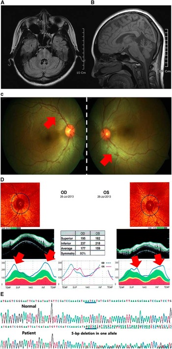
3-T brain magnetic resonance imaging (MRI) showed horizontal pontine hypointense stripes, vertical hyperintensities in lateral pons, and atrophy of the superior cerebellar vermis as well as cervical spinal cord (Figure 1A, B). Subcortical atrophy including basal ganglia was not found, and cortical atrophy was only suspicious in the bilateral parietal lobe. One neuro-ophthalmologist (KH) revealed unremarkable findings in visual acuity as well as the perimetry test. However, peripapillary swelling with striation on funduscopy was present (Figure 1C). Optical coherence tomography documented an abnormal thickening of the retinal nerve fiber layer (Figure 1D). Genetic analysis for Friedreich ataxia was negative, whereas Sanger sequencing of whole coding regions of the SACS gene (the gene responsible for ARSACS), based on a fragment analysis, revealed a heterozygote state with a novel frame shift mutation, c.[4756_4760del] (p.Asn1586Tyrfx*3), at giant exon 9 in one allele (Department of Human Genetics, Radboud University Nijmegen Medical Centre, Nijmegen, The Netherlands) (Figure 1E).Reference Bouhlal, Amouri, El Euch-Fayeche and Hentati 1

Figure 1 Evidence supporting the diagnosis of ARSACS in a Korean patient. A, Axial fluid-attenuated inversion recovery images reveal horizontal hypointense stripes as well as vertical hyperintensities in the pons. B, Sagittal T1-weighted images exhibit the atrophy of not only the superior cerebellar vermis but also the cervical spinal cord. In ophthalmological evaluation, increased thickness (red arrows) of the retinal nerve fiber layer is demonstrated by funduscopic photographs (C) as well as optic coherence tomography assessment (D). E, In addition, mutation screening for whole exons of ARSACS results in heterozygote with a novel mutation of frame shift, c.[4756_4760del] (p.Asn1586Tyrfx*3), at giant exon 9 in one allele.
Discussion
Classical characteristics of ARSACS consist of cerebellar ataxia, spasticity, and peripheral neuropathy.Reference Bouhlal, Amouri, El Euch-Fayeche and Hentati 1 Our patient revealed the classical symptoms of early-onset spastic ataxia, dysarthria, and distal amyotrophy combined with sensorimotor polyneuropathy. Recent advances in comprehensive analysis of brain MRI as well as retinal examination allow early identification of ARSACS. Most patients showed characteristic MRI findings of horizontal hypointense stripes and/or vertical hyperintensities in pons, in addition to the atrophy of superior cerebellar vermis or cervical spinal cord.Reference Prodi, Grisoli and Panzeri 2 , Reference Synofzik, Soehn and Gburek-Augustat 3 Furthermore, ophthalmological evaluation has added a distinct feature of increased thickness of the retinal nerve fiber layer regarding hyperplasia or hypertrophy of the retinal fibers.Reference Garcia-Martin, Pablo and Gazulla 4 Based on the specific findings of brain MRI and ophthalmological examination in addition to clinical manifestations, this patient was clinically diagnosed with ARSACS. The patient was clinically distinguishable from Friedreich's ataxia (FRDA):Reference Storey 5 (1) cardiac abnormality is frequently observed in FRDA, but not in ARSACS; (2) ARSACS shows a sensorimotor polyneuropathy with combined axonal and demyelinating type, whereas FRDA primarily reveals a sensory dominant polyneuropathy; and (3) there has been no report of such retinal as well as the brain MRI findings in FRDA.
Until now, more than 100 mutations of SACS gene have been investigated that represent variable phenotypes.Reference Bouhlal, Amouri, El Euch-Fayeche and Hentati 1 , Reference Synofzik, Soehn and Gburek-Augustat 3 Although the majority of ARSACS patients showed diverse mutations in protein coding regions, some were reported with mutations in noncoding regions.Reference Bouhlal, Amouri, El Euch-Fayeche and Hentati 1 The patient was heterozygote for a novel frame shift mutation at giant exon 9 in one allele, suggesting that she might have another mutation in noncoding region or intron affecting gene transcription or translation in the other allele. Besides, because array CGH was not performed, we could not rule out the possibility of large deletion as well.
We report here the first probable Korean case of ARSACS showing the characteristic neuroimaging and ophthalmologic findings. Our case will help to raise more awareness of ARSACS, especially for Asian medical doctors. Careful evaluations of brain MRI as well as ophthalmological examinations are required in young patients with progressive ataxic syndrome.
Statement of Authorship
Research project concept: SK, KBK; organization: SCK; execution: SK, KK, KH, BE, HY, EK, HS. Manuscript preparation writing of the first draft: KK, SK; review and critique: KH, BE, HY, EK, HS, KK, SK.
Disclosures
None.


