Hostname: page-component-cd9895bd7-gxg78 Total loading time: 0 Render date: 2024-12-30T22:04:18.908Z Has data issue: false hasContentIssue false

Silver-decorated ZnO hexagonal nanoplate arrays as SERS-active substrates: An experimental and simulation study

Published online by Cambridge University Press:  13 December 2013

Kun Liu
Affiliation:
Institute of Near-field Optics and Nanotechnology, School of Physics and Optoelectronic Technology, Dalian University of Technology, Ganjingzi District, Dalian 116024, People’s Republic of China
Dawei Li
Affiliation:
Institute of Near-field Optics and Nanotechnology, School of Physics and Optoelectronic Technology, Dalian University of Technology, Ganjingzi District, Dalian 116024, People’s Republic of China
Rui Li
Affiliation:
Institute of Near-field Optics and Nanotechnology, School of Physics and Optoelectronic Technology, Dalian University of Technology, Ganjingzi District, Dalian 116024, People’s Republic of China
Qiao Wang
Affiliation:
Institute of Near-field Optics and Nanotechnology, School of Physics and Optoelectronic Technology, Dalian University of Technology, Ganjingzi District, Dalian 116024, People’s Republic of China
Shi Pan
Affiliation:
Institute of Near-field Optics and Nanotechnology, School of Physics and Optoelectronic Technology, Dalian University of Technology, Ganjingzi District, Dalian 116024, People’s Republic of China
Wei Peng
Affiliation:
Institute of Near-field Optics and Nanotechnology, School of Physics and Optoelectronic Technology, Dalian University of Technology, Ganjingzi District, Dalian 116024, People’s Republic of China
Maodu Chen*
Affiliation:
Institute of Near-field Optics and Nanotechnology, School of Physics and Optoelectronic Technology, Dalian University of Technology, Ganjingzi District, Dalian 116024, People’s Republic of China
*
a)Address all correspondence to this author. e-mail: [email protected]
Get access

Abstract

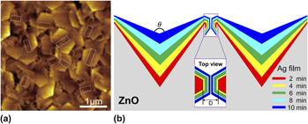
We have fabricated Ag-decorated ZnO nanoplate arrays by combining water-bath heating toward ZnO hexagonal nanoplate arrays and subsequent decoration of Ag films or nanoparticles on the ZnO surfaces by magnetron sputtering or photoreduction. Experimental surface-enhanced Raman scattering (SERS) results show that Ag-film–ZnO hybrid substrates with different Ag sputtering times exhibit a large difference in enhanced SERS signals for Rhodamine 6G (10−7 M). Atomic force microscope analysis reveals that two kinds of positions create abundant “hot spots” in this SERS substrate: one is located at the gap between adjacent separate Ag-film–ZnO hybrid nanoplates, and the other is located at the V-grooves formed by two adjacent interlaced Ag-film–ZnO hybrid nanoplates. The effects of simultaneous changes in interplate spacing and groove wall angle are considered to be the key factors affecting the SERS of our prepared Ag-film–ZnO hybrid substrates, which have also been evaluated by finite-difference time-domain simulation.

Type
Articles
Copyright
Copyright © Materials Research Society 2013 

Access options

Get access to the full version of this content by using one of the access options below. (Log in options will check for institutional or personal access. Content may require purchase if you do not have access.)

References

REFERENCES

Fleischmann, M., Hendra, P.J., and McQuillan, A.J.: Raman spectra of pyridine adsorbed at a silver electrode. Chem. Phys. Lett. 26(2), 163 (1974).CrossRefGoogle Scholar
Li, D., Wu, S., Wang, Q., Wu, Y., Peng, W., and Pan, L.: Ag@C core–shell colloidal nanoparticles prepared by the hydrothermal route and the low temperature heating–stirring method and their application in surface enhanced Raman scattering. J. Phys. Chem. C 116(22), 12283 (2012).CrossRefGoogle Scholar
Shen, C., Hui, C., Yang, T., Xiao, C., Tian, J., Bao, L., Chen, S., Ding, H., and Gao, H.: Monodisperse noble-metal nanoparticles and their surface enhanced Raman scattering properties. Chem. Mater. 20(22), 6939 (2008).CrossRefGoogle Scholar
Nikoobakht, B. and El-Sayed, M.A.: Surface-enhanced Raman scattering studies on aggregated gold nanorods. J. Phys. Chem. A 107(18), 3372 (2003).CrossRefGoogle Scholar
Lee, S.J., Morrill, A.R., and Moskovits, M.: Hot spots in silver nanowire bundles for surface-enhanced Raman spectroscopy. J. Am. Chem. Soc. 128(7), 2200 (2006).CrossRefGoogle ScholarPubMed
Wang, T., Hu, X., and Dong, S.: Surfactantless synthesis of multiple shapes of gold nanostructures and their shape-dependent SERS spectroscopy. J. Phys. Chem. B 110(34), 16930 (2006).CrossRefGoogle ScholarPubMed
Cao, L.Y., Nabet, B., and Spanier, J.E.: Enhanced Raman scattering from individual semiconductor nanocones and nanowires. Phys. Rev. Lett. 96(15), 157402 (2006).CrossRefGoogle ScholarPubMed
Yang, L.B., Jiang, X., Ruan, W.D., Zhao, B., Xu, W.Q., and Lombardi, J.R.: Observation of enhanced Raman scattering for molecules adsorbed on TiO2 nanoparticles: Charge-transfer contribution. J. Phys. Chem. C 112(50), 20095 (2008).CrossRefGoogle Scholar
Prokes, S.M., Glembocki, O.J., Livenere, J.E., Tumkur, T.U., Kitur, J.K., Zhu, G., Wells, B., Podolskiy, V.A., and Noginov, M.A.: Hyperbolic and plasmonic properties of silicon/Ag aligned nanowire arrays. Opt. Express 21(12), 14962 (2013).CrossRefGoogle ScholarPubMed
Liao, F., Cheng, L., Li, J., Shao, M.W., Wang, Z.H., and Lee, S.T.: An effective oxide shell-protected surface-enhanced Raman scattering (SERS) substrate: The easy route to Ag@AgxO-silicon nanowire films via surface doping. J. Mater. Chem. C 1(8), 1628 (2013).CrossRefGoogle Scholar
Wu, Y., Liu, K., Li, X., and Pan, S.: Integrate silver colloids with silicon nanowire arrays for surface-enhanced Raman scattering. Nanotechnology 22(21), 215701 (2011).CrossRefGoogle ScholarPubMed
Peng, M.F., Gao, J., Zhang, P.P., Li, Y., Sun, X.H., and Lee, S.T.: Reductive self-assembling of Ag nanoparticles on germanium nanowires and their application in ultrasensitive surface-enhanced Raman spectroscopy. Chem. Mater. 23(14), 3296 (2011).CrossRefGoogle Scholar
Yang, L.B., Jiang, X., Ruan, W.D., Yang, J.X., Zhao, B., Xu, W.Q., and Lombardi, J.R.: Charge-transfer-induced surface-enhanced Raman scattering on Ag-TiO2 nanocomposites. J. Phys. Chem. C 113(36), 16226 (2009).CrossRefGoogle Scholar
Mills, A., Hill, G., Stewart, M., Graham, D., Smith, W.E., Hodgen, S., Halfpenny, P.J., Faulds, K., and Robertson, P.: Characterization of novel Ag on TiO2 films for surface-enhanced Raman scattering. Appl. Spectrosc. 58(8), 922 (2004).CrossRefGoogle ScholarPubMed
Li, D., Pan, L., Li, S., Liu, K., Wu, S., and Peng, W.: Controlled preparation of uniform TiO2-catalyzed silver nanoparticle films for surface-enhanced Raman scattering. J. Phys. Chem. C 117(13), 6861 (2013).CrossRefGoogle Scholar
Es-Souni, M., Es-Souni, M., Habouti, S., Pfeiffer, N., Lahmar, A., Dietze, M., and Solterbeck, C-H.: Brookite formation in TiO2 Ag nanocomposites and visible-light-induced templated growth of Ag nanostructures in TiO2. Adv. Funct. Mater. 20(3), 377 (2010).CrossRefGoogle Scholar
Chen, L., Luo, L., Chen, Z., Zhang, M., Zapien, J.A., Lee, C.S., and Lee, S.T.: ZnO/Au composite nanoarrays as substrates for surface-enbanced Raman scattering detection. J. Phys. Chem. C 114(1), 93 (2010).CrossRefGoogle Scholar
Song, W., Wang, Y., Hu, H., and Zhao, B.: Fabrication of surface-enhanced Raman scattering-active ZnO/Ag composite microspheres. J. Raman Spectrosc. 38(10), 1320 (2007).CrossRefGoogle Scholar
Cheng, C., Yan, B., Wong, S.M., Li, X., Zhou, W., Yu, T., Shen, Z., Yu, H., and Fan, H.J.: Fabrication and SERS performance of silver-nanoparticle-decorated Si/ZnO nanotrees in ordered arrays. ACS Appl. Mater. Interfaces 2(7), 1824 (2010).CrossRefGoogle ScholarPubMed
Georgekutty, R., Seery, M.K., and Pillai, S.C.: A highly efficient Ag-ZnO photocatalyst: Synthesis, properties, and mechanism. J. Phys. Chem. C 112(35), 13563 (2008).CrossRefGoogle Scholar
Ozgur, U., Alivov, Y.I., Liu, C., Teke, A., Reshchikov, M.A., Dogan, S., Avrutin, V., Cho, S.J., and Morkoc, H.: A comprehensive review of ZnO materials and devices. J. Appl. Phys. 98(4), 041301 (2005).CrossRefGoogle Scholar
Emanetoglu, N.W., Gorla, C., Liu, Y., Liang, S., and Lu, Y.: Epitaxial ZnO piezoelectric thin films for saw filters. Mater. Sci. Semicond. Process. 2(3), 247 (1999).CrossRefGoogle Scholar
He, J-H., Hsin, C.L., Liu, J., Chen, L.J., and Wang, Z.L.: Piezoelectric gated diode of a single ZnO nanowire. Adv. Mater. 19(6), 781 (2007).CrossRefGoogle Scholar
Dalcorso, A., Posternak, M., Resta, R., and Baldereschi, A.: Ab initio study of piezoelectricity and spontaneous polarization in ZnO. Phys. Rev. B. 50(15), 10715 (1994).CrossRefGoogle Scholar
Gardeniers, J.G.E., Rittersma, Z.M., and Burger, G.J.: Preferred orientation and piezoelectricity in sputtered ZnO films. J. Appl. Phys. 83(12), 7844 (1998).CrossRefGoogle Scholar
Xiang, H.J., Yang, J., Hou, J.G., and Zhu, Q.: Piezoelectricity in ZnO nanowires: A first-principles study. Appl. Phys. Lett. 89(22), 223111 (2006).CrossRefGoogle Scholar
Suzuki, A., Matsushita, T., Wada, N., Sakamoto, Y., and Okuda, M.: Transparent conducting Al-doped ZnO thin films prepared by pulsed laser deposition. Jpn. J. Appl. Phys., Part 2 35(1A), L56 (1996).CrossRefGoogle Scholar
Wang, R.P., King, L.L.H., and Sleight, A.W.: Highly conducting transparent thin films based on zinc oxide. J. Mater. Res. 11(7), 1659 (1996).CrossRefGoogle Scholar
Chen, M., Pei, Z.L., Wang, X., Sung, C., and Wen, L.S.: Structural, electrical, and optical properties of transparent conductive oxide ZnO: Al films prepared by dc magnetron reactive sputtering. J. Vac. Sci. Technol., A 19(3), 963 (2001).CrossRefGoogle Scholar
Dehuff, N.L., Kettenring, E.S., Hong, D., Chiang, H.Q., Wager, J.F., Hoffman, R.L., Park, C.H., and Keszler, D.A.: Transparent thin-film transistors with zinc indium oxide channel layer. J. Appl. Phys. 97(6), 064505 (2005).CrossRefGoogle Scholar
Mitra, P., Chatterjee, A.P., and Maiti, H.S.: ZnO thin film sensor. Mater. Lett. 35(1–2), 33 (1998).CrossRefGoogle Scholar
Wan, Q., Li, Q.H., Chen, Y.J., Wang, T.H., He, X.L., Li, J.P., and Lin, C.L.: Fabrication and ethanol sensing characteristics of ZnO nanowire gas sensors. Appl. Phys. Lett. 84(18), 3654 (2004).CrossRefGoogle Scholar
Wang, J.X., Sun, X.W., Yang, Y., Huang, H., Lee, Y.C., Tan, O.K., and Vayssieres, L.: Hydrothermally grown oriented ZnO nanorod arrays for gas sensing applications. Nanotechnology 17(19), 4995 (2006).CrossRefGoogle Scholar
Lee, C.J., Lee, T.J., Lyu, S.C., Zhang, Y., Ruh, H., and Lee, H.J.: Field emission from well-aligned zinc oxide nanowires grown at low temperature. Appl. Phys. Lett. 81(19), 3648 (2002).CrossRefGoogle Scholar
Xu, C.X. and Sun, X.W.: Field emission from zinc oxide nanopins. Appl. Phys. Lett. 83(18), 3806 (2003).CrossRefGoogle Scholar
Zhu, Y.W., Zhang, H.Z., Sun, X.C., Feng, S.Q., Xu, J., Zhao, Q., Xiang, B., Wang, R.M., and Yu, D.P.: Efficient field emission from ZnO nanoneedle arrays. Appl. Phys. Lett. 83(1), 144 (2003).CrossRefGoogle Scholar
Premkumar, T., Zhou, Y.S., Lu, Y.F., and Baskar, K.: Optical and field-emission properties of ZnO nanostructures deposited using high-pressure pulsed laser deposition. ACS Appl. Mater. Interfaces 2(10), 2863 (2010).CrossRefGoogle ScholarPubMed
Yin, J., Zang, Y., Yue, C., Wu, Z., Wu, S., Li, J., and Wu, Z.: Ag nanoparticle/ZnO hollow nanosphere arrays: Large scale synthesis and surface plasmon resonance effect induced Raman scattering enhancement. J. Mater. Chem. 22(16), 7902 (2012).CrossRefGoogle Scholar
Deng, S., Fan, H.M., Zhang, X., Loh, K.P., Cheng, C.L., Sow, C.H., and Foo, Y.L.: An effective surface-enhanced Raman scattering template based on a Ag nanocluster-ZnO nanowire array. Nanotechnology 20(17), 175705 (2009).CrossRefGoogle Scholar
Tang, H., Meng, G., Huang, Q., Zhang, Z., Huang, Z., and Zhu, C.: Arrays of cone-shaped ZnO nanorods decorated with Ag nanoparticles as 3D surface-enhanced Raman scattering substrates for rapid detection of trace polychlorinated biphenyls. Adv. Funct. Mater. 22(1), 218 (2012).CrossRefGoogle Scholar
Liu, J., Xu, L., Wei, B., Lv, W., Gao, H., and Zhang, X.: One-step hydrothermal synthesis and optical properties of aluminium doped ZnO hexagonal nanoplates on a zinc substrate. CrystEngComm 13(5), 1283 (2011).CrossRefGoogle Scholar
Xu, F., Yuan, Z.Y., Du, G.H., Halasa, M., and Su, B.L.: High-yield synthesis of single-crystalline ZnO hexagonal nanoplates and accounts of their optical and photocatalytic properties. Appl. Phys. A 86(2), 181 (2007).CrossRefGoogle Scholar
Magonov, S.N., Elings, V., and Whangbo, M.H.: Phase imaging and stiffness in tapping-mode atomic force microscopy. Surf. Sci. 375(2–3), L385 (1997).CrossRefGoogle Scholar
Wang, Z.L., Kong, X.Y., and Zuo, J.M.: Induced growth of asymmetric nanocantilever arrays on polar surfaces. Phys. Rev. Lett. 91(18), 185502 (2003).CrossRefGoogle ScholarPubMed
Hildebrandt, P. and Stockburger, M.: Surface-enhanced resonance Raman spectroscopy of Rhodamine 6G adsorbed on colloidal silver. J. Phys. Chem. 88(24), 5935 (1984).CrossRefGoogle Scholar
Li, D., Pan, L., Wu, S., and Li, S.: An active surface enhanced Raman scattering substrate using carbon nanocoils. J. Mater. Res. 28(16), 2113 (2013).CrossRefGoogle Scholar
Le Ru, E.C., Blackie, E., Meyer, M., and Etchegoin, P.G.: Surface enhanced Raman scattering enhancement factors: A comprehensive study. J. Phys. Chem. C 111(37), 13794 (2007).CrossRefGoogle Scholar
Oubre, C. and Nordlander, P.: Finite-difference time-domain studies of the optical properties of nanoshell dimers. J. Phys. Chem. B 109(20), 10042 (2005).CrossRefGoogle ScholarPubMed
Li, J.F., Huang, Y.F., Ding, Y., Yang, Z.L., Li, S.B., Zhou, X.S., Fan, F.R., Zhang, W., Zhou, Z.Y., Wu, D.Y., Ren, B., Wang, Z.L., and Tian, Z.Q.: Shell-isolated nanoparticle-enhanced Raman spectroscopy. Nature 464(7287), 392 (2010).CrossRefGoogle ScholarPubMed
Li, S., Pedano, M.L., Chang, S-H., Mirkin, C.A., and Schatz, G.C.: Gap structure effects on surface-enhanced Raman scattering intensities for gold gapped rods. Nano Lett. 10(5), 1722 (2010).CrossRefGoogle ScholarPubMed
Bozhevolnyi, S.I.: Effective-index modeling of channel plasmon polaritons. Opt. Express 14(20), 9467 (2006).CrossRefGoogle ScholarPubMed
Vernon, K.C., Davis, T.J., Scholes, F.H., Gomez, D.E., and Lau, D.: Physical mechanisms behind the SERS enhancement of pyramidal pit substrates. J. Raman Spectrosc. 41(10), 1106 (2010).CrossRefGoogle Scholar