Hostname: page-component-586b7cd67f-dlnhk Total loading time: 0 Render date: 2024-11-24T22:28:42.561Z Has data issue: false hasContentIssue false

Mono and dialkoxysilane surface modification of superparamagnetic iron oxide nanoparticles for application as magnetic resonance imaging contrast agents

Published online by Cambridge University Press:  03 July 2012

Brian A. Larsen
Affiliation:
Department of Mechanical Engineering, University of Colorado, Boulder, Colorado 80401
Kendall M. Hurst
Affiliation:
Department of Chemical Engineering, Auburn University, Auburn, Alabama 36849
W. Robert Ashurst
Affiliation:
Department of Chemical Engineering, Auburn University, Auburn, Alabama 36849
Natalie J. Serkova
Affiliation:
Department of Anesthesiology, University Cancer of Colorado Center Imaging Core, Anschutz Medical Center, Aurora, Colorado 80045
Conrad R. Stoldt*
Affiliation:
Department of Mechanical Engineering, University of Colorado, Boulder, Colorado 80401
*
a)Address all correspondence to this author. e-mail: [email protected]
Get access

Abstract

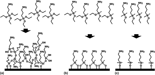
In this study, we have developed and characterized two previously unstudied alkoxysilane surface chemistries for use with superparamagnetic iron oxide (SPIO) nanoparticles as a magnetic resonance imaging contrast agent. We modified superparamagnetic iron oxide nanoparticles (SPIO) using aminopropyl triethoxysilane and two analogous alkoxysilanes, aminopropyl dimethylethoxysilane and aminopropyl methyldiethoxysilane, to compare a mono- and dialkoxysilane, respectively, to a more commonly used trialkoxysilane as two new SPIO surface chemistries capable of forming ultrathin functional surface coatings. The ligand densities of the mono- and dialkoxysilane-modified SPIO produced in this study are consistent with near monolayers of ligands on the SPIO surface. We studied the chemical stability of the mono-, di-, and trialkoxysilane-modified SPIO in neutral and acidic media to evaluate the viability of these surface chemistries for use in long-term intracellular applications. The mono- and dialkoxysilane-modified SPIO demonstrate comparable chemical stability to the trialkoxysilane-modified SPIO, indicating that the mono- and dialkoxysilane are both viable new SPIO surface chemistries for future applications requiring minimally thick alkoxysilane surface coatings.

Type
Articles
Copyright
Copyright © Materials Research Society 2012

Access options

Get access to the full version of this content by using one of the access options below. (Log in options will check for institutional or personal access. Content may require purchase if you do not have access.)

References

REFERENCES

1.Anzai, Y. and Prince, M.R.: Iron oxide-enhanced MR lymphography: The evaluation of cervical lymph node metastases in head and neck cancer. J. Magn. Reson. Imaging 7(1), 7581 (1997).CrossRefGoogle ScholarPubMed
2.Moore, A., Weissleder, R., and Bogdanov, A.: Uptake of dextran-coated monocrystalline iron oxides in tumor cells and macrophages. J. Magn. Reson. Imaging 7(6), 11401145 (1997).CrossRefGoogle Scholar
3.Petri-Fink, A., Chastellain, M., Juillerat-Jeanneret, L., Ferrari, A., and Hofmann, H.: Development of functionalized superparamagnetic iron oxide nanoparticles for interaction with human cancer cells. Biomaterials 26(15), 26852694 (2005).CrossRefGoogle ScholarPubMed
4.Lee, J., Isobe, T., and Senna, M.: Preparation of ultrafine Fe3O4 particles by precipitation in the presence of PVA at high pH. J. Colloid Interface Sci. 177(2), 490494 (1996).CrossRefGoogle Scholar
5.Lee, S.J., Jeong, J.R., Shina, S.C., Kimb, J.C., Chang, Y.H., Chang, Y.M., and Kim, J.D.: Nanoparticles of magnetic ferric oxides encapsulated with poly(D,L latide-co-glycolide) and their applications to magnetic resonance imaging contrast agent. J. Magn. Magn. Mater. 272276(3), 24322433 (2004).CrossRefGoogle Scholar
6.Moffat, B.A., Reddy, G.R., McConville, P., Hall, D.E., Chenevert, T.L., Kopelman, R.R., Philbert, M., Weissleder, R., Rehemtulla, A., and Ross, B.D.: A novel polyacrylamide magnetic nanoparticle contrast agent for molecular imaging using MRI. Mol. Imaging 2(4), 324332 (2003).CrossRefGoogle ScholarPubMed
7.Zhang, C., Wängler, B., Morgenstern, B., Zentgraf, H., Eisenhut, M., Untenecker, H., Krüger, R., Huss, R., Seliger, C., Semmler, W., and Kiessling, F.: Silica- and alkoxysilane-coated ultrasmall superparamagnetic iron oxide particles: A promising tool to label cells for magnetic resonance imaging. Langmuir 23(3), 14271434 (2007).CrossRefGoogle ScholarPubMed
8.Mikhaylova, M., Kim, D.K., Berry, C.C., Zagorodni, A., Toprak, M., Curtis, A.S., and Muhammed, M.: BSA immobilization on amine-functionalized superparamagnetic iron oxide nanoparticles. Chem. Mater. 16(12), 23442354 (2004).CrossRefGoogle Scholar
9.Koh, I., Wang, X., Varughese, B., Isaacs, L., Ehrman, S.H., and English, D.S.: Magnetic iron oxide nanoparticles for biorecognition: Evaluation of surface coverage and activity. J. Phys. Chem. B 110(4), 15531558 (2006).CrossRefGoogle ScholarPubMed
10.Kim, K.D., Kim, S.S., and Kim, H.T.: Formation and characterization of silica-coated magnetic nanoparticles by sol-gel method. J. Ind. Eng. Chem. 11(4), 584589 (2005).Google Scholar
11.Kim, D.K., Mikhaylova, M., Zhang, Y., and Muhammed, M.: Protective coating of superparamagnetic iron oxide nanoparticles. Chem. Mater. 15(8), 16171627 (2003).CrossRefGoogle Scholar
12.Yamaura, M., Camilo, R.L., Sampaio, L.C., Macedo, M.A., Nakamura, M., and Toma, H.E.: Preparation and characterization of (3-aminopropyl) triethoxysilane-coated magnetite nanoparticles. J. Magn. Magn. Mater. 279(2–3), 210217 (2004).CrossRefGoogle Scholar
13.Kohler, N., Sun, C., Wang, J., and Zhang, M.: Methotrexate-modified superparamagnetic nanoparticles and their intracellular uptake into human cancer cells. Langmuir 21(19), 88588864 (2005).CrossRefGoogle ScholarPubMed
14.Bruce, I.J. and Sen, T.: Surface modification of magnetic nanoparticles with alkoxysilanes and their application in magnetic bioseparations. Langmuir 21(15), 70297035 (2005).CrossRefGoogle ScholarPubMed
15.De Palma, R., Trekker, J., Peeters, S., Van Bael, M.J., Bonroy, K., Wirix-Speetjens, R., Reekmans, G., Laureyn, W., Borghs, G., and Maes, G.: Surface modification gamma-Fe2O3@SiO2 magnetic nanoparticles for the controlled interaction with biomolecules. J. Nanosci. Nanotechnol. 7(12), 46264641 (2007).CrossRefGoogle ScholarPubMed
16.De Palma, R., Peeters, S., Van Bael, M.J., Van den Rul, H., Bonroy, K., Laureyn, W., Mullens, J., Borghs, G., and Maes, G.: Silane ligand exchange to make hydrophobic superparamagnetic nanoparticles water-dispersible. Chem. Mater. 19(7), 18211831 (2007).CrossRefGoogle Scholar
17.Moon, J.H., Kim, J.H., Kim, K., Kang, T., Kim, B., Kim, C., Hahn, J.H., and Park, J.W.: Absolute surface density of the amine group of the aminosilylated thin layers: Ultraviolet-visible spectroscopy, second harmonic generation, and synchrotron-radiation photoelectron spectroscopy study. Langmuir 13(16), 43054310 (1997).CrossRefGoogle Scholar
18.Miyoshi, S., Flexman, J.A., Cross, D.J., Maravilla, K.R., Kim, Y., Anzai, Y., Oshima, J., and Minoshima, S.: Transfection of neuroprogenitor cells with iron nanoparticles for magnetic resonance imaging tracking: Cell viability, differentiation, and intracellular localization. Mol. Imaging Biol. 7(4), 286295 (2005).CrossRefGoogle ScholarPubMed
19.Sykova, E. and Jendelova, P.: In vivo tracking of stem cells in brain and spinal cord injury. Prog Brain Res. 161, 367383 (2007).CrossRefGoogle ScholarPubMed
20.Shapiro, E.M., Sharer, K., Skrtic, S., and Koretsky, A.P.: In vivo detection of single cells by MRI. Magn. Reson. Med. 55(2), 242249 (2006).CrossRefGoogle ScholarPubMed
21.Kircher, M.F., Allport, J.R., Graves, E.E., Love, V., Josephson, L., Lichtman, A.H., and Weissleder, R.: In vivo high-resolution three-dimensional imaging of antigen-specific cytotoxic T-lymphocyte trafficking to tumors. Cancer Res. 63(20), 68386846 (2003).Google ScholarPubMed
22.Valable, S., Barbiera, E.L., Bernaudinc, M., Roussel, S., Segebartha, C., Petit, E., and Rémy, C.: In vivo MRI tracking of exogenous monocytes/macrophages targeting brain tumors in a rat model of glioma. Neuroimage 37, S47S58 (2007).CrossRefGoogle Scholar
23.Mukherjee, S., Ghosh, R.N., and Maxfield, F.R.: Endocytosis. Physiol. Rev. 77(3), 759803 (1997).CrossRefGoogle ScholarPubMed
24.Lu, C.W., Hung, Y., Hsiao, J.K., Yao, M., Chung, T.H., Lin, Y.S., Wu, S.H., Hsu, S.C., Liu, H.M., Mou, C.Y., Yang, C.S., Huang, D.M., and Chen, Y.C.: Bifunctional magnetic silica nanoparticles for highly efficient human stem cell labeling. Nano Lett. 7(1), 149154 (2007).CrossRefGoogle ScholarPubMed
25.Arbab, A.S., Wilson, L.B., Ashari, P., Jordan, E.K., Lewis, B.K., and Frank, J.A.: A model of lysosomal metabolism of dextran-coated superparamagnetic iron oxide (SPIO) nanoparticles: Implications for cellular magnetic resonance imaging. NMR Biomed. 18(6), 383389 (2005).CrossRefGoogle Scholar
26.Skotland, T., Sontum, P.C., and Oulie, I.: In vitro stability analyses as a model for metabolism of ferromagnetic particles (ClariscanTM), a contrast agent for magnetic resonance imaging. J. Pharm. Biomed. Anal. 28(2), 323329 (2002).CrossRefGoogle Scholar
27.Barker, A.J., Cage, B., Russek, S., and Stoldt, C.R.: Ripening during magnetite nanoparticle synthesis: Resulting interfacial defects and magnetic properties. J. Appl. Phys. 98(6), 63528 (2005).CrossRefGoogle Scholar
28.Larsen, B.A., Haag, M.A., Serkova, N.J., Shroyer, K.R., and Stoldt, C.R.: Controlled aggregation of superparamagnetic iron oxide nanoparticles for the development of molecular magnetic resonance imaging probes. Nanotechnology 19, 265102 (2008).CrossRefGoogle ScholarPubMed