Published online by Cambridge University Press: 21 May 2018

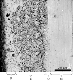
To increase the corrosion prevention of stainless steel implant and fast recovery due to new bone-cell formation at the orthopedic implant site, in the present investigation, a trilayered (with bioceramic interlayer sandwiched between innermost passivated surface and outermost polymer coating) 316L stainless steel (SS) implant was designed and investigated. It was inferred that this new designed implant invokes faster and more bone-cell formation than uncoated commercially available 316L SS implants. Faster bone-cell formation at the coated implant site reduces the initial threat of implant corrosion. The electrochemical corrosion study proved that this model of coated implants is able to prevent corrosion up to 90% better than uncoated commercially available 316L SS. Subsequently, preclinical studies in the rabbit bone defect model (which included histology, radiology, fluorochrome labeling, push-out test, and scanning electron microscopy taken after 45 and 90 days) proved higher rate of new bone tissue formation and better push-out strength between tissue in contact and the coated implant. The toxicological study of vital organs like liver, kidney, and heart also exhibited no abnormality. The outcome of the experimentations indicates suitability of this trilayered 316L SS implant for bone repair and healing.