Hostname: page-component-586b7cd67f-tf8b9 Total loading time: 0 Render date: 2024-11-24T12:49:58.189Z Has data issue: false hasContentIssue false

In vitro evaluation of a novel multiwalled carbon nanotube/nanohydroxyapatite/polycaprolactone composite for bone tissue engineering

Published online by Cambridge University Press:  21 February 2019
Huixiao Yang
Affiliation:
Key Laboratory of Oral Medicine, Guangzhou Institute of Oral Disease, Stomatological Hospital of Guangzhou Medical University, Guangzhou, Guangdong 510140, People’s Republic of China
Jieqing Li
Affiliation:
Department of Breast Surgery, Tianjin Central Hospital of Gynecology and Obstetrics, Tianjin 300100, People’s Republic of China; and Division of Cancer Research and Training, Department of Internal Medicine, Charles R. Drew University of Medicine and Science, Los Angeles, California 90059, USA
Qiong Liao
Affiliation:
Key Laboratory of Birth Defects and Related Diseases of Women and Children, Ministry of Education (Sichuan University) China, West China Second University Hospital, Sichuan University, Chengdu, Sichuan 610041, People’s Republic of China
Hua Guo
Affiliation:
Shriners Hospitals Pediatric Research Center, Lewis Katz School of Medicine, Temple University, Philadelphia, Pennsylvania 19140, USA
Huishan Chen
Affiliation:
Key Laboratory of Oral Medicine, Guangzhou Institute of Oral Disease, Stomatological Hospital of Guangzhou Medical University, Guangzhou, Guangdong 510140, People’s Republic of China
Yuting Zhu
Affiliation:
Key Laboratory of Oral Medicine, Guangzhou Institute of Oral Disease, Stomatological Hospital of Guangzhou Medical University, Guangzhou, Guangdong 510140, People’s Republic of China
Meijuan Cai
Affiliation:
Key Laboratory of Oral Medicine, Guangzhou Institute of Oral Disease, Stomatological Hospital of Guangzhou Medical University, Guangzhou, Guangdong 510140, People’s Republic of China
Huling Lv*
Affiliation:
Key Laboratory of Oral Medicine, Guangzhou Institute of Oral Disease, Stomatological Hospital of Guangzhou Medical University, Guangzhou, Guangdong 510140, People’s Republic of China; and State Key Laboratory of Oral Diseases, Sichuan University West China College of Stomatology, Sichuan University, Chengdu, Sichuan 610041, People’s Republic of China
*
a)Address all correspondence to this author. e-mail: [email protected]
Get access

Abstract

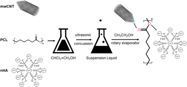
In this study, a three-phased multiwalled scaffold, composed of carbon nanotube (mwCNT), nanocrystalline hydroxyapatite (nHA), and polycaprolactone (PCL), was fabricated by the solvent evaporation technique. The structure character, mechanical properties, and degradation activity in simulated body fluid (SBF), along with osteoproductive ability in human osteosarcoma cell MG63, were investigated thoroughly. Results showed that the three phases in mwCNT/nHA/PCL composite presented excellent miscibility and stronger interfacial force when the weight content was 1/15/84 (wt%). Simultaneously, the composite had smaller porosity and slower degradation rate, and there was massive crystallized hydroxyapatite formed on the surface after being soaked in SBF. With regard to bioactivity, MG63s on this scaffolds presented good proliferation performance and differentiated into the osteogenic lineage by expressing high levels of ALP. It was concluded that mwCNTs/nHA/PCL composite scaffolds might be beneficial for bone tissue engineering at a relatively low concentration of mwCNTs and nHA.

Type
Article
Copyright
Copyright © Materials Research Society 2019 

Access options

Get access to the full version of this content by using one of the access options below. (Log in options will check for institutional or personal access. Content may require purchase if you do not have access.)

Footnotes

b)

These authors contributed equally to this work.

References

Kneser, U., Schaefer, D.J., Munder, B., Klemt, C., Andree, C., and Stark, G.B.: Tissue engineering of bone. Minimally Invasive Ther. Allied Technol. 11, 107 (2004).CrossRefGoogle Scholar
Dean, D., Min, K.J., and Bond, A.: Computer aided design of large-format prefabricated cranial plates. J. Craniofacial Surg. 14, 819 (2003).CrossRefGoogle ScholarPubMed
Siu, T.L., Rogers, J.M., Lin, K., Thompson, R., and Owbridge, M.: Custom-made titanium 3D printed interbody cages for treatment of osteoporotic fracture related spinal deformity. World Neurosurg. 111, 1 (2018).CrossRefGoogle ScholarPubMed
Hutmacher, D.W.: Scaffolds in tissue engineering bone and cartilage. Biomaterials 21, 2529 (2000).CrossRefGoogle ScholarPubMed
Hollister, S.J.: Porous scaffold design for tissue engineering. Nat. Mater. 4, 518 (2005).CrossRefGoogle ScholarPubMed
Armentano, I., Dottori, M., Fortunati, E., Mattioli, S., and Kenny, J.M.: Biodegradable polymer matrix nanocomposites for tissue engineering: A review. Polym. Degrad. Stab. 95, 2126 (2010).CrossRefGoogle Scholar
Abu Bakar, M., Cheang, P., and Khor, K.: Mechanical properties of injection molded hydroxyapatite-polyetheretherketone biocomposites. Compos. Sci. Technol. 63, 421 (2003).CrossRefGoogle Scholar
Li, K. and Tjong, S.C.: Preparation and characterization of isotactic polypropylene reinforced with hydroxyapatite nanorods. J. Macromol. Sci., Phys. 50, 1983 (2011).CrossRefGoogle Scholar
Tasis, D., Tagmatarchis, N., Bianco, A., and Prato, M.: Chemistry of carbon nanotubes. Chem. Rev. 106, 1105 (2006).CrossRefGoogle ScholarPubMed
Sahithi, K., Swetha, M., Ramasamy, K., Srinivasan, N., and Selvamurugan, N.: Polymeric composites containing carbon nanotubes for bone tissue engineering. Int. J. Biol. Macromol. 46, 281 (2010).CrossRefGoogle ScholarPubMed
Cheng, Q., Rutledge, K., and Jabbarzadeh, E.: Carbon nanotube–poly(lactide-co-glycolide) composite scaffolds for bone tissue engineering applications. Ann. Biomed. Eng. 41, 904 (2013).CrossRefGoogle ScholarPubMed
Liao, C.Z., Li, K., Wong, H.M., Tong, W.Y., Yeung, K.W.K., and Tjong, S.C.: Novel polypropylene biocomposites reinforced with carbon nanotubes and hydroxyapatite nanorods for bone replacements. Mater. Sci. Eng., C 33, 1380 (2013).CrossRefGoogle ScholarPubMed
Shokuhfar, T., Makradi, A., Titus, E., Cabral, G., Ahzi, S., Sousa, A.C., Belouettar, S., and Gracio, J.: Prediction of the mechanical properties of hydroxyapatite/polymethyl methacrylate/carbon nanotubes nanocomposite. J. Nanosci. Nanotechnol. 8, 4279 (2008).CrossRefGoogle ScholarPubMed
Zhang, J., Wen, Z., Zhao, M., and Dai, C.: Effect of the addition CNTs on performance of CaP/chitosan/coating deposited on magnesium alloy by electrophoretic deposition. Mater. Sci. Eng., C 58, 992 (2016).CrossRefGoogle ScholarPubMed
Liao, S., Xu, G.F., Wang, W., Watari, F., Cui, F.Z., Ramakrishna, S., and Chan, C.K.: Self-assembly of nano-hydroxyapatite on multi-walled carbon nanotubes. Acta Biomater. 3, 669 (2007).CrossRefGoogle ScholarPubMed
Woodruff, M.A. and Hutmacher, D.W.: The return of a forgotten polymer—polycaprolactone in the 21st century. Prog. Polym. Sci. 35, 1217 (2010).CrossRefGoogle Scholar
Huang, J., Best, S.M., Bonfield, W., Brooks, R.A., Rushton, N., Jayasinghe, S.N., and Edirisinghe, M.J.: In vitro assessment of the biological response to nano-sized hydroxyapatite. J. Mater. Sci.: Mater. Med. 15, 441 (2004).Google ScholarPubMed
Dorj, B., Won, J.E., Kim, J.H., Choi, S.J., Shin, U.S., and Kim, H.W.: Robocasting nanocomposite scaffolds of poly(caprolactone)/hydroxyapatite incorporating modified carbon nanotubes for hard tissue reconstruction. J. Biomed. Mater. Res., Part A 101, 1670 (2013).CrossRefGoogle ScholarPubMed
Goncalves, E.M., Oliveira, F.J., Silva, R.F., Neto, M.A., Fernandes, M.H., Amaral, M., Regí, M.V., and Vila, M.: Three-dimensional printed PCL-hydroxyapatite scaffolds filled with CNTs for bone cell growth stimulation. J. Biomed. Mater. Res., Part B 104, 1210 (2015).CrossRefGoogle ScholarPubMed
Baji, A., Wong, S.C., Liu, T., Li, T., and Srivatsan, T.S.: Morphological and X-ray diffraction studies of crystalline hydroxyapatite-reinforced polycaprolactone. J. Biomed. Mater. Res., Part B 81, 343 (2007).CrossRefGoogle ScholarPubMed
Kim, H.W., Knowles, J.C., and Kim, H.E.: Hydroxyapatite/poly(ε-caprolactone) composite coatings on hydroxyapatite porous bone scaffold for drug delivery. Biomaterials 25, 1279 (2004).CrossRefGoogle ScholarPubMed
Cadek, M., Coleman, J.N., Barron, V., Hedicke, K., and Blau, W.J.: Morphological and mechanical properties of carbon-nanotube-reinforced semicrystalline and amorphous polymer composites. Appl. Phys. Lett. 81, 5123 (2003).CrossRefGoogle Scholar
Kokubo, T., Kim, H.M., and Kawashita, M.: Novel bioactive materials with different mechanical properties. Biomaterials 24, 2161 (2003).CrossRefGoogle ScholarPubMed
Boyd, D., Towler, M.R., Wren, A.W., Clarkin, O.M., and Tanner, D.A.: TEM analysis of apatite surface layers observed on zinc based glass polyalkenoate cements. J. Mater. Sci. 43, 1170 (2008).CrossRefGoogle Scholar
Zhong, X., Lu, Z.F., Valtchev, P., Wei, H., Zreiqat, H., and Dehghani, F.: Surface modification of poly(propylene carbonate) by aminolysis and layer-by-layer assembly for enhanced cytocompatibility. Colloids Surf., B 93, 75 (2012).CrossRefGoogle ScholarPubMed
Hwang, Y.S., Sangaj, N., and Varghese, S.: Interconnected macroporous poly(ethylene glycol) cryogels as a cell scaffold for cartilage tissue engineering. Tissue Eng., Part A 16, 3033 (2010).CrossRefGoogle ScholarPubMed
Anselme, K., Linez, P., Bigerelle, M., Le Maguer, D., Hardouin, P., Hildebrand, H.F., Iost, A., and Leroy, J.M.: The relative influence of the topography and chemistry of TiAl6V4 surfaces on osteoblastic cell behaviour. Biomaterials 21, 1567 (2000).CrossRefGoogle ScholarPubMed
Tucker, B. and Lardelli, M.: A rapid apoptosis assay measuring relative acridine orange fluorescence in zebrafish embryos. Zebrafish 4, 113 (2007).CrossRefGoogle ScholarPubMed
Vega-Avila, E. and Pugsley, M.K.: An overview of colorimetric assay methods used to assess survival or proliferation of mammalian cells. Proc. West. Pharmacol. Soc. 54, 10 (2011).Google ScholarPubMed
Anselme, K.: Osteoblast adhesion on biomaterials. Biomaterials 21, 667 (2000).CrossRefGoogle ScholarPubMed
Neuhoff, V., Stamm, R., and Eibl, H.: Clear background and highly sensitive protein staining with Coomassie Blue dyes in polyacrylamide gels: A systematic analysis. Electrophoresis 6, 427 (1985).CrossRefGoogle Scholar
Li, C.Y. and Chou, T.W.: Modeling of elastic buckling of carbon nanotubes by molecular structural mechanics approach. Mech. Mater. 36, 1047 (2004).CrossRefGoogle Scholar
Zhang, Y.Z., Venugopal, J.R., El-Turki, A., Ramakrishna, S., Su, B., and Lim, C.T.: Electrospun biomimetic nanocomposite nanofibers of hydroxyapatite/chitosan for bone tissue engineering. Biomaterials 29, 4314 (2008).CrossRefGoogle ScholarPubMed
Fu, S.Z., Ni, P.Y., Wang, B.Y., Chu, B.Y., Peng, J.R., Zheng, L., Zhao, X., Luo, F., Wei, Y.Q., and Qian, Z.Y.: In vivo biocompatibility and osteogenesis of electrospun poly(ε-caprolactone)–poly(ethylene glycol)–poly(ε-caprolactone)/nano-hydroxyapatite composite scaffold. Biomaterials 33, 8363 (2012).CrossRefGoogle ScholarPubMed
Zhang, Y.B., Leblanc-Boily, V., Zhao, Y., and Prud’homme, R.E.: Wide angle X-ray diffraction investigation of crystal orientation in miscible blend of poly(ε-caprolactone)/poly(vinyl chloride) crystallized under strain. Polymer 46, 8141 (2005).CrossRefGoogle Scholar
Cho, K., Saheb, D.N., Yang, H., Kang, B., Kim, J., and Lee, S.: Real time in situ X-ray diffraction studies on the melting memory effect in the crystallization of β-isotactic polypropylene. Polymer 44, 4053 (2003).CrossRefGoogle Scholar
McCarthy, B., Coleman, J.N., Czerw, R., Dalton, A.B., Panhuis, M.I.H., Maiti, A., Drury, A., Bernier, P., Nagy, J.B., Lahr, B., Byrne, H.J., Carroll, D.L., and Blau, W.J.: A microscopic and spectroscopic study of interactions between carbon nanotubes and a conjugated polymer. J. Phys. Chem. B 106, 2210 (2002).CrossRefGoogle Scholar
Pan, L.L., Pei, X.B., He, R., Wan, Q.B., and Wang, J.: Multiwall carbon nanotubes/polycaprolactone composites for bone tissue engineering application. Colloids Surf., B 93, 226 (2012).CrossRefGoogle ScholarPubMed
Luo, F., Pan, L.L., Hong, G., Wang, T., Pei, X.B., Wang, J., and Wan, Q.B.: In vitro and in vivo characterization of multi-walled carbon nanotubes/polycaprolactone composite scaffolds for bone tissue engineering applications. J. Biomater. Tissue Eng. 7, 787 (2017).CrossRefGoogle Scholar
Babensee, J.E., Anderson, J.M., McIntire, L.V., and Mikos, A.G.: Host response to tissue engineered devices. Adv. Drug Delivery Rev. 33, 111 (1998).CrossRefGoogle Scholar
Persenaire, O., Alexandre, M., Degee, P., and Dubois, P.: Mechanisms and kinetics of thermal degradation of poly(ε-caprolactone). Biomacromolecules 2, 288 (2001).CrossRefGoogle Scholar
Lam, C.X.F., Savalani, M.M., Teoh, S.H., and Hutmacher, D.W.: Dynamics of in vitro polymer degradation of polycaprolactone-based scaffolds: Accelerated versus simulated physiological conditions. Biomed. Mater. 3, 4108 (2008).CrossRefGoogle ScholarPubMed
Meseguer-Duenas, J.M., Mas-Estelles, J., Castilla-Cortazar, I., Escobar Ivirico, J.L., and Vidaurre, A.: Alkaline degradation study of linear and network poly(ε-caprolactone). J. Mater. Sci.: Mater. Med. 22, 11 (2011).Google Scholar
Lei, Y., Rai, B., Ho, K.H., and Teoh, S.H.: In vitro degradation of novel bioactive polycaprolactone—20% tricalcium phosphate composite scaffolds for bone engineering. Mater. Sci. Eng., C 27, 293 (2007).CrossRefGoogle Scholar
Cadek, M., Coleman, J.N., Barron, V., Hedicke, K., and Blau, W.J.: Morphological and mechanical properties of carbon-nanotube-reinforced semicrystalline and amorphous polymer composites. Appl. Phys. Lett. 81, 5123 (2002).CrossRefGoogle Scholar
Kokubo, T. and Takadama, H.: How useful is SBF in predicting in vivo bone bioactivity? Biomaterials 27, 2907 (2006).CrossRefGoogle ScholarPubMed
Thalji, G., Gretzer, C., and Cooper, L.F.: Comparative molecular assessment of early osseointegration in implant-adherent cells. Bone 52, 444 (2013).CrossRefGoogle ScholarPubMed
Costa, D.O., Prowse, P.D.H., Chrones, T., Sims, S.M., Hamilton, D.W., Rizkalla, A.S., and Jeffrey Dixon, S.: The differential regulation of osteoblast and osteoclast activity by surface topography of hydroxyapatite coatings. Biomaterials 34, 7215 (2013).CrossRefGoogle ScholarPubMed
Freed, L.E., Vunjak-Novakovic, G., Biron, R.J., Eagles, D.B., Lesnoy, D.C., Barlow, S.K., and Langer, R.: Biodegradable polymer scaffolds for tissue engineering. Biotechnology 12, 689 (1994).Google ScholarPubMed
Yamashita, D., Machigashira, M., Miyamoto, M., Takeuchi, H., Noguchi, K., Izumi, Y., and Ban, S.: Effect of surface roughness on initial responses of osteoblast-like cells on two types of zirconia. Dent. Mater. J. 28, 461 (2009).CrossRefGoogle ScholarPubMed
Mata, D., Oliveira, F.J., Ferro, M., Gomes, P.S., Fernandes, M.H., Lopes, M.A., and Silva, R.F.: Multifunctional carbon nanotube/bioceramics modulate the directional growth and activity of osteoblastic cells. J. Biomed. Nanotechnol. 10, 725 (2014).CrossRefGoogle ScholarPubMed
Behring, J., Junker, R., Walboomers, X.F., Chessnut, B., and Jansen, J.A.: Toward guided tissue and bone regeneration: Morphology, attachment, proliferation, and migration of cells cultured on collagen barrier membranes. A systematic review. Odontology 96, 1 (2008).CrossRefGoogle ScholarPubMed
Raffaini, G. and Ganazzoli, F.: Surface topography effects in protein adsorption on nanostructured carbon allotropes. Langmuir 29, 4883 (2013).CrossRefGoogle ScholarPubMed
Lee, H.U., Jeong, Y.S., Jeong, S.Y., Park, S.Y., Bae, J.S., Kim, H.G., and Cho, C.R.: Role of reactive gas in atmospheric plasma for cell attachment and proliferation on biocompatible poly ε-caprolactone film. Appl. Surf. Sci. 254, 5700 (2008).CrossRefGoogle Scholar
Marom, R., Shur, I., Solomon, R., and Benayahu, D.: Characterization of adhesion and differentiation markers of osteogenic marrow stromal cells. J. Cell. Physiol. 202, 41 (2005).CrossRefGoogle ScholarPubMed
Morelli, S., Salerno, S., Holopainen, J., Ritala, M., and Bartolo, L.D.: Osteogenic and osteoclastogenic differentiation of co-cultured cells in polylactic acid–nanohydroxyapatite fiber scaffolds. J. Biotechnol. 204, 53 (2015).CrossRefGoogle ScholarPubMed
Abarrategi, A., Gutierrez, M.C., Moreno-Vicente, C., Hortiguela, M.J., Ramos, V., Lopez-Lacomba, J.L., Ferrer, M.L., and Monte, F.D.: Multiwall carbon nanotube scaffolds for tissue engineering purposes. Biomaterials 29, 94 (2008).CrossRefGoogle ScholarPubMed
Im, O., Li, J., Wang, M., Zhang, L.G., and Keidar, M.: Biomimetic three-dimensional nanocrystalline hydroxyapatite and magnetically synthesized single-walled carbon nanotube chitosan nanocomposite for bone regeneration. Int. J. Nanomed. 7, 2087 (2012).Google ScholarPubMed