Hostname: page-component-78c5997874-xbtfd Total loading time: 0 Render date: 2024-11-16T13:24:31.891Z Has data issue: false hasContentIssue false

Graphene/chitosan-functionalized iron oxide nanoparticles for biomedical applications

Published online by Cambridge University Press:  20 September 2019

Suresh Bandi
Affiliation:
Department of Metallurgical and Materials Engineering, VNIT Nagpur, Maharashtra 440010, India
Vikram Hastak
Affiliation:
Department of Metallurgical and Materials Engineering, VNIT Nagpur, Maharashtra 440010, India
Chokkakula L.P. Pavithra
Affiliation:
Department of Materials Science and Metallurgical Engineering, IIT Hyderabad, Telangana 502285, India
Sanjay Kashyap
Affiliation:
School of Physics and Materials Science, Thapar Institute of Engineering and Technology, Patiala, Punjab 147004, India
Dhananjay Kumar Singh
Affiliation:
Department of Molecular Bioprospection, CSIR-Central Institute of Medicinal and Aromatic Plants, Lucknow, Uttar Pradesh 226002, India; and Department of Human Molecular Genetics and Biochemistry, Sackler School of Medicine, Tel Aviv University, Tel Aviv699780, Israel
Suaib Luqman
Affiliation:
Department of Molecular Bioprospection, CSIR-Central Institute of Medicinal and Aromatic Plants, Lucknow, Uttar Pradesh 226002, India
Dilip R. Peshwe
Affiliation:
Department of Metallurgical and Materials Engineering, VNIT Nagpur, Maharashtra 440010, India
Ajeet K. Srivastav*
Affiliation:
Department of Metallurgical and Materials Engineering, VNIT Nagpur, Maharashtra 440010, India
*
a)Address all correspondence to this author. e-mail: [email protected], [email protected]
Get access

Abstract

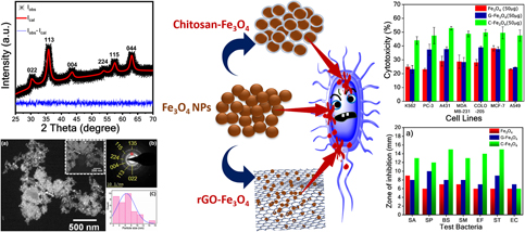
Superparamagnetic iron oxide nanoparticles are well known for biomedical applications. The particle size, morphology, surface area, and functionalization are the key parameters that affect their bioactivity properties. Inline to this, the superparamagnetic Fe3O4 nanoparticles were prepared via chemical coprecipitation method with an average particle size of 6 ± 3 nm. The particles were surface-functionalized with chitosan and in-house prepared reduced graphene oxide (rGO) to obtain chitosan-coated Fe3O4 nanoparticles (C-Fe3O4) and rGO-Fe3O4 nanocomposites (G-Fe3O4), respectively. Upon functionalization, the physicochemical properties of the materials were characterized thoroughly using X-ray diffraction, transmission electron microscopy, vibrating sample magnetometer, Raman Spectroscopy, and thermal gravimetric analysis. Furthermore, they have subjected to cytotoxicity assay, agar two-fold broth dilution test, and disc diffusion assay experiments for the determination of cytotoxicity and antibacterial activities. The effect of surface functionalization on their bioactivity was investigated thoroughly. The surface functionalization with chitosan and rGO has enhanced the bioactivity of the Fe3O4 nanoparticles.

Type
Article
Copyright
Copyright © Materials Research Society 2019 

Access options

Get access to the full version of this content by using one of the access options below. (Log in options will check for institutional or personal access. Content may require purchase if you do not have access.)

Footnotes

b)

These authors contributed equally to this work.

References

Nazarenus, M., Zhang, Q., Soliman, M.G., del Pino, P., Pelaz, B., Carregal-Romero, S., Rejman, J., Rothen-Rutishauser, B., Clift, M.J.D., Zellner, R., Nienhaus, G.U., Delehanty, J.B., Medintz, I.L., and Parak, W.J.: In vitro interaction of colloidal nanoparticles with mammalian cells: What have we learned thus far? Beilstein J. Nanotechnol. 5, 1477 (2014).CrossRefGoogle ScholarPubMed
Baldi, G., Bonacchi, D., Innocenti, C., Lorenzi, G., and Sangregorio, C.: Cobalt ferrite nanoparticles: The control of the particle size and surface state and their effects on magnetic properties. J. Magn. Magn. Mater. 311, 10 (2007).CrossRefGoogle Scholar
Song, Q. and Zhang, Z.J.: Shape control and associated magnetic properties of spinel cobalt ferrite nanocrystals. J. Am. Chem. Soc. 126, 6164 (2004).CrossRefGoogle ScholarPubMed
Kucheryavy, P., He, J., John, V.T., Maharjan, P., Spinu, L., Goloverda, G.Z., and Kolesnichenko, V.L.: Superparamagnetic iron oxide nanoparticles with variable size and an iron oxidation state as prospective imaging agents. Langmuir 29, 710 (2013).CrossRefGoogle ScholarPubMed
Frey, N.A., Peng, S., Cheng, K., and Sun, S.: Magnetic nanoparticles: Synthesis, functionalization, and applications in bioimaging and magnetic energy storage. Chem. Soc. Rev. 27, 2532 (2009).CrossRefGoogle Scholar
Polshettiwar, V., Luque, R., Fihri, A., Zhu, H., Bouhrara, M., and Basset, J.M.: Magnetically recoverable nanocatalysts. Chem. Rev. 111, 3036 (2011).CrossRefGoogle ScholarPubMed
Mangrulkar, P.A., Polshettiwar, V., Labhsetwar, N.K., Varma, R.S., and Rayalu, S.S.: Nano-ferrites for water splitting: Unprecedented high photocatalytic hydrogen production under visible light. Nanoscale 4, 5202 (2012).CrossRefGoogle ScholarPubMed
Goncalves, R.H. and Leite, E.R.: Nanostructural characterization of mesoporous hematite thin film photoanode used for water splitting. J. Mater. Res. 29, 47 (2014).CrossRefGoogle Scholar
Kumar, C.S.S.R. and Mohammad, F.: Magnetic nanomaterials for hyperthermia-based therapy and controlled drug delivery. Adv. Drug Delivery Rev. 63, 789 (2011).CrossRefGoogle ScholarPubMed
Salunkhe, A.B., Khot, V.M., and Pawar, S.H.: Magnetic hyperthermia with magnetic nanoparticles: A status review. Curr. Top. Med. Chem. 14, 572 (2014).CrossRefGoogle ScholarPubMed
Qiao, R., Yang, C., and Gao, M.: Superparamagnetic iron oxide nanoparticles: From preparations to in vivo MRI applications. J. Mater. Chem. 19, 6274 (2009).CrossRefGoogle Scholar
Macher, T., Totenhagen, J., Sherwood, J., Qin, Y., Gurler, D., Bolding, M.S., and Bao, Y.: Ultrathin iron oxide nanowhiskers as positive contrast agents for magnetic resonance imaging. Adv. Funct. Mater. 25, 490 (2015).CrossRefGoogle Scholar
Iv, M., Telischak, N., Feng, D., Holdsworth, S., Yeom, K., and Daldrup-Link, H.: Clinical applications of iron oxide nanoparticles for magnetic resonance imaging of brain tumors. Nanomedicine 10, 993 (2015).CrossRefGoogle ScholarPubMed
Goodwill, P.W. and Conolly, S.M.: Experimental demonstration of X-space magnetic particle imaging. SPIE Proc. Med. Imaging 7965, 79650U (2011).CrossRefGoogle Scholar
Ferguson, R.M., Khandhar, A.P., and Krishnan, K.M.: Tracer design for magnetic particle imaging (invited). J. Appl. Phys. 111, 07B318 (2012).CrossRefGoogle Scholar
Tomitaka, A., Arami, H., Gandhi, S., and Krishnan, K.M.: Lactoferrin conjugated iron oxide nanoparticles for targeting brain glioma cells in magnetic particle imaging. Nanoscale 7, 16890 (2015).CrossRefGoogle ScholarPubMed
Marszałł, M.P.: Application of magnetic nanoparticles in pharmaceutical sciences. Pharm. Res. 28, 480 (2011).CrossRefGoogle ScholarPubMed
Zhang, G., Qie, F., Hou, J., Luo, S., Luo, L., Sun, X., and Tan, T.: One-pot solvothermal method to prepare functionalized Fe3O4 nanoparticles for bioseparation. J. Mater. Res. 27, 1006 (2012).CrossRefGoogle Scholar
Widder, K.J., Senyei, A.E., and Scarpelli, D.G.: Magnetic microspheres: A model system for site specific drug delivery in vivo. Exp. Biol. Med. 158, 141 (1978).CrossRefGoogle ScholarPubMed
Laurent, S., Dutz, S., Häfeli, U.O., and Mahmoudi, M.: Magnetic fluid hyperthermia: Focus on superparamagnetic iron oxide nanoparticles. Adv. Colloid Interface Sci. 166, 8 (2011).CrossRefGoogle ScholarPubMed
Assa, F., Jafarizadeh-Malmiri, H., Ajamein, H., Vaghari, H., Anarjan, N., Ahmadi, O., and Berenjian, A.: Chitosan magnetic nanoparticles for drug delivery systems. Crit. Rev. Biotechnol. 37, 492 (2017).CrossRefGoogle ScholarPubMed
Salehizadeh, H., Hekmatian, E., Sadeghi, M., and Kennedy, K.: Synthesis and characterization of core–shell Fe3O4–gold–chitosan nanostructure. J. Nanobiotechnol. 10, 3 (2012).CrossRefGoogle ScholarPubMed
Roper, P.R. and Drewinko, B.: Comparison of in vitro methods to determine drug-induced cell lethality. Cancer Res. 36, 2182 (1976).Google ScholarPubMed
Valgas, C., Machado de Souza, S., A Smânia, E.F., and Smânia, A. Jr: Screening methods to determine antibacterial activity of natural products. Braz. J. Microbiol. 38, 369 (2007).CrossRefGoogle Scholar
Perreault, F., De Faria, A.F., Nejati, S., and Elimelech, M.: Antimicrobial properties of graphene oxide nanosheets: Why size matters. ACS Nano 9, 7226 (2015).CrossRefGoogle ScholarPubMed
Gurunathan, S., Woong Han, J., Eppakayala, V., and Kim, J.: Green synthesis of graphene and its cytotoxic effects in human breast cancer cells. Int. J. Nanomed. 8, 1015 (2013).CrossRefGoogle ScholarPubMed
Santos, C.M., Mangadlao, J., Ahmed, F., Leon, A., Advincula, R.C., and Rodrigues, D.F.: Graphene nanocomposite for biomedical applications: Fabrication, antimicrobial and cytotoxic investigations. Nanotechnology 23, 395101 (2012).CrossRefGoogle ScholarPubMed
Pelin, M., Fusco, L., León, V., Martín, C., Criado, A., Sosa, S., Vázquez, E., Tubaro, A., and Prato, M.: Differential cytotoxic effects of graphene and graphene oxide on skin keratinocytes. Sci. Rep. 7, 1 (2017).CrossRefGoogle ScholarPubMed
Hastak, V., Bandi, S., Kashyap, S., Singh, S., Luqman, S., Lodhe, M., Peshwe, D.R., and Srivastav, A.K.: Antioxidant efficacy of chitosan/graphene functionalized superparamagnetic iron oxide nanoparticles. J. Mater. Sci.: Mater. Med. 29, 154 (2018).Google ScholarPubMed
Yang, Z., Hao, X., Chen, S., Ma, Z., Wang, W., Wang, C., Yue, L., Sun, H., Shao, Q., Murugadoss, V., and Guo, Z.: Long-term antibacterial stable reduced graphene oxide nanocomposites loaded with cuprous oxide nanoparticles. J. Colloid Interface Sci. 533, 13 (2019).CrossRefGoogle ScholarPubMed
Jedrzejczak-Silicka, M.: Cytotoxicity and genotoxicity of GO–Fe3O4 hybrid in cultured mammalian cells. Pol. J. Chem. Technol. 19, 27 (2017).CrossRefGoogle Scholar
Gade, N.E., Dar, R.M., Mishra, O.P., Khan, J.R., Kumar, V., and Patyal, A.: Evaluation of dose-dependent cytotoxic effects of graphene oxide-iron oxide nanocomposite on caprine Wharton’s jelly derived mesenchymal stem cells. J. Anim. Res. 5, 415 (2015).CrossRefGoogle Scholar
Gatta, G.D., Kantor, I., Ballaran, T.B., Dubrovinsky, L., and Mccammon, C.: Effect of non-hydrostatic conditions on the elastic behaviour of magnetite: An in situ single-crystal X-ray diffraction study. Phys. Chem. Miner. 34, 627 (2007).CrossRefGoogle Scholar
Prince, E., Prince, E., and Stalick, J.K.: Accuracy in Powder Diffraction II, Vol. 846 (NIST Special Publicaiton, Gaithersburg, MD, 1992).Google Scholar
Srivastav, A.K., Panindre, A.M., and Murty, B.S.: XRD characterization of microstructural evolution during mechanical alloying of W–20 wt% Mo. Trans. Indian Inst. Met. 66, 409 (2013).CrossRefGoogle Scholar
Bandi, S., Hastak, V., Peshwe, D.R., and Srivastav, A.K.: In situ TiO2–rGO nanocomposites for CO gas sensing. Bull. Mater. Sci. 41, 115 (2018).CrossRefGoogle Scholar
Balzar, D. and Ledbetter, H.: Voigt-function modeling in Fourier analysis of size-and strain-broadened X-ray diffraction peaks. J. Appl. Crystallogr. 26, 97 (1993).CrossRefGoogle Scholar
Srivastav, A.K., Basu, J., Kashyap, S., Chawake, N., Yadav, D., and Murty, B.S.: Crystallographic-shear-phase-driven W18O49 nanowires growth on nanocrystalline W surfaces. Scr. Mater. 115, 28 (2016).CrossRefGoogle Scholar
Cullity, B.D. and Graham, C.D.: Introduction to Magnetic Materials (John Wiley & Sons, Inc., Hoboken, New Jersey, 2008). ISBN: 978-0-471-47741-9 47741.CrossRefGoogle Scholar
Padalia, D., Johri, U.C., and Zaidi, M.G.H.: Study of cerium doped magnetite (Fe3O4:Ce)/PMMA nanocomposites. Phys. B 407, 838 (2012).CrossRefGoogle Scholar
Pham, X.N., Nguyen, T.P., Pham, T.N., Tran, T.T.N., and Tran, T.V.T.: Synthesis and characterization of chitosan-coated magnetite nanoparticles and their application in curcumin drug delivery. Adv. Nat. Sci. Nanosci. Nanotechnol. 7, 045010 (2016).CrossRefGoogle Scholar
Safari, J. and Javadian, L.: Chitosan decorated Fe3O4 nanoparticles as a magnetic catalyst in the synthesis of phenytoin derivatives. RSC Adv. 4, 48973 (2014).CrossRefGoogle Scholar
Karimzadeh, I., Aghazadeh, M., Doroudi, T., Ganjali, M.R., and Kolivand, P.H.: Electrochemical preparation and characterization of chitosan-coated superparamagnetic iron oxide (Fe3O4) nanoparticles. Mater. Res. Innovations 1 (2017).Google Scholar
Bin Wu, J., Lin, M.L., Cong, X., Liu, H.N., and Tan, P.H.: Raman spectroscopy of graphene-based materials and its applications in related devices. Chem. Soc. Rev. 47, 1822 (2018).Google Scholar
Mura, S., Jiang, Y., Vassalini, I., Gianoncelli, A., Alessandri, I., Granozzi, G., Calvillo, L., Senes, N., Enzo, S., Innocenzi, P., and Malfatti, L.: Graphene oxide/iron oxide nanocomposites for water remediation. ACS Appl. Nano Mater. 1, 6724 (2018).CrossRefGoogle Scholar
Wall, M.: The Raman spectroscopy of graphene and the determination of layer thickness. Thermo Sci. Appl., 52252 (2011). Available at: http://tools.thermofisher.com/content/sfs/brochures/AN52252_E%201111%20LayerThkns_H_1.pdf.Google Scholar
Bandi, S., Ravuri, S., Peshwe, D.R., and Srivastav, A.K.: Graphene from discharged dry cell battery electrodes. J. Hazard. Mater. 366, 358 (2019).CrossRefGoogle ScholarPubMed
Wang, H., Wang, Y., Cao, X., Feng, M., and Lan, G.: Vibrational properties of graphene and graphene layers. J. Raman Spectrosc. 40, 1791 (2009).CrossRefGoogle Scholar
Corazzari, I., Nisticò, R., Turci, F., Faga, M.G., Franzoso, F., Tabasso, S., and Magnacca, G.: Advanced physico-chemical characterization of chitosan by means of TGA coupled on-line with FTIR and GCMS: Thermal degradation and water adsorption capacity. Polym. Degrad. Stab. 112, 1 (2015).CrossRefGoogle Scholar
Hasan, A., Waibhaw, G., Tiwari, S., Dharmalingam, K., Shukla, I., and Pandey, L.M.: Fabrication and characterization of chitosan, polyvinylpyrrolidone, and cellulose nanowhiskers nanocomposite films for wound healing drug delivery application. J. Biomed. Mater. Res., Part A 105, 2391 (2017).CrossRefGoogle ScholarPubMed
Tayyebi, A. and Outokesh, M.: Supercritical synthesis of a magnetite-reduced graphene oxide hybrid with enhanced adsorption properties toward cobalt & strontium ions. RSC Adv. 6, 13898 (2016).CrossRefGoogle Scholar
Shi, S.F., Jia, J.F., Guo, X.K., Zhao, Y.P., Chen, D.S., Guo, Y.Y., Cheng, T., and Zhang, X.L.: Biocompatibility of chitosan-coated iron oxide nanoparticles with osteoblast cells. Int. J. Nanomedicine 7, 5593 (2012).Google ScholarPubMed
Kavinkumar, T., Varunkumar, K., Ravikumar, V., and Manivannan, S.: Anticancer activity of graphene oxide-reduced graphene oxide–silver nanoparticle composites. J. Colloid Interface Sci. 505, 1125 (2017).CrossRefGoogle ScholarPubMed
Andrews, J.M.: Determination of minimum inhibitory concentrations. J. Antimicrob. Chemother. 48, 5 (2001).CrossRefGoogle ScholarPubMed
Uygur, B., Craig, G., Mason, M.D., and Ng, A.K.: Cytotoxicity and Genotoxicity of Silver Nanoparticles in Human Cells. Tech. Proc. 2009 NSTI Nanotechnol. Conf. Expo, NSTI-Nanotech 2009 2, 383 (2009).Google Scholar
Hussain, S.M., Hess, K.L., Gearhart, J.M., Geiss, K.T., and Schlager, J.J.: In vitro toxicity of nanoparticles in BRL 3A rat liver cells. Toxicol. In Vitro 19, 975 (2005).CrossRefGoogle ScholarPubMed
Padhi, D.K., Panigrahi, T.K., Parida, K., Singh, S.K., and Mishra, P.M.: Green synthesis of Fe3O4/RGO nanocomposite with enhanced photocatalytic performance for Cr(VI) reduction, phenol degradation, and antibacterial activity. ACS Sustainable Chem. Eng. 5, 10551 (2017).CrossRefGoogle Scholar
Nayamadi Mahmoodabadi, A., Kompany, A., and Mashreghi, M.: Characterization, antibacterial and cytotoxicity studies of graphene–Fe3O4 nanocomposites and Fe3O4 nanoparticles synthesized by a facile solvothermal method. Mater. Chem. Phys. 213, 285 (2018).CrossRefGoogle Scholar
Lee, C., Kim, J.Y., Il Lee, W., Nelson, K.L., Yoon, J., and Sedlak, D.L.: Bactericidal effect of zero-Valent iron nanoparticles on Escherichia coli. Environ. Sci. Technol. 42, 4927 (2008).CrossRefGoogle ScholarPubMed
Srivastava, V., Darokar, M.P., Fatima, A., Kumar, J.K., Chowdhury, C., Saxena, H.O., Dwivedi, G.R., Shrivastava, K., Gupta, V., Chattopadhyay, S.K., Luqman, S., Gupta, M.M., Negi, A.S., and Khanuja, S.P.S.: Synthesis of diverse analogues of Oenostacin and their antibacterial activities. Bioorg. Med. Chem. 15, 518 (2007).CrossRefGoogle ScholarPubMed
Luqman, S., Dwivedi, G.R., Darokar, M.P., Kalra, A., and Khanuja, S.P.S.: Antimicrobial activity of Eucalyptus citriodora essential oil. Int. J. Essent. Oil Ther. 2, 69 (2008).Google Scholar