Hostname: page-component-cd9895bd7-gxg78 Total loading time: 0 Render date: 2024-12-27T06:46:02.312Z Has data issue: false hasContentIssue false

Formation of aligned core/sheath microfiber scaffolds with a poly-L-lactic acid (PLLA) sheath and a conductive poly(3,4-ethylenedioxythiophene) (PEDOT) core

Published online by Cambridge University Press:  04 June 2019

Rachel A. Martin
Affiliation:
Chemical Engineering, Michigan Technological University, Houghton, Michigan 49931, USA
Marie Wendling
Affiliation:
Bachelor’s Degree Student, Biomedical Engineering, Michigan Technological University, Houghton, Michigan 49931, USA
Bailey Mohrenweiser
Affiliation:
Bachelor’s Degree Student, Chemical Engineering, Michigan Technological University, Houghton, Michigan 49931, USA
Zichen Qian
Affiliation:
Ph.D. Student, Biomedical Engineering, Michigan Technological University, Houghton, Michigan 49931, USA
Feng Zhao
Affiliation:
Associate Professor, Ph.D., Biomedical Engineering, Michigan Technological University, Houghton, Michigan 49931, USA
Michael E. Mullins*
Affiliation:
Professor, Ph.D., Chemical Engineering, Michigan Technological University, Houghton, Michigan 49931, USA
*
a)Address all correspondence to this author. e-mail: [email protected]
Get access

Abstract

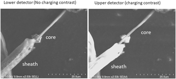
Electrospun coaxial fibers are used to create core/sheath fiber structures to act as growth-promoting scaffolds for in vitro dorsal root ganglia (DRG) cell cultures. The core was a conducting polymer, poly(3,4-ethylenedioxythiophene):poly(styrene sulfonate) (PEDOT:PSS), and the sheath was poly-L-lactic acid (PLLA), which created coaxial fibers with a conductive core and an insulating sheath. SEM analysis confirmed the conductivity of the core and insulation of the sheath. Several coaxial spinneret designs were tested with the best results obtained by using various annular spinning needle combinations. Using a 22G/16G and 22G/17G combination, fibers with diameters of 6.1 ± 2.4 µm and 3.3 ± 0.9 µm were spun, respectively. The fibers showed a Young’s modulus and hardness of 0.16 ± 0.13 and 0.02 ± 0.01 GPa for the larger diameters, and 0.7 ± 0.4 and 0.03 ± 0.03 GPa for the smaller diameter fibers. In vitro test cultures showed the fibers successfully directed chick DRG axonal outgrowth with low biotoxicity.

Type
Article
Copyright
Copyright © Materials Research Society 2019 

Access options

Get access to the full version of this content by using one of the access options below. (Log in options will check for institutional or personal access. Content may require purchase if you do not have access.)

References

Ahuja, C.S., Jefferson, R.W., Nori, S., Kotter, M.R.N., Druschel, C., Curt, A., and Fehlings, M.G.: Traumatic spinal cord injury. Nat. Rev. Dis. Primers 3, 17018 (2017).CrossRefGoogle ScholarPubMed
Ingavle, G.C. and Leach, J.K.: Advancements in electrospinning of polymeric nanofibrous scaffolds for tissue engineering. Tissue Eng., Part B 20, 277293 (2014).CrossRefGoogle ScholarPubMed
Raspa, A., Marchini, A., Pugliese, R., Mauri, M., Maleki, M., Vasita, R., and Gelain, F.: A biocompatibility study of new nanofibrous scaffolds for nervous system regeneration. Nanoscale 8, 253265 (2016).CrossRefGoogle ScholarPubMed
Schaub, N.J., Johnson, C.D., Cooper, B., and Gilbert, R.J.: Electrospun fibers for spinal cord injury research and regeneration. J. Neurotrauma 33, 14051415 (2016).CrossRefGoogle ScholarPubMed
Hurtado, A., Cregg, J.M., Wang, H.B., Wendell, D.F., Oudega, M., Gilbert, R.J., and McDonald, J.W.: Robust CNS regeneration after complete spinal cord transection using aligned poly-L-lactic acid microfibers. Biomaterials 32, 60686079 (2011).CrossRefGoogle ScholarPubMed
Corey, J.M., Lin, D.Y., Mycek, K.B., Chen, Q., Samuel, S., Feldman, E.L., and Martin, D.C.: Aligned electrospun nanofibers specify the direction of dorsal root ganglia neurite growth. J. Biomed. Mater. Res. 83, 636645 (2007).CrossRefGoogle ScholarPubMed
Wang, H.B., Mullins, M.E., Cregg, J.M., Hurtado, A., Oudega, M., Trombley, M.T., and Gilbert, R.J.: Creation of highly aligned electrospun poly-L-lactic acid fibers for nerve regeneration applications. J. Neural Eng. 6, 016001 (2009).CrossRefGoogle ScholarPubMed
He, L., Tang, S., Prabhakaran, M.P., Liao, S., Tian, L., Zhang, Y., Xue, W., and Ramakrishna, S.: Surface modification of PLLA nano-scaffolds with laminin multilayer by LbL assembly for enhancing neurite outgrowth. Macromol. Biosci. 13, 16011609 (2013).CrossRefGoogle ScholarPubMed
Zhang, K., Zheng, H., Liang, S., and Gao, C.: Aligned PLLA nanofibrous scaffolds coated with graphene oxide for promoting neural cell growth. Acta Biomater. 37, 131142 (2016).CrossRefGoogle ScholarPubMed
Koppes, A.N., Zaccor, N.W., Rivet, C.J., Williams, L.A., Piselli, J.M., Gilbert, R.J., and Thompson, D.M.: Neurite outgrowth on electrospun PLLA fibers is enhanced by exogenous electrical stimulation. J. Neural Eng. 11, 046002 (2014).CrossRefGoogle ScholarPubMed
Mohtaram, N.K., Ko, J., Agbay, A., Rattray, D., Neill, P.O., Rajwani, A., Vasandani, R., Thu, H.L., Jun, M.B.G., and Willerth, S.M.: Development of a glial cell-derived neurotrophic factor-releasing artificial dura for neural tissue engineering applications. J. Mater. Chem. B 3, 79747985 (2015).CrossRefGoogle Scholar
Xie, J., Liu, W., MacEwan, M.R., Bridgman, P.C., and Younan, X.: Neurite outgrowth on electrospun nanofibers with uniaxial alignment. ACS Nano (2014).CrossRefGoogle ScholarPubMed
Huang, C., Ouyang, Y., Niu, H., He, N., Ke, Q., Jin, X., Li, D., Fang, J., Liu, W., Fan, C., and Lin, T.: Nerve guidance conduits from aligned nanofibers: Improvement of nerve regeneration through longitudinal nanogrooves on a fiber surface. ACS Appl. Mater. Interfaces 7, 71897196 (2015).CrossRefGoogle ScholarPubMed
Xu, T., Miszuk, J.M., Zhao, Y., Sun, H., and Fong, H.: Electrospun polycaprolactone 3D nanofibrous scaffold with interconnected and hierarchically structured pores for bone tissue engineering. Adv. Healthcare Mater. 4, 22382246 (2015).CrossRefGoogle ScholarPubMed
Entekhabi, E., Haghbin Nazarpak, M., Moztarzadeh, F., and Sadeghi, A.: Design and manufacture of neural tissue engineering scaffolds using hyaluronic acid and polycaprolactone nanofibers with controlled porosity. Mater. Sci. Eng., C 69, 380387 (2016).CrossRefGoogle ScholarPubMed
Jenkins, P.M., Laughter, M.R., Lee, D.J., Lee, Y.M., Freed, C.R., and Park, D.: A nerve guidance conduit with topographical and biochemical cues: Potential application using human neural stem cells. Nanoscale Res. Lett. 10, 972 (2015).CrossRefGoogle ScholarPubMed
Pawar, K., Cummings, B.J., Thomas, A., Shea, L.D., Levine, A., Pfaff, S., and Anderson, A.J.: Biomaterial bridges enable regeneration and re-entry of corticospinal tract axons into the caudal spinal cord after SCI: Association with recovery of forelimb function. Biomaterials 65, 112 (2015).CrossRefGoogle ScholarPubMed
Nune, M., Krishnan, U.M., and Sethuraman, S.: PLGA nanofibers blended with designer self-assembling peptides for peripheral neural regeneration. Mater. Sci. Eng., C 62, 329337 (2016).CrossRefGoogle ScholarPubMed
Kijenska, E., Prabhakaran, M.P., Swieszkowski, W., Kurzydlowski, K.J., and Ramakrishna, S.: Electrospun bio-composite P(LLA-CL)/collagen I/collagen III scaffolds for nerve tissue engineering. J. Biomed. Mater. Res., Part B 100, 10931102 (2012).CrossRefGoogle ScholarPubMed
Bhutto, M.A., Zhang, J., Sun, B., El-Hamshary, H., Al-Deyab, S.S., and Mo, X.: Development of poly(L-lactide-co-caprolactone) multichannel nerve conduit with aligned electrospun nanofibers for Schwann cell proliferation. Int. J. Polym. Mater. Polym. Biomater. 65, 323329 (2016).CrossRefGoogle Scholar
Zhang, Y., Huang, J., Huang, L., Liu, Q., Shao, H., Hu, X., and Song, L.: Silk fibroin-based scaffolds with controlled delivery order of VEGF and BDNF for cavernous nerve regeneration. ACS Biomater. Sci. Eng. 2, 20182025 (2016).CrossRefGoogle Scholar
Hurtado, A., Gilbert, R.J., Wang, H.B., Cregg, J.M., Mullins, M.E., and Oudega, M.: Three-dimensional scaffolds, methods for fabricating the same, and methods of treating a peripheral nerve or spinal cord injury. Google Patents, 2013.Google Scholar
Zuidema, J.M., Provenza, C., Caliendo, T., Dutz, S., and Gilbert, R.J.: Magnetic NGF-releasing PLLA/iron oxide nanoparticles direct extending neurites and preferentially guide neurites along aligned electrospun microfibers. ACS Chem. Neurosci. 6, 17811788 (2015).CrossRefGoogle ScholarPubMed
Kuihua, Z., Chunyang, W., Cunyi, F., and Xiumei, M.: Aligned SF/P(LLA-CL)-blended nanofibers encapsulating nerve growth factor for peripheral nerve regeneration. J. Biomed. Mater. Res. 102, 26802691 (2014).CrossRefGoogle ScholarPubMed
Lu, Y., Huang, J., Yu, G., Cardenas, R., Wei, S., Wujcik, E.K., and Guo, Z.: Coaxial electrospun fibers: Applications in drug delivery and tissue engineering. Wiley Interdiscip. Rev.: Nanomed. Nanobiotechnol. 8, 654677 (2016).Google ScholarPubMed
Nguyen, T.T., Ghosh, C., Hwang, S.G., Chanunpanich, N., and Park, J.S.: Porous core/sheath composite nanofibers fabricated by coaxial electrospinning as a potential mat for drug release system. Int. J. Pharm. 439, 296306 (2012).CrossRefGoogle ScholarPubMed
Wang, C.Y., Liu, J.J., Fan, C.Y., Mo, X.M., Ruan, H.J., and Li, F.F.: The effect of aligned core–shell nanofibres delivering NGF on the promotion of sciatic nerve regeneration. J. Biomater. Sci., Polym. Ed. 23, 167184 (2012).CrossRefGoogle ScholarPubMed
Anderson, M., Shelke, N.B., Manoukian, O.S., Yu, X., McCullough, L.D., and Kumbar, S.G.: Peripheral nerve regeneration strategies: Electrically stimulating polymer based nerve growth conduits. Crit. Rev. Biomed. Eng. 43, 131159 (2015).CrossRefGoogle ScholarPubMed
Lee, J.Y., Bashur, C.A., Goldstein, A.S., and Schmidt, C.E.: Polypyrrole-coated electrospun PLGA nanofibers for neural tissue applications. Biomaterials 30, 43254335 (2009).CrossRefGoogle ScholarPubMed
Guex, A.G., Spicer, C.D., Armgarth, A., and Glemi, A.: Electrospun aniline-tetramer-co-polycaprolactone fibers for conductive, biodegradable scaffolds. MRS Commun. 7, 375382 (2017).CrossRefGoogle Scholar
Al-Majed, A.A., Neumann, C.M., Brushart, T.M., and Gordon, T.: Brief electrical stimulation promotes the speed and accuracy of motor axonal regeneration. J. Neurosci. 20, 26022608 (2000).CrossRefGoogle ScholarPubMed
Wood, M.D. and Willits, R.K.: Applied electric field enhances DRG neurite growth: Influence of stimulation media, surface coating and growth supplements. J. Neural Eng. 6, 046003 (2009).CrossRefGoogle ScholarPubMed
Elzinga, K., Tyreman, N., Ladak, A., Savaryn, B., Olson, J., and Gordon, T.: Brief electrical stimulation improves nerve regeneration after delayed repair in Sprague Dawley rats. Exp. Neurol. 269, 142153 (2015).CrossRefGoogle ScholarPubMed
Schmidt, C.E., Shastri, V.R., Vacanti, J.P., and Langer, R.: Stimulation of neurite outgrowth using an electrically conducting polymer. Proc. Natl. Acad. Sci. U. S. A. 94, 89488953 (1997).CrossRefGoogle ScholarPubMed
Xu, C., Kou, Y., Zhang, P., Han, N., Yin, X., Deng, J., Chen, B., and Jiang, B.: Electrical stimulation promotes regeneration of defective peripheral nerves after delayed repair intervals lasting under one month. PLoS One 9 (2014).Google ScholarPubMed
Moreno-Cortez, I.E., Alvarado-Castañeda, A., Garcia-Gutierrez, D.F., Garcia-Gomez, N.A., Sepulveda-Guzman, S., and Garcia-Gutierrez, D.I.: Core–shell PEDOT:PSS—PVP nanofibers containing PbS nanoparticles through coaxial electrospinning. Synth. Met. 220, 255262 (2016).CrossRefGoogle Scholar
Jin, L., Wang, T., Feng, Z-Q., Leach, M.K., Wu, J., Mo, S., and Jiang, Q.: A facile approach for the fabrication of core–shell PEDOT nanofiber mats with superior mechanical properties and biocompatibility. J. Mater. Chem. B 1, 1818 (2013).CrossRefGoogle Scholar
Kiristi, M., Oksuz, A.U., Oksuz, L., and Ulusoy, S.: Electrospun chitosan/PEDOT nanofibers. Mater. Sci. Eng., C 33, 38453850 (2013).CrossRefGoogle ScholarPubMed
Yamato, H., Ohwa, M., and Wernet, W.: Stability of polypyrrole and poly(3,4-ethylenedioxythiophene) for biosensor application. J. Electroanal. Chem. 397, 163170 (1995).CrossRefGoogle Scholar
Asplund, M., Thaning, E., Lundberg, J., Sandberg-Nordqvist, A.C., Kostyszyn, B., Inganas, O., and von Holst, H.: Toxicity evaluation of PEDOT/biomolecular composites intended for neural communication electrodes. Biomed. Mater. 4, 045009 (2009).CrossRefGoogle ScholarPubMed
Martin, R., Mullins, M.E., Zhao, F., and Qian, Z.: Electrospinning 3D scaffolds for use in neural tissue engineering. MRS Proc. 1798 (2015).CrossRefGoogle Scholar
Lim, C.H.: Production of liquid core–polymer shell microcapsules. Ph.D. dissertation, Michigan Technological University, Houghton, Michigan, 2011; p. 182.Google Scholar
Cong, Y., Liu, S., and Chen, H.: Fabrication of conductive polypyrrole nanofibers by electrospinning. J. Nanomater. 2013, 16 (2013).CrossRefGoogle Scholar
Seo, J.M.: Solution property and characteristic study for ultra fine fibers via electrospinning. M.S. thesis, Michigan Technological University, Houghton, MI, 2005.Google Scholar
Pharr, G.M., Oliver, W.C., and Brotzen, F.R.: On the generality of the relationship among contact stiffness, contact area, and elastic modulus during indentation. J. Mater. Res. 7, 613617 (1992).CrossRefGoogle Scholar
Wang, H.B., Mullins, M.E., Cregg, J.M., McCarthy, C.W., and Gilbert, R.J.: Varying the diameter of aligned electrospun fibers alters neurite outgrowth and Schwann cell migration. Acta Biomater. 6, 29702978 (2010).CrossRefGoogle ScholarPubMed
Reilly, G.C. and Engler, A.J.: Intrinsic extracellular matrix properties regulate stem cell differentiation. J. Biomech. 43, 5562 (2010).CrossRefGoogle ScholarPubMed