Hostname: page-component-78c5997874-mlc7c Total loading time: 0 Render date: 2024-11-05T03:57:13.334Z Has data issue: false hasContentIssue false

Effect of Ti6Al4V surface morphology on the osteogenic differentiation of human embryonic stem cells

Published online by Cambridge University Press:  26 October 2017

Leonardo Marasca Antonini*
Affiliation:
Laboratório de Pesquisa em Corrosão/Programa de Pós-Graduação em Engenharia de Minas, Metalúrgica e de Materiais (LAPEC/PPGE3M), Universidade Federal do Rio Grande do Sul, Porto Alegre, RS 91501-970, Brasil
Vinícius Kothe
Affiliation:
Laboratório de Pesquisa em Corrosão/Programa de Pós-Graduação em Engenharia de Minas, Metalúrgica e de Materiais (LAPEC/PPGE3M), Universidade Federal do Rio Grande do Sul, Porto Alegre, RS 91501-970, Brasil
Gwendolen C. Reilly
Affiliation:
Institute for in silico Medicine (INSIGNEO), University of Sheffield, Sheffield S1 3JD, U.K.
Robert Owen
Affiliation:
Institute for in silico Medicine (INSIGNEO), University of Sheffield, Sheffield S1 3JD, U.K.
Jossano Saldanha Marcuzzo
Affiliation:
Instituto Nacional de Pesquisas Espaciais (INPE), São José dos Campos, SP 12228-970, Brasil
Célia de Fraga Malfatti
Affiliation:
Laboratório de Pesquisa em Corrosão/Programa de Pós-Graduação em Engenharia de Minas, Metalúrgica e de Materiais (LAPEC/PPGE3M), Universidade Federal do Rio Grande do Sul, Porto Alegre, RS 91501-970, Brasil
*
a)Address all correspondence to this author. e-mail: [email protected]
Get access

Abstract

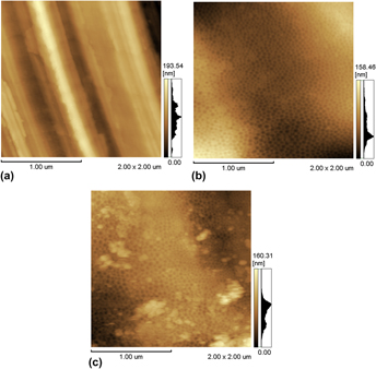
Ti6Al4V alloys usually need to undergo some kind of surface treatment to enable good bone growth and implant integration. In this work, three treatments that modify the titanium alloy surface were studied with the aim of promoting osteogenic differentiation of human-embryonic-stem-cell-derived mesenchymal progenitors (hESC-MPs). The surface treatments used were mechanical polishing and electropolishing for 4 or 12 min in an H2SO4/HF/glycerine solution. The samples were characterised by atomic force microscopy, profilometry, X-ray photoelectron spectroscopy, and wettability. Samples were seeded with hESC-MPs in osteogenic media, and the cell number and alkaline phosphatase activity were measured. The electropolishing surface treatments influenced the nanometric morphology and wettability. However, the electropolished surfaces contributed in the same way as the mechanically polished surface to osteogenic differentiation, indicating that differentiation was strongly influenced by microroughness, which did not differ among the treatments used in the present work.

Type
Articles
Copyright
Copyright © Materials Research Society 2017 

Access options

Get access to the full version of this content by using one of the access options below. (Log in options will check for institutional or personal access. Content may require purchase if you do not have access.)

Footnotes

Contributing Editor: Lakshmi Nair

References

REFERENCES

Huang, C.A., Hsu, F., and Yu, C.H.: Electropolishing behaviour of pure titanium in sulphuric acid–ethanol electrolytes with an addition of water. Corros. Sci. 53(2), 589 (2011).Google Scholar
Van, E.L., Rodriguez, I., Low, H.Y., Elmouelhi, N., Lowenhaupt, B., Natarajan, S., Lim, C.T., Prajapati, R., Vyakarnam, M., and Cooper, K.: Review: Micro- and nanostructured surface engineering for biomedical applications. J. Mater. Res. 28(2), 165 (2013).Google Scholar
Nazneen, F., Galvin, P., Arrigan, D.W.M., Thompson, M., Benvenuto, P., and Herzog, G.: Electropolishing of medical-grade stainless steel in preparation for surface nano-texturing. J. Solid State Electrochem. 16, 1389 (2012).Google Scholar
Bettinger, C., Zhang, Z., Gerecht, S., Borenstein, J.T., and Langer, R.: Enhancement of in vitro capillary tube formation by substrate nanotopography. Adv. Mater. 20, 99 (2008).Google Scholar
Lu, J., Rao, M., MacDonald, N., Khang, D., and Webster, T.: Improved endothelial cell adhesion and proliferation on patterned titanium surfaces with rationally designed, micrometer to nanometer features. Acta Biomater. 4, 192 (2008).Google Scholar
Drensler, S., Neelakantan, L., Somsen, C., Eggeler, G., and Hassel, A.H.: Electropolishing of a nickel–titanium–copper shape memory alloy in methanolic sulfuric acid. Electrochem. Solid-State Lett. 12, C1 (2009).CrossRefGoogle Scholar
Hao, Y., Li, S., Han, X., Hao, Y., and Ai, H.: Effects of the surface characteristics of nanoporous titanium oxide films on Ti–24Nb–4Zr–8Sn alloy on the initial adhesion of osteoblast-like MG-63 cells. Exp. Ther. Med. 6, 241 (2013).Google Scholar
Kim, K-H., Kwon, T-Y., Kim, S-Y., Kang, I-K., Kim, S., Yang, Y., and Ong, J.L.: Preparation and characterization of anodized titanium surface and their effect on osteoblast responses. J. Oral Implantol. 32, 8 (2006).Google Scholar
Cydzik, E.K., Kowalski, K., and Kaczmarek, A.: Anodic and nanostructural layers on titanium and its alloys for medical applications. Inz. Mater. 5, 425 (2009).Google Scholar
Karmarker, S., Yu, W., and Kyung, H-M.: Effect of surface anodization on stability of orthodontic microimplant. Korean J. Orthod 42, 4 (2012).Google Scholar
Xie, J. and Luan, B.L.: Microstructural and electrochemical characterization of hydroxyapatite-coated Ti6Al4V alloy for medical implants. J. Mater. Res. 23(3), 768 (2008).Google Scholar
Landis, W.J. and Silver, F.H.: Mineral deposition in the extracellular matrices of vertebrate tissues: Identification of possible apatite nucleation sites on type I collagen. Cells Tissues Organs 189, 20 (2009).Google Scholar
Tomsia, A.P., Lee, J.S., Wegst, U.G., and Saiz, E.: Nanotechnology for dental implants. Int. J. Oral Maxillofac. Implants 28, e535 (2013).Google Scholar
Gao, C., Zhuang, J., Li, P., Shuai, C., and Peng, S.: Preparation of micro/nanometer-sized porous surface structure of calcium phosphate scaffolds and the influence on biocompatibility. J. Mater. Res. 29(10), 1144 (2014).Google Scholar
Martinez, E., Engel, E., Planell, J.A., and Samitier, J.: Effects of artificial micro- and nano-structured surfaces on cell behaviour. Ann. Anat. 191, 126 (2009).Google Scholar
Lord, M., Foss, M., and Besenbacher, F.: Influence of nanoscale surface topography on protein adsorption and cellular response. Nano Today 5, 66 (2010).Google Scholar
Wang, P., Zhao, L., Liu, J., Weir, M.D., Zhou, X., and Xu, H.H.K.: Bone tissue engineering via nanostructured calcium phosphate biomaterials and stem cells. Bone Res. 2, 1 (2014).Google Scholar
Zhao, L., Liu, L., Wu, Z., Zhang, Y., and Chu, P.K.: Effects of micropitted/nanotubular titania topographies on bone mesenchymal stem cell osteogenic differentiation. Biomaterials 33, 2629 (2012).Google Scholar
Liao, S., Nguyen, L.T., Ngiam, M., Wang, C., Cheng, Z., Chan, C.K., and Ramakrishna, S.: Biomimetic nanocomposites to control osteogenic differentiation of human mesenchymal stem cells. Adv. Healthcare Mater. 3, 737 (2014).Google Scholar
Lock, J. and Liu, H.: Nanomaterials enhance osteogenic differentiation of human mesenchymal stem cells similar to a short peptide of BMP-7. Int. J. Nanomed. 6, 2769 (2011).Google Scholar
Polini, A., Pisignano, D., Parodi, M., Quarto, R., and Scaglione, S.: Osteoinduction of human mesenchymal stem cells by bioactive composite scaffolds without supplemental osteogenic growth factors. PLoS One 6, e26211 (2011).Google Scholar
Koegler, P., Clayton, A., Thissen, H., Santos, G.N., and Kingshott, P.: The influence of nanostructured materials on biointerfacial interactions. Adv. Drug Delivery Rev. 64, 1820 (2012).Google Scholar
Fricain, J.C., Schlaubitz, S., Le Visage, C., Arnault, I., Derkaoui, S.M., Siadous, R., Catros, S., Lalande, C., Bareille, R., Renard, M., Fabre, T., Cornet, S., Durand, M., Léonard, A., Sahraoui, N., Letourneur, D., and Amédéd, J.: A nano-hydroxyapatite–pullulan/dextran polysaccharide composite macroporous material for bone tissue engineering. Biomaterials 34, 2947 (2013).Google Scholar
Nikukar, H., Reid, S., Tsimbouri, P.M., Riehle, M.O., Curtis, A.S., and Dalby, M.J.: Osteogenesis of mesenchymal stem cells by nanoscale mechanotransduction. ACS Nano 7, 2758 (2013).Google Scholar
Mendonca, G., Mendonca, D.B., Simoes, L.G., Araújo, A.L., Leite, E.R., Duarte, W.R., Aragão, F.J., and Cooper, L.F.: The effects of implant surface nanoscale features on osteoblast-specific gene expression. Biomaterials 30, 4053 (2009).Google Scholar
Marini, F., Luzi, E., Fabbri, S., Ciuffi, S., Sorace, S., Tognarini, I., Galli, G., Zonefrati, R., Sbaiz, F., and Brandi, M.L.: Osteogenic differentiation of adipose tissue-derived mesenchymal stem cells on nanostructured Ti6Al4V and Ti13Nb13Zr. Clin. Cases Miner. Bone Metab. 12, 224 (2015).Google Scholar
Tognarini, I., Sorace, S., Zonefrati, R., Galli, G., Gozzini, A., Carbonell, S.S., Thyrion, G.D., Carossino, A.M., Tanini, A., Mavilia, C., Azzari, C., Sbaiz, F., Facchini, A., Capanna, R., and Brandi, M.L.: In vitro differentiation of human mesenchymal stem cells on Ti6Al4V surfaces. Biomaterials 29, 809 (2008).Google Scholar
Gittens, R.A., Olivares-Navarrete, R., McLachlan, T., Cai, Y., Hyzy, S.L., Schneider, J.M., Schwartz, Z., Sandhage, H.K., and Boyn, B.D.: Differential responses of osteoblast lineage cells to nanotopographically-modified, microroughened titanium–aluminium–vanadium alloy surfaces. Biomaterials 33, 8986 (2012).Google Scholar
Jiang, P., Liang, J., and Lin, C.: Construction of micro-nano network structure on titanium surface for improving bioactivity. Appl. Surf. Sci. 280, 373 (2013).CrossRefGoogle Scholar
Deligianni, D.D., Katsala, N., Ladas, S., Sotiropoulou, D., Amedee, J., and Missirlis, Y.F.: Effect of surface roughness of the titanium alloy Ti–6A1–4V on human bone marrow cell response and on protein adsorption. Biomaterials 22, 1241 (2001).CrossRefGoogle Scholar
Zhu, X., Chen, J., Scheideler, L., Altebaeumer, T., Geis-Gerstorfer, J., and Kern, D.: Cellular reactions of osteoblasts to micron- and submicron-scale porous structures of titanium surfaces. Cells Tissues Organs 178, 13 (2004).Google Scholar
Takahashi, K. and Yamanaka, S.: Induction of pluripotent stem cells from mouse embryonic and adult fibroblast cultures by defined factors. Cell 126, 663 (2006).Google Scholar
Chou, L., Firth, J.D., Uitto, V.J., and Brunette, D.M.: Substratum surface topography alters cell shape and regulates fibronectin mRNA level, mRNA stability, secretion and assembly in human fibroblasts. J. Cell Sci. 108, 1563 (1995).CrossRefGoogle ScholarPubMed
Bab, I., Passi-Even, L., Gazit, D., Sekeles, E., Ashton, B.A., Peylan-Ramu, N., Ziv, I., and Uimansky, M.: Osteogenesis in in vivo diffusion chamber cultures of human marrow cells. Bone Miner. 4, 373 (1988).Google Scholar
Pittenger, M.F., Mackay, A.M., Beck, S.C., Jaiswal, R.K., Douglas, R., Mosca, J.D., Moorman, M.A., Simonetti, D.W., Craiq, S., and Marshak, D.R.: Multilineage potential of adult human mesenchymal stem cells. Science 284, 143 (1999).CrossRefGoogle ScholarPubMed
Mamalis, A. and Silvestros, S.: Modified titanium surfaces alter osteogenic differentiation: A comparative microarray-based analysis of human mesenchymal cell response to commercial titanium surfaces. J. Oral Implantol. 39, 591 (2013).Google Scholar
Olivares-Navarrete, R., Hyzy, S.L., Park, J.H., Dunn, G., Haithcock, D., Wasilewski, C., Boyan, B., and Schwartz, Z.: Mediation of osteogenic differentiation of human mesenchymal stem cells on titanium surfaces by a Wnt-integrin feedback loop. Biomaterials 32, 6399 (2011).Google Scholar
Zhao, G., Raines, A.L., Wieland, M., Schwartz, Z., and Boyan, B.D.: Requirement for both micron- and submicron scale structure for synergistic responses of osteoblasts to substrate surface energy and topography. Biomaterials 28, 2821 (2007).Google Scholar
Yang, Y., Tian, J., Deng, L., and Ong, J.L.: Morphological behaviour of osteoblast-like cells on surface modified titanium in vitro. Biomaterials 23, 1383 (2002).CrossRefGoogle ScholarPubMed
Antonini, L.M.: Superfícies nanoestruturadas de titânio e tratamento superficial com filmes diamond like carbon (DLC). Master's Dissertation in Mining Engineering, Metallurgy and Materials, School of Engineering, Universidade Federal do Rio Grande do Sul, Porto Alegre, Brazil, 1125 (2012).Google Scholar
Sulka, G.D. and Parkola, K.G.: Anodising potential influence on well-ordered nanostructures formed by anodisation of aluminium in sulphuric acid. Thin Solid Films 515, 338 (2006).Google Scholar
Kutes, Y., Vyas, V., and Huey, B.D.: Nano and micro scale analysis of dentin with in vitro and high speed atomic force microscopy. J. Mater. Res. 28(17), 2000 (2013).CrossRefGoogle Scholar
O’Brien, J., Wilson, I., Orton, T., and Pognan, F.: Investigation of the Alamar Blue (resazurin) fluorescent dye for the assessment of mammalian cell cytotoxicity. Eur. J. Biochem. 267(17), 5421 (2000).CrossRefGoogle ScholarPubMed
Laitinen, M., Halttunen, T., Jortikka, L., Teronen, O., Sorsa, T., and Lindholm, T.S.: The role of transforming growth factor-beta on retarded osteoblastic differentiation in vitro. Life Sci. 64, 847 (1999).Google Scholar
Farley, J.R., Wergedal, J.E., and Bavlink, D.J.: Fluoride directly stimulates proliferation and alkaline phosphatase activity of bone-forming cells. Science 222(4621), 330 (1983).Google Scholar
Hoemann, C.D., El-Gabalawy, H., and McKee, M.D.: In vitro osteogenesis assays: Influence of the primary cell source on alkaline phosphatase activity and mineralization. Pathol. Biol. 57(4), 318 (2009).Google Scholar
Albu, P.S., Ghicov, A., Aldabergenova, S., Drechsel, P., LeClere, D., Thompson, G.E., Macak, J.M., and Schmuki, P.: Formation of double-walled TiO2 nanotubes and robust anatase membranes. Adv. Mater. 20, 4135 (2008).Google Scholar
Bismarck, A., Tahhan, R., Springer, J., Schulz, A., Klapötke, T.M., Zell, H., and Michaeli, W.: Influence of fluorination on the properties of carbon fibres. J. Fluorine Chem. 84, 127 (1997).Google Scholar
Cai, K., Bossert, J., and Jandt, K.D.: Does the nanometre scale topography of titanium influence protein adsorption and cell proliferation? Colloids Surf., B 49, 136 (2006).Google Scholar
Winkelmann, M., Gold, J., Hauert, R., Kasemo, B., Spencer, D.M., Brunette, D.M., and Textor, M.: Chemically patterned, metal oxide based surfaces produced by photolithographic techniques for studying protein-and cell-surfaces interactions I: Microfabrication and surface characterization. Biomaterials 24, 1133 (2003).Google Scholar
Rouxhet, P.G. and Genet, M.J.: XPS analysis of bio-organic systems. Surf. Interface Anal. 43(12), 1453 (2011).CrossRefGoogle Scholar
Cui, D-Z., Park, K-D., Lee, K-K., Jung, Y-S., Lee, B-A., Lee, Y-J., Kim, O-S., Chung, H-J., and Kim, Y-J.: Surface characteristics and osteoblastic cell response to titanium–8tantalum–3neobium alloy. Appl. Surf. Sci. 262, 107 (2012).Google Scholar
Lin, C. and Hu, C.C.: Electropolishing of 304 stainless steel: Surface roughness control using experimental design strategies and a summarized electropolishing model. Electrochim. Acta 53, 3356 (2008).CrossRefGoogle Scholar
Kim, H-S., Kim, Y-J., Jang, J-H., and Park, J-W.: Surface engineering of nanostructured titanium implants with bioactive ions. J. Dent. Res. 95, 558 (2016).CrossRefGoogle ScholarPubMed
Li, H.F., Wang, Y.B., Zheng, Y.F., and Lin, J.P.: Osteoblast response on Ti- and Zr-based bulk metallic glass surfaces after sand blasting modification. J. Biomed. Mater. Res., Part B 100, 1721 (2012).CrossRefGoogle ScholarPubMed
Mendonça, D.B.S., Miguez, P.A., Mendonça, G., Yamauchi, M., Aragão, F.J.L., and Cooper, L.F.: Titanium surface topography affects collagen biosynthesis of adherent cells. Bone 49, 463 (2011).CrossRefGoogle ScholarPubMed
Olivares-Navarrete, R., Hyzy, S.L., Berg, M.E., Schneider, J.M., Hotchkiss, K., Schwartz, Z., and Boyan, B.D.: Osteoblast lineage cells can discriminate microscale topographic features on titanium–aluminum–vanadium surfaces. Ann. Biomed. Eng. 42, 2551 (2014).Google Scholar
Vandrovcová, M. and Bačáková, L.: Adhesion, growth and differentiation of osteoblasts on surface-modified materials developed for bone implants. Physiol. Res. 60, 403 (2011).CrossRefGoogle ScholarPubMed
Yang, W.E. and Huang, H.H.: Improving the biocompatibility of titanium surface through formation of a TiO2 nano-mesh layer. Thin Solid Films 518, 7545 (2010).CrossRefGoogle Scholar
Liu, X., Li, M., Zhu, Y., Yeung, K.W.K., Chu, P.K., and Wu, S.: The modulation of stem cell behaviors by functionalized nanoceramic coatings on Ti-based implants. Bioact. Mater. 1, 65 (2016).Google Scholar