Hostname: page-component-745bb68f8f-f46jp Total loading time: 0 Render date: 2025-01-26T01:35:53.223Z Has data issue: false hasContentIssue false

Cytotoxicity and antibacterial efficacy of silver deposited onto titanium plates by low-energy ion implantation

Published online by Cambridge University Press:  12 July 2018
Tatiana P. Soares
Affiliation:
Área do Conhecimento de Ciências Exatas e Engenharias, Universidade de Caxias do Sul, Caxias do Sul, RS 95070-560, Brazil
Charlene S.C. Garcia
Affiliation:
Instituto de Biotecnologia, Universidade de Caxias do Sul, Caxias do Sul, RS 95070-560, Brazil
Mariana Roesch-Ely
Affiliation:
Instituto de Biotecnologia, Universidade de Caxias do Sul, Caxias do Sul, RS 95070-560, Brazil
Marcelo E.H. Maia da Costa
Affiliation:
Departamento de Física, Pontifícia Universidade Católica, Rio de Janeiro, RJ 22453-900, Brazil
Marcelo Giovanela
Affiliation:
Área do Conhecimento de Ciências Exatas e Engenharias, Universidade de Caxias do Sul, Caxias do Sul, RS 95070-560, Brazil
Cesar Aguzzoli*
Affiliation:
Área do Conhecimento de Ciências Exatas e Engenharias, Universidade de Caxias do Sul, Caxias do Sul, RS 95070-560, Brazil
*
a)Address all correspondence to this author. e-mail: [email protected]
Get access

Abstract

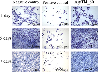
Contamination by bacterial biofilms has a strong negative impact, especially on the surface of prostheses, implants, pins, and other medical-surgical devices. To prevent their formation, one of the alternatives is the modification of the metal surface incorporating silver by low-energy ion implantation, thus avoiding initial bacteria adhesion to the modified surface and further development of the biofilm. The bactericidal properties of silver atoms incorporated on commercially pure titanium surfaces by low-energy ion implantation (4 keV) were evaluated. The surface modifications were analyzed by Rutherford backscattering spectrometry, glow discharge-optical emission spectroscopy, contact angle measure, optical profilometry, and X-ray photoelectron spectroscopy. The microbiological assays were conducted by using Escherichia coli (E. coli). The results demonstrated a reduction on bacterial counting. No toxic effect of silver was detected on human MG-63 cells. The choice of parameters to obtain a bactericidal and nontoxic biomaterial for human cells should consider the ideal combination “energy + silver concentration”. Therefore, it can be considered for industrial application.

Type
REVIEW
Copyright
Copyright © Materials Research Society 2018 

Access options

Get access to the full version of this content by using one of the access options below. (Log in options will check for institutional or personal access. Content may require purchase if you do not have access.)

Footnotes

This section of Journal of Materials Research is reserved for papers that are reviews of literature in a given area.

References

REFERENCES

Liu, X., Chu, P.K., and Ding, C.: Surface modification of titanium, titanium alloys, and related materials for biomedical application. Mater. Sci. Eng., R 47, 49 (2004).CrossRefGoogle Scholar
Topolski, K., Bochniak, W., Lagoda, M., Ostachowski, P., and Garbacz, H.: Structure and properties of titanium produced by a new method of chip recycling. J. Mater. Process Technol. 248, 80 (2017).CrossRefGoogle Scholar
Oldani, C. and Dominguez, A.: Titanium as a biomaterial for implants. In Recent Advances in Arthroplasty, Fokter, S.K., ed. (InTech, London, U.K., 2012); pp. 149162. Available at: https://www.intechopen.com/books/recent-advances-in-arthroplasty/titanium-as-a-biomaterial-for-implants.Google Scholar
Zhu, C., Bao, N-R., Chen, S., and Zhao, J-N.: Antimicrobial design of titanium surface that kill sessile bacteria but support stem cells adhesion. Appl. Surf. Sci. 389, 7 (2016).CrossRefGoogle Scholar
Feng, H., Wu, J., Chen, G.Q., Cuiz, F.Z., Kim, T.N., and Kim, J.O.: A mechanistic study of the antibacterial effect of silver ions on Escherichia coli and Staphylococcus aureus. J. Biomed. Mater. Res. 52, 662 (2000).3.0.CO;2-3>CrossRefGoogle ScholarPubMed
Wan, Y.Z., Raman, S., Hea, F., and Huang, Y.: Surface modification of medical metals by ion implantation of silver and copper. Vacuum 81, 1114 (2007).CrossRefGoogle Scholar
Kurtz, S.M., Lau, E., Watsom, H., Schmier, J.K., and Parvizi, J.: Economic burden of periprosthetic joint infection in the United States. J. Arthroplasty 27, 61 (2012).CrossRefGoogle ScholarPubMed
Inman, R.D., Gallegos, K.V., Brause, B.D., Redecha, P.B., and Christian, C.L.: Clinical and microbial features of prosthetic joint infection. Am. J. Med. 77, 47 (1984).CrossRefGoogle ScholarPubMed
Zmistowski, B., Karam, J.A., Durinka, J.B., Casper, D.S., and Parvizi, J.: Periprosthetic joint infection increases the risk of one-year mortality. J. Bone Jt. Surg. 95, 2177 (2013).CrossRefGoogle ScholarPubMed
Lentino, J.R.: Prosthetic joint infections: Bane of orthopedists, challenge for infectious disease specialists. Clin. Infect. Dis. 36, 1157 (2003).CrossRefGoogle ScholarPubMed
Brady, M.J., Lisay, C.M., Yurkovetskiy, A.V., and Sawan, S.P.: Persistent silver disinfectant for the environmental control of pathogenic bacteria. Am. J. Infect. Control. 31, 208 (2003).CrossRefGoogle ScholarPubMed
Ferrara, M.S., Courson, R., and Paulson, D.S.: Evaluation of persistent antimicrobial effects of an antimicrobial formulation. J. Athl. Train. 46, 629 (2011).CrossRefGoogle ScholarPubMed
Liu, J., Sonshine, D.A., Shervani, S., and Hurt, R.H.: Controlled release of biologically active silver from nanosilver surfaces. ACS Nano 4, 6903 (2010).CrossRefGoogle ScholarPubMed
Dunn, K. and Edwards-Jones, V.: The role of Acticoat™ with nanocrystalline silver in the management of burns. Burns 30, S1 (2004).CrossRefGoogle Scholar
Zarpelon, F., Galiotto, D., Aguzzoli, C., Carli, L.N., Figueroa, C.A., Baumvol, I.J.R., Machado, G., Crespo, J.S., and Giovanela, M.: Removal of coliform bacteria from industrial wastewaters using polyelectrolytes/silver nanoparticles self-assembled thin films. J. Environ. Chem. Eng. 4, 137 (2016).CrossRefGoogle Scholar
Wang, G., Jin, W., Qasim, A.M., Gao, A., Peng, X., Li, W., Feng, H., and Chu, P.K.: Antibacterial effects of titanium embedded with silver nanoparticles based on electron-transfer-induced reactive oxygen species. Biomaterials 124, 25 (2017).CrossRefGoogle ScholarPubMed
Ballottin, D., Fulaz, S., Cabrini, F., Tsukamoto, J., Durán, N., Alves, O.L., and Tasic, L.: Antimicrobial textiles: Biogenic silver nanoparticles against Candida and Xanthomonas. Mater. Sci. Eng., C 75, 582 (2017).CrossRefGoogle ScholarPubMed
Boateng, J. and Matthews, K.: Wound healing dressings and drug delivery systems: A review. J. Pharm. Sci. 97, 2892 (2008).CrossRefGoogle ScholarPubMed
Innes, M.E., Umraw, N., Fish, J.S., Gomez, M., and Cartotto, R.C.: The use of silver coated dressings on donor site wounds: A prospective, controlled matched pair study. Burns 27, 621 (2001).CrossRefGoogle ScholarPubMed
Saint, S., Elmore, J.G., Sullivan, S.D., Emerson, S.S., and Koepsell, T.D.: The efficacy of silver alloy-coated urinary catheters in preventing urinary tract infection: A meta-analysis. Am. J. Med. 105, 236 (1998).CrossRefGoogle ScholarPubMed
Perelshtein, I., Applerot, G., Perkas, N., Wehrschuetz-Sigl, E., Hasmann, A., Guebitz, G., and Gedanken, A.: CuO–cotton nanocomposite: Formation, morphology, and antibacterial activity. Surf. Coat. Technol. 204, 54 (2009).CrossRefGoogle Scholar
Knetsch, M.L.W. and Koole, L.H.: New strategies in the sevelopment of sntimicrobial coatings: The example of increasing usage of silver and silver nanoparticles. Polymers 3, 340 (2011).CrossRefGoogle Scholar
Liao, K-H., Ou, K-L., Cheng, H-C., Lin, C-T., and Peng, P-W.: Effect of silver on antibacterial properties of stainless steel. Appl. Surf. Sci. 256, 3642 (2010).CrossRefGoogle Scholar
Lemire, J.A., Harrison, J.J., and Turner, R.J.: Antimicrobial activity of metals: Mechanisms, molecular targets and applications. Nat. Rev. Microbiol. 11, 371 (2013).CrossRefGoogle ScholarPubMed
Politano, A.D., Campbell, K.T., Rosenberger, L.H., and Sawyer, R.G.: Use of silver in the prevention and treatment of infections: Silver review. Surg. Infect. 14, 8 (2013).CrossRefGoogle ScholarPubMed
Sioshansi, P. and Tobin, E.J.: Surface treatment of biomaterials by ion beam processes. Surf. Coat. Technol. 83, 175 (1996).CrossRefGoogle Scholar
Hirvonen, J.K.: Ion beam processing for industrial applications. Mater. Sci. Eng., A 116, 167 (1989).CrossRefGoogle Scholar
Pezzagna, S. and Meijer, J.: High-resolution ion implantation from keV to MeV. In Ion Implantation, Goorsky, M., ed. (Rubion, Ruhr-Universität Bochum, London, U.K., 2012); pp. 324.Google Scholar
Poon, V.K. and Burd, A.: In vitro cytotoxicity of silver: Implication for clinical wound care. Burns 30, 140 (2004).CrossRefGoogle Scholar
Wataha, J.C., Lockwood, P.E., and Schedle, A.: Effect of silver, copper, mercury, and nickel ions on cellular proliferation during extended, low-dose exposures. J. Biomed. Mater. Res. 52, 360 (2000).3.0.CO;2-B>CrossRefGoogle ScholarPubMed
Zhang, T., Wang, L., Chen, Q., and Chen, C.: Cytotoxic potential of silver nanoparticles. Yonsei Med. J. 55, 283 (2014).CrossRefGoogle ScholarPubMed
Echeverrigaray, F.G., Echeverrigaray, S., Delamare, A.P.L., Wanke, C.H., Figueroa, C.A., Baumvol, I.J.R., and Aguzzoli, C.: Antibacterial properties obtained by low-energy silver implantation in stainless steel surfaces. Surf. Coat. Technol. 307, 345 (2016).CrossRefGoogle Scholar
ASTM F 67: Standard Specification for Unalloyed Titanium for Surgical Implant Applications (ASTM International, West Conshohocken, Pennsylvania, 2006).Google Scholar
Duraccio, D., Mussano, F., and Faga, M.G.: Biomaterials for dental implants: Current and future trends. J. Mater. Sci. 50, 4779 (2015).CrossRefGoogle Scholar
Ziegler, J.P., Ziegler, M.D., and Biersack, J.P.: The Program Stopping and Range of Ion in Matter (SRIM) 2013 Pro, 2018. Available at: http://www.srim.org.Google Scholar
Strnad, G., Petrovan, C., and Russu, O.: Contact angle measurement on medical implant titanium based biomaterials. Proc. Technol. 22, 946 (2016).CrossRefGoogle Scholar
Zeraik, A.N. and Nitschke, M.: Biosurfactants as agents to reduce adhesion of pathogenic bacteria to polystyrene surfaces: Effect of temperature and hydrophobicity. Curr. Microbiol. 61, 554 (2010).CrossRefGoogle ScholarPubMed
Zareidoost, A., Yousefpour, M., Ghaseme, B., and Amanzadeh, A.: The relationship of surface roughness and cell response of chemical surface modification of titanium. J. Mater. Sci.: Mater. Med. 23, 1479 (2012).Google ScholarPubMed
Gadelmawla, E.S., Koura, M.M., Maksoud, T.M.A., Elewa, I.M., and Soliman, H.H.: Roughness parameters. J. Mater. Process. Technol. 123, 133 (2002).CrossRefGoogle Scholar
Calderon, V.S., Cavaleiro, A., and Carvalho, S.: Chemical and structural characterization of ZrCNAg coatings: XPS, XRD, and Raman spectroscopy. Appl. Surf. Sci. 346, 240 (2015).CrossRefGoogle Scholar
Ni, H-W., Zhang, H-S., Chen, R-S., Zhan, W-T., Huo, K-F., and Zuo, Z-Y.: Antibacterial properties and corrosion resistance of AISI 420 stainless steels implanted by silver and copper ions. Int. J. Miner., Metall. Mater. 19, 322 (2012).CrossRefGoogle Scholar
Benzo, P., Cattaneo, L., Farcau, C., Andreozzi, A., Perego, M., Benassayag, G., Pécassou, B., Carles, R., and Bonafos, C.: Stability of Ag nanocrystals synthesized by ultra-low energy ion implantation in SiO2 matrices. J. Appl. Phys. 109, 103524 (2011).CrossRefGoogle Scholar
Rai, M., Yadav, A., and Gade, A.: Silver nanoparticles as a new generation of antimicrobials. Biotechnol. Adv. 27, 76 (2009).CrossRefGoogle ScholarPubMed
Fontenoy, C. and Kamel, S.O.: Silver in the medical devices/equipaments: Marketing or real clinical interest? Pharm. Hosp. 46, e1 (2011).Google Scholar
Chang, H-I. and Wang, Y.: Cell responses to surface and architecture of tissue engineering scaffolds. In Regenerative Medicine and Tissue Engineering—Cells and Biomaterials, Daniel, E., ed. (InTech, London, U.K., 2011); p. 571. http://dx.doi.org/10.5772/21983. Retrieve from: https://www.intechopen.com/books/regenerative-medicine-and-tissue-engineering-cells-and-biomaterials/cell-responses-to-surface-and-architecture-of-tissue-engineering-scaffolds.Google Scholar