Hostname: page-component-cd9895bd7-gbm5v Total loading time: 0 Render date: 2024-12-30T20:59:09.707Z Has data issue: false hasContentIssue false

An active surface enhanced Raman scattering substrate using carbon nanocoils

Published online by Cambridge University Press:  06 August 2013
Dawei Li
Affiliation:
School of Physics and Optoelectronic Technology, Dalian University of Technology, Ganjingzi District, Dalian 116024, PR China
Lujun Pan*
Affiliation:
School of Physics and Optoelectronic Technology, Dalian University of Technology, Ganjingzi District, Dalian 116024, PR China
Shifa Wu
Affiliation:
School of Physics and Optoelectronic Technology, Dalian University of Technology, Ganjingzi District, Dalian 116024, PR China
Shuai Li
Affiliation:
School of Physics and Optoelectronic Technology, Dalian University of Technology, Ganjingzi District, Dalian 116024, PR China
*
a)Address all correspondence to this author. e-mail: [email protected]
Get access

Abstract

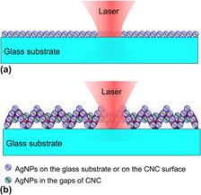
A novel surface enhanced Raman scattering (SERS) substrate was produced by combining Ag nanoparticles (AgNPs) and carbon nanocoils (CNCs). Three different methods were developed for loading AgNPs on CNCs, which include (i) direct deposition of AgNPs on CNCs by radio-frequency magnetron sputtering (RFMS) to form an Ag–CNC hybrid, (ii) deposition of a TiO2 film on CNCs by RFMS, followed by photoinduced growth of AgNPs to form an Ag–TiO2–CNC hybrid (called A-substrate), and (iii) deposition of a TiO2 film on CNCs by spin coating and then photoinduced growth of AgNPs to form an Ag–TiO2–CNC hybrid (called B-substrate). Experimental SERS results showed that B-substrates exhibited the highest SERS enhancement with an enhancement factor of over 107 for rhodamine 6G. The as-prepared Ag–TiO2–CNC substrates also showed much higher Raman signal enhancement than ordinary planar SERS substrates in our system. This was mainly due to the unique three-dimensional structure where the large surface area was available for loading more densely packed AgNPs which contribute to abundant Raman hot spots.

Type
Articles
Copyright
Copyright © Materials Research Society 2013 

Access options

Get access to the full version of this content by using one of the access options below. (Log in options will check for institutional or personal access. Content may require purchase if you do not have access.)

References

REFERENCES

Fleischmann, M., Hendra, P.J., and McQuillan, A.J.: Raman spectra of pyridine adsorbed at a silver electrode. Chem. Phys. Lett. 26(2), 163 (1974).CrossRefGoogle Scholar
Aroca, R.: Surface Enhanced Vibrational Spectroscopy (Wiley, Chichester, 2006).CrossRefGoogle Scholar
Gunnarsson, L., Bjerneld, E.J., Xu, H., Petronis, S., Kasemo, B., and Kall, M.: Interparticle coupling effects in nanofabricated substrates for surface-enhanced Raman scattering. Appl. Phys. Lett. 78(6), 802 (2001).CrossRefGoogle Scholar
Sun, Y., Liu, K., Miao, J., Wang, Z., Tian, B., Zhang, L., Li, Q., Fan, S., and Jiang, K.: Highly sensitive surface-enhanced Raman scattering substrate made from superaligned carbon nanotubes. Nano Lett. 10(5), 1747 (2010).CrossRefGoogle ScholarPubMed
Liu, G.L. and Lee, L.P.: Nanowell surface enhanced Raman scattering arrays fabricated by soft-lithography for label-free biomolecular detections in integrated microfluidics. Appl. Phys. Lett. 87(7), 074101 (2005).CrossRefGoogle Scholar
Lee, S.J., Morrill, A.R., and Moskovits, M.: Hot spots in silver nanowire bundles for surface-enhanced Raman spectroscopy. J. Am. Chem. Soc. 128(7), 2200 (2006).CrossRefGoogle ScholarPubMed
Xu, H., Aizpurua, J., Käll, M., and Apell, P.: Electromagnetic contributions to single-molecule sensitivity in surface-enhanced Raman scattering. Phys. Rev. E 62(3), 4318 (2000).CrossRefGoogle ScholarPubMed
Shen, C., Hui, C., Yang, T., Xiao, C., Tian, J., Bao, L., Chen, S., Ding, H., and Gao, H.: Monodisperse noble-metal nanoparticles and their surface enhanced Raman scattering properties. Chem. Mater. 20(22), 6939 (2008).CrossRefGoogle Scholar
Wang, H., Kundu, J., and Halas, N.J.: Plasmonic nanoshell arrays combine surface-enhanced vibrational spectroscopies on a single substrate. Angew. Chem. Int. Ed. 46(47), 9040 (2007).CrossRefGoogle ScholarPubMed
Wang, T., Hu, X., and Dong, S.: Surfactantless synthesis of multiple shapes of gold nanostructures and their shape-dependent SERS spectroscopy. J. Phys. Chem. B 110(34), 16930 (2006).CrossRefGoogle ScholarPubMed
Nikoobakht, B. and El-Sayed, M.A.: Surface-enhanced Raman scattering studies on aggregated gold nanorods. J. Phys. Chem. A 107(18), 3372 (2003).CrossRefGoogle Scholar
Li, D., Wu, S., Wang, Q., Wu, Y., Peng, W., and Pan, L.: Ag@C core–shell colloidal nanoparticles prepared by the hydrothermal route and the low temperature heating–stirring method and their application in surface enhanced Raman scattering. J. Phys. Chem. C 116(22), 12283 (2012).CrossRefGoogle Scholar
Zhao, X., Zhang, B., Ai, K., Zhang, G., Cao, L., Liu, X., Sun, H., Wang, H., and Lu, L.: Monitoring catalytic degradation of dye molecules on silver-coated ZnO nanowire arrays by surface-enhanced Raman spectroscopy. J. Mater. Chem. 19(31), 5547 (2009).CrossRefGoogle Scholar
Song, W., Wang, Y., Hu, H., and Zhao, B.: Fabrication of surface-enhanced Raman scattering-active ZnO/Ag composite microspheres. J. Raman Spectrosc. 38(10), 1320 (2007).CrossRefGoogle Scholar
Kim, K., Kim, H.S., and Park, H.K.: Facile method to prepare surface enhanced Raman scattering active Ag nanostructures on silica spheres. Langmuir 22(19), 8083 (2006).CrossRefGoogle ScholarPubMed
Mubeen, S., Zhang, S., Kim, N., Lee, S., Krämer, S., Xu, H., and Moskovits, M.: Plasmonic properties of gold nanoparticles separated from a gold mirror by an ultrathin oxide. Nano Lett. 12(4), 2088 (2012).CrossRefGoogle Scholar
Chen, L.M. and Liu, Y.N.: Surface-enhanced Raman detection of melamine on silver-nanoparticle-decorated silver/carbon nanospheres: Effect of metal ions. ACS Appl. Mater. Interfaces 3(8), 3091 (2011).CrossRefGoogle ScholarPubMed
Chen, Y.C., Young, R.J., Macpherson, J.V., and Wilson, N.R.: Silver-decorated carbon nanotube networks as SERS substrates. J. Raman Spectrosc. 42(6), 1255 (2011).CrossRefGoogle Scholar
Lu, G., Li, H., Liusman, C., Yin, Z., Wu, S., and Zhang, H.: Surface enhanced Raman scattering of Ag or Au nanoparticle-decorated reduced graphene oxide for detection of aromatic molecules. Chem. Sci. 2(9), 1817 (2011).CrossRefGoogle Scholar
Ling, X., Xie, L., Fang, Y., Xu, H., Zhang, H., Kong, J., Dresselhaus, M.S., Zhang, J., and Liu, Z.: Can graphene be used as a substrate for Raman enhancement? Nano Lett. 10(2), 553 (2009).CrossRefGoogle Scholar
Li, D., Pan, L., Qian, J., and Liu, D.: Highly efficient synthesis of carbon nanocoils by catalyst particles prepared by a sol–gel method. Carbon 48(1), 170 (2010).CrossRefGoogle Scholar
Li, D.W., Pan, L.J., Liu, D.P., and Yu, N.S.: Relationship between geometric structures of catalyst particles and growth of carbon nanocoils. Chem. Vap. Deposition 16(4–6), 166 (2010).CrossRefGoogle Scholar
Li, D. and Pan, L.: Growth of carbon nanocoils using Fe–Sn–O catalyst film prepared by a spin-coating method. J. Mater. Res. 26(16), 2024 (2011).CrossRefGoogle Scholar
Treacy, M.M.J., Ebbesen, T.W., and Gibson, J.M.: Exceptionally high Young's modulus observed for individual carbon nanotubes. Nature 381(6584), 678 (1996).CrossRefGoogle Scholar
Hayashida, T., Pan, L., and Nakayama, Y.: Mechanical and electrical properties of carbon tubule nanocoils. Phys. B Condens. Matter. 323(1–4), 352 (2002).CrossRefGoogle Scholar
Park, K.W., Sung, Y.E., Han, S., Yun, Y., and Hyeon, T.: Origin of the enhanced catalytic activity of carbon nanocoil-supported PtRu alloy electrocatalysts. J. Phys. Chem. B 108(3), 939 (2003).CrossRefGoogle Scholar
Hokushin, S., Pan, L., Konishi, Y., Tanaka, H., and Nakayama, Y.: Field emission properties and structural changes of a stand-alone carbon nanocoil. Jpn. J. Appl. Phys. 46(23), L565 (2007).CrossRefGoogle Scholar
Tang, N., Yang, Y., Lin, K., Zhong, W., Au, C., and Du, Y.: Synthesis of plait-like carbon nanocoils in ultrahigh yield, and their microwave absorption properties. J. Phys. Chem. C 112(27), 10061 (2008).CrossRefGoogle Scholar
Sai, V.V.R., Gangadean, D., Niraula, I., Jabal, J.M.F., Corti, G., McIlroy, D.N., Eric Aston, D., Branen, J.R., and Hrdlicka, P.J.: Silica nanosprings coated with noble metal nanoparticles: Highly active SERS substrates. J. Phys. Chem. C 115(2), 453 (2010).CrossRefGoogle Scholar
Hildebrandt, P. and Stockburger, M.: Surface-enhanced resonance Raman spectroscopy of rhodamine 6G adsorbed on colloidal silver. J. Phys. Chem. 88(24), 5935 (1984).CrossRefGoogle Scholar
Mills, A., Hill, G., Stewart, M., Graham, D., Smith, W.E., Hodgen, S., Halfpenny, P.J., Faulds, K., and Robertson, P.: Characterization of novel Ag on TiO2 films for surface-enhanced Raman scattering. Appl. Spectrosc. 58(8), 922 (2004).CrossRefGoogle ScholarPubMed
Ahmed, M.H., Keyes, T.E., Byrne, J.A., Blackledge, C.W., and Hamilton, J.W.: Adsorption and photocatalytic degradation of human serum albumin on TiO2 and Ag–TiO2 films. J. Photochem. Photobiol., A 222(1), 123 (2011).CrossRefGoogle Scholar
Li, D., Pan, L., Li, S., Liu, K., Wu, S., and Peng, W.: Controlled preparation of uniform TiO2-catalyzed silver nanoparticle films for surface-enhanced Raman scattering. J. Phys. Chem. C 117(13), 6861 (2013).CrossRefGoogle Scholar
Le Ru, E.C., Blackie, E., Meyer, M., and Etchegoin, P.G.: Surface enhanced Raman scattering enhancement factors: A comprehensive study. J. Phys. Chem. C 111(37), 13794 (2007).CrossRefGoogle Scholar
Yang, L.B., Jiang, X., Ruan, W.D., Yang, J.X., Zhao, B., Xu, W.Q., and Lombardi, J.R.: Charge-transfer-induced surface-enhanced Raman scattering on Ag-TiO2 nanocomposites. J. Phys. Chem. C 113(36), 16226 (2009).CrossRefGoogle Scholar
Supplementary material: File

Li Supplementary Material

Figures S1-S4

Download Li Supplementary Material(File)
File 3.6 MB