Hostname: page-component-586b7cd67f-2brh9 Total loading time: 0 Render date: 2024-11-28T01:32:24.245Z Has data issue: false hasContentIssue false

Knockdown of vitamin D receptor genes impairs touch-evoked escape behavior in zebrafish

Subject: Life Science and Biomedicine

Published online by Cambridge University Press:  25 October 2021

Hye-Joo Kwon*
Affiliation:
Department of Biology, University of Utah Asia Campus, Incheon21985, Korea
*
Corresponding author. Email: [email protected]

Abstract

Vitamin D is a steroid hormone well-known for its role in calcium homeostasis and bone health. Biological actions of vitamin D are mediated through the vitamin D receptor (VDR) present in various cells and tissues. Vitamin D has been implicated in multiple aspects of neuromuscular functions. This study aimed to investigate the role of VDR signaling during early stage of locomotor development utilizing a gene knockdown approach. Zebrafish larvae deficient in VDR showed severe motor impairment and no obvious response to touch. These results indicate that VDR signaling is indispensable for the correct neuromuscular development and touch-evoked escape swimming behavior in zebrafish.

Type
Research Article
Information
Result type: Novel result, Supplementary result
Creative Commons
Creative Common License - CCCreative Common License - BY
This is an Open Access article, distributed under the terms of the Creative Commons Attribution licence (http://creativecommons.org/licenses/by/4.0), which permits unrestricted re-use, distribution and reproduction, provided the original article is properly cited.
Copyright
© The Author(s), 2021. Published by Cambridge University Press

1. Introduction

Vitamin D has been known to play an essential role in regulating bone metabolism and maintaining calcium and phosphate homeostasis. The active form of vitamin D exerts its biological effects mainly by binding to the vitamin D receptor (VDR). VDR is expressed in most of the cells (Bouillon et al., Reference Bouillon, Carmeliet, Verlinden, van Etten, Verstuyf, Luderer, Lieben, Mathieu and Demay2008), which explains why VDR signaling has been implicated in a variety of physiological processes, including neuromuscular function. Indeed, the VDR has been found in the sensory and motor areas of the nervous system and muscles in both humans and rodent models (Bischoff et al., Reference Bischoff, Borchers, Gudat, Duermueller, Theiler, Stähelin and Dick2001; Eyles et al., Reference Eyles, Smith, Kinobe, Hewison and McGrath2005; Girgis et al., Reference Girgis, Mokbel, Cha, Houweling, Abboud, Fraser, Mason, Clifton-Bligh and Gunton2014; Prüfer et al., Reference Prüfer, Veenstra, Jirikowski and Kumar1999). In addition, many studies indicate that vitamin D is related to various neurological and neuromuscular disorders (Di Somma et al., Reference Di Somma, Scarano, Barrea, Zhukouskaya, Savastano, Mele, Scacchi, Aimaretti, Colao and Marzullo2017; Dodig et al., Reference Dodig, Tarnopolsky and Currie2017). VDR null mice display motor deficits and muscular impairments (Burne et al., Reference Burne, McGrath, Eyles and Mackay-Sim2005; Kalueff et al., Reference Kalueff, Lou, Laaksi and Tuohimaa2004). It has been suggested that Schwann cells and the neuromuscular junctions (NMJs) are a target of VDR signaling (Sakai et al., Reference Sakai, Suzuki, Tashiro, Tanaka, Takeda, Aizawa, Hirata, Yogo and Endo2015).

Zebrafish have become an effective animal model for studying neuromuscular functions since muscle activity can be assessed easily in the early stages of development, the first few days after fertilization (Sztal et al., Reference Sztal, Ruparelia, Williams and Bryson-Richardson2016). In zebrafish embryos, two paralogs for VDR genes (vdra and vdrb) have been identified (Kollitz et al., Reference Kollitz, Hawkins, Whitfield and Kullman2014; Lin et al., Reference Lin, Su, Tseng, Ding and Hwang2012). Although previous studies have demonstrated that VDR signaling in zebrafish regulates heart development (Han et al., Reference Han, Chen, Umansky, Oonk, Choi, Dickson, Ou, Cigliola, Yifa, Cao, Tornini, Cox, Tzahor and Poss2019; Kwon, Reference Kwon2016), ocular angiogenesis (Merrigan & Kennedy, Reference Merrigan and Kennedy2017), and hematopoiesis (Cortes et al., Reference Cortes, Chen, Stachura, Liu, Kwan, Wright, Vo, Theodore, Esain, Frost, Schlaeger, Goessling, Daley and North2016), its function in neuromuscular development is largely unknown.

2. Objective

The objective of the present study was to investigate whether loss of VDR affects neuromuscular activity in zebrafish embryo and larvae using touch-evoked response behavior analysis.

3. Methods

3.1. Animal care and use

The author asserts that all procedures contributing to this work comply with the ethical standards of the relevant national and institutional guides on the care and use of laboratory animals. Zebrafish (Danio rerio) were reared and maintained at 28.5°C in accordance with the Animal Care and Use Committee at Texas A&M University. Larvae were staged as hours postfertilization (hpf) or days postfertilization (dpf) according to the established guidelines (Kimmel et al., Reference Kimmel, Ballard, Kimmel, Ullmann and Schilling1995).

3.2. The knockdown of VDR genes

The VDR gene knockdown experiment was carried out using antisense morpholino oligonucleotides (MOs) as described previously (Kwon, Reference Kwon2016; Lin et al., Reference Lin, Su, Tseng, Ding and Hwang2012). To knockdown vdra, a translation blocker (5′-AAC GGC ACT ATT TTC CGT AAG CAT C-3′) was used. To knockdown vdrb, a splice blocker (5′-TCC ATC ACT AGC AGA CGA GGG AAG A-3′) targeting the intron2-exon3 (I2E3) junction was used. Zebrafish embryos were co-injected with 5 ng of vdra MOs and 5 ng of vdrb MOs at the one-cell stage. All MOs used here were obtained from Gene Tools, LLC (Philomath, OR). The experiments were conducted at least three times.

3.3. Touch-evoked response behavior analysis

Touch-evoked zebrafish movements were recorded using a CCD camera mounted on a dissecting microscope at 40 hpf and 6 dpf. For touch responses at 40 hpf, tactile stimuli were generated by the manual dechorionation using fine forceps. For touch responses at 6 dpf, tactile stimuli were elicited by touching the tail region of the larvae with forceps. Images were captured, converted into a video file, and analyzed individual frames of time-lapse. To assess behavioral phenotypes, at least 10 embryos were examined in each group. The phenotypes described in this study were completely penetrant.

4. Results

To investigate the effects of the loss of VDRs on motor development, antisense MOs against vdra and vdrb (vdra/b MO) were co-injected into the one-cell embryo. During the hatching period, over 94% of the wild-type control embryos hatched by 3 dpf and 100% of them hatched on the next day (n = 17). It was noticeable that vdra/b MO-injection resulted in a reduction of hatching rate, 70% at 3 dpf. At 5 dpf, the hatching rate of vdra/b MO-injected larvae was still less than 81% (n = 31). At 40 hpf, 100% of control embryos showed rapid escape swimming behaviors in response to tactile stimuli generated in the manual dechorionation (Figure 1ae; Supplementary Video S1). In contrast, 100% vdra/b morphants exhibited either a very weak or no muscle contraction and all of them failed to escape in response to touch during or after the dechorionation (Figure 1fj; Supplementary Video S2).

Figure 1. Knockdown of VDRs impairs touch-evoked behaviors. Video frames showing touch-evoked escape response at 40 hpf of wild-type control (a–e) but not of vdra/b MO-injected (f–j) embryos. The time of the frame is shown in the top right portion. The mechanosensory stimulation during the manual dechorionation causes control embryos to escape rapidly and exit the field of view (d and e). In contrast, vdra/b MO-injected embryos do not exhibit any escape response and fail to exit the field of view at the same time frames (i and j). Scale bar = 500 μm.

To determine whether the impairment of touch-evoked escape in vdra/b morphants was due to the delayed onset of touch-responsiveness, the tactile response of zebrafish larvae at 6 dpf was examined. Upon touch at the tail, 100% of vdra/b-depleted larvae at 6 dpf displayed no obvious muscle contraction and none of them showed the escape response observed in 100% of control larvae (Figure 2; Supplementary Videos S3 and S4). vdra/b knockdown caused significantly reduced locomotor activity and perturbed development of the swim bladder. All of the vdra/b MO-injected larvae at 6 dpf had severe defects in free-swimming.
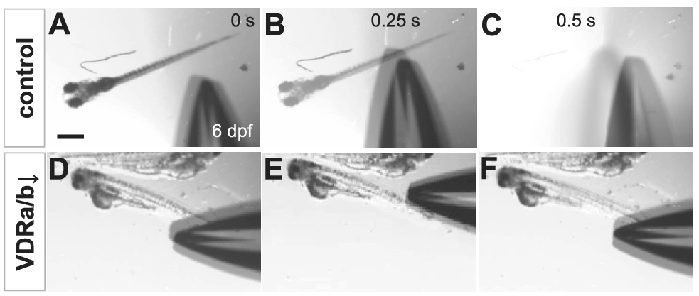
Figure 2. Loss-of-VDRs results in loss of touch-evoked escape swimming behaviors. Video frames showing touch-evoked response at 6 dpf of wild-type control (a–c) but not of vdra/b MO-injected (d–f) larvae. While the stimulation by forceps causes the wild-type larva to swim rapidly away and fully exit the field of view (c), vdra/b MO-injected larvae exhibit no touch-evoked response and remain in the same field of view (f). Scale bar = 500 μm.

5. Discussion

In the current study, VDR signaling was found to be involved in neuromuscular development and touch-evoked escape swimming behavior. In zebrafish embryos, spontaneous muscle contractions start to be noted around 17 hpf (Saint-Amant & Drapeau, Reference Saint-Amant and Drapeau1998). Even before hatching, the zebrafish embryos acquire the ability to respond to touch by 24–27 hpf (Carmean & Ribera, Reference Carmean and Ribera2010). The twitch contraction reaches the peak frequency, which is a driving force of releasing embryos from the chorion during the hatching period around 3 dpf. Delay in hatching found in vdra/b morphants could be due to the impaired muscle activity (Skobo et al., Reference Skobo, Benato, Grumati, Meneghetti, Cianfanelli, Castagnaro, Chrisam, Di Bartolomeo, Bonaldo, Cecconi and Dalla Valle2014).

Together with the muscular and motor impairments observed in VDR knockout mice (Burne et al., Reference Burne, McGrath, Eyles and Mackay-Sim2005; Kalueff et al., Reference Kalueff, Lou, Laaksi and Tuohimaa2004), these findings indicate that VDRs play an essential role in neuromuscular activity and locomotor behavior. The specific mechanism remains to be elucidated, but it may be explained by the effects of vitamin D on calcium homeostasis or maintenance of peripheral nerve axons and acetylcholine receptor clusters in NMJs (Lin et al., Reference Lin, Su, Tseng, Ding and Hwang2012; Sakai et al., Reference Sakai, Suzuki, Tashiro, Tanaka, Takeda, Aizawa, Hirata, Yogo and Endo2015).

6. Conclusion

This study demonstrates that knockdown of VDRs causes impairment of muscle performance and elimination of touch-evoked escape swimming response in zebrafish embryos and larvae. Thus, the VDR signaling is required for the correct neuromuscular activity during early development. These results suggest the role of VDR signaling in locomotor behavior appears to be well-conserved between mammals and fish.

Acknowledgments

The author wishes to thank Bruce B. Riley (Texas A&M University) for providing zebrafish lab facilities and valuable comments.

Funding Statement

This research received no specific grant from any funding agency, commercial or not-for-profit sectors.

Conflict of Interest

The author declares no conflicts of interest.

Data Availability Statement

The data that support the findings of this study are available as Supplementary Material.

Supplementary Materials

To view supplementary material for this article, please visit http://dx.doi.org/10.1017/exp.2021.22.

References

Bischoff, H. A., Borchers, M., Gudat, F., Duermueller, U., Theiler, R., Stähelin, H. B., & Dick, W. (2001). In situ detection of 1,25-dihydroxyvitamin D3 receptor in human skeletal muscle tissue. The Histochem Journal, 33, 1924. https://doi.org/10.1023/A:1017535728844CrossRefGoogle ScholarPubMed
Bouillon, R., Carmeliet, G., Verlinden, L., van Etten, E., Verstuyf, A., Luderer, H. F., Lieben, L., Mathieu, C., & Demay, M. (2008). Vitamin D and human health: Lessons from vitamin D receptor null mice. Endocrine Reviews, 29, 726776. https://doi.org/10.1210/er.2008-0004CrossRefGoogle ScholarPubMed
Burne, T. H., McGrath, J. J., Eyles, D. W., & Mackay-Sim, A. (2005). Behavioural characterization of vitamin D receptor knockout mice. Behavioural Brain Research, 157, 299308. https://doi.org/10.1016/j.bbr.2004.07.008CrossRefGoogle ScholarPubMed
Carmean, V., & Ribera, A. B. (2010). Genetic analysis of the touch response in zebrafish (Danio rerio). International Journal of Comparative Psychology, 23, 91.Google Scholar
Cortes, M., Chen, M. J., Stachura, D. L., Liu, S. Y., Kwan, W., Wright, F., Vo, L. T., Theodore, L. N., Esain, V., Frost, I. M., Schlaeger, T. M., Goessling, W., Daley, G. Q., & North, T. E. (2016). Developmental vitamin D availability impacts hematopoietic stem cell production. Cell Reports, 17, 458468. https://doi.org/10.1016/j.celrep.2016.09.012CrossRefGoogle ScholarPubMed
Di Somma, C., Scarano, E., Barrea, L., Zhukouskaya, V. V., Savastano, S., Mele, C., Scacchi, M., Aimaretti, G., Colao, A., & Marzullo, P. (2017). Vitamin D and neurological diseases: An endocrine view. International Journal of Molecular Sciences, 18, 2482. https://doi.org/10.3390/ijms18112482CrossRefGoogle ScholarPubMed
Dodig, D., Tarnopolsky, M., & Currie, S. (2017). Vitamin deficiencies in patients with myopathies and other neuromuscular conditions. Neurology, 88, P2.115.Google Scholar
Eyles, D. W., Smith, S., Kinobe, R., Hewison, M., & McGrath, J. J. (2005). Distribution of the vitamin D receptor and 1 alpha-hydroxylase in human brain. Journal of Chemical Neuroanatomy, 29, 2130. https://doi.org/10.1016/j.jchemneu.2004.08.006CrossRefGoogle ScholarPubMed
Girgis, C. M., Mokbel, N., Cha, K. M., Houweling, P. J., Abboud, M., Fraser, D. R., Mason, R. S., Clifton-Bligh, R. J., & Gunton, J. E. (2014). The vitamin D receptor (VDR) is expressed in skeletal muscle of male mice and modulates 25-hydroxyvitamin D (25OHD) uptake in myofibers. Endocrinology, 155, 32273237. https://doi.org/10.1210/en.2014-1016CrossRefGoogle ScholarPubMed
Han, Y., Chen, A., Umansky, K. B., Oonk, K. A., Choi, W. Y., Dickson, A. L., Ou, J., Cigliola, V., Yifa, O., Cao, J., Tornini, V. A., Cox, B. D., Tzahor, E., & Poss, K. D. (2019). Vitamin D stimulates cardiomyocyte proliferation and controls organ size and regeneration in zebrafish. Developmental Cell, 48, 853863.e5. https://doi.org/10.1016/j.devcel.2019.01.001CrossRefGoogle ScholarPubMed
Kalueff, A. V., Lou, Y. R., Laaksi, I., & Tuohimaa, P. (2004). Impaired motor performance in mice lacking neurosteroid vitamin D receptors. Brain Research Bulletin, 64, 2529. https://doi.org/10.1016/j.brainresbull.2004.04.015CrossRefGoogle ScholarPubMed
Kimmel, C. B., Ballard, W. W., Kimmel, S. R., Ullmann, B., & Schilling, T. F. (1995). Stages of embryonic development of the zebrafish. Developmental Dynamics, 203, 253310. https://doi.org/10.1002/aja.1002030302CrossRefGoogle ScholarPubMed
Kollitz, E. M., Hawkins, M. B., Whitfield, G. K., & Kullman, S. W. (2014). Functional diversification of vitamin D receptor paralogs in teleost fish after a whole genome duplication event. Endocrinology, 155, 46414654. https://doi.org/10.1210/en.2014-1505CrossRefGoogle ScholarPubMed
Kwon, H.-J. (2016). Vitamin D receptor signaling is required for heart development in zebrafish embryo. Biochemical and Biophysical Research Communications, 470, 575578. https://doi.org/10.1016/j.bbrc.2016.01.103CrossRefGoogle ScholarPubMed
Lin, C. H., Su, C. H., Tseng, D. Y., Ding, F. C., & Hwang, P. P. (2012). Action of vitamin D and the receptor, VDRa, in calcium handling in zebrafish (Danio rerio). PLoS One, 7, e45650. https://doi.org/10.1371/journal.pone.0045650CrossRefGoogle Scholar
Merrigan, S. L., & Kennedy, B. N. (2017). Vitamin D receptor agonists regulate ocular developmental angiogenesis and modulate expression of dre-miR-21 and VEGF. British Journal of Pharmacology, 174, 26362651. https://doi.org/10.1111/bph.13875CrossRefGoogle ScholarPubMed
Prüfer, K., Veenstra, T. D., Jirikowski, G. F., & Kumar, R. (1999). Distribution of 1,25-dihydroxyvitamin D3 receptor immunoreactivity in the rat brain and spinal cord. Journal of Chemical Neuroanatomy, 16, 135145. https://doi.org/10.1016/S0891-0618(99)00002-2CrossRefGoogle ScholarPubMed
Saint-Amant, L., & Drapeau, P. (1998). Time course of the development of motor behaviors in the zebrafish embryo. Journal of Neurobiology, 37, 622632. https://doi.org/10.1002/(SICI)1097-4695(199812)37:4<622::AID-NEU10>3.0.CO;2-S3.0.CO;2-S>CrossRefGoogle ScholarPubMed
Sakai, S., Suzuki, M., Tashiro, Y., Tanaka, K., Takeda, S., Aizawa, K., Hirata, M., Yogo, K., & Endo, K. (2015). Vitamin D receptor signaling enhances locomotive ability in mice. Journal of Bone and Mineral Research, 30, 128136. https://doi.org/10.1002/jbmr.2317CrossRefGoogle ScholarPubMed
Skobo, T., Benato, F., Grumati, P., Meneghetti, G., Cianfanelli, V., Castagnaro, S., Chrisam, M., Di Bartolomeo, S., Bonaldo, P., Cecconi, F., & Dalla Valle, L. (2014). Zebrafish ambra1a and ambra1b knockdown impairs skeletal muscle development. PLoS One, 9, e99210. https://doi.org/10.1371/journal.pone.0099210CrossRefGoogle ScholarPubMed
Sztal, T. E., Ruparelia, A. A., Williams, C., & Bryson-Richardson, R. J. (2016). Using touch-evoked response and locomotion assays to assess muscle performance and function in zebrafish. Journal of Visualized Experiments: JoVE, 116, 54431. https://doi.org/10.3791/54431Google Scholar
Figure 0

Figure 1. Knockdown of VDRs impairs touch-evoked behaviors. Video frames showing touch-evoked escape response at 40 hpf of wild-type control (a–e) but not of vdra/b MO-injected (f–j) embryos. The time of the frame is shown in the top right portion. The mechanosensory stimulation during the manual dechorionation causes control embryos to escape rapidly and exit the field of view (d and e). In contrast, vdra/b MO-injected embryos do not exhibit any escape response and fail to exit the field of view at the same time frames (i and j). Scale bar = 500 μm.

Figure 1

Figure 2. Loss-of-VDRs results in loss of touch-evoked escape swimming behaviors. Video frames showing touch-evoked response at 6 dpf of wild-type control (a–c) but not of vdra/b MO-injected (d–f) larvae. While the stimulation by forceps causes the wild-type larva to swim rapidly away and fully exit the field of view (c), vdra/b MO-injected larvae exhibit no touch-evoked response and remain in the same field of view (f). Scale bar = 500 μm.

Supplementary material: PDF

Kwon supplementary material

Kwon supplementary material

Download Kwon supplementary material(PDF)
PDF 194.7 KB
Reviewing editor:  Mariana Bexiga University of Coimbra Center for Neuroscience and Cell Biology, Coimbra, Portugal, 3004-504
This article has been accepted because it is deemed to be scientifically sound, has the correct controls, has appropriate methodology and is statistically valid, and has been sent for additional statistical evaluation and met required revisions.

Review 1: Knockdown of vitamin D receptor (VDR) genes impairs touch-evoked escape behavior in zebrafish

Conflict of interest statement

Reviewer declares none

Comments

Comments to the Author: This is a concise and interesting work. I watched the videos and they clearly shows the difference in the tactile response. The difference is dramatic. I think this would be a beneficial finding for zebrafish researchers.

Presentation

Overall score 4 out of 5
Is the article written in clear and proper English? (30%)
4 out of 5
Is the data presented in the most useful manner? (40%)
4 out of 5
Does the paper cite relevant and related articles appropriately? (30%)
4 out of 5

Context

Overall score 4 out of 5
Does the title suitably represent the article? (25%)
4 out of 5
Does the abstract correctly embody the content of the article? (25%)
4 out of 5
Does the introduction give appropriate context? (25%)
4 out of 5
Is the objective of the experiment clearly defined? (25%)
4 out of 5

Analysis

Overall score 4 out of 5
Does the discussion adequately interpret the results presented? (40%)
4 out of 5
Is the conclusion consistent with the results and discussion? (40%)
4 out of 5
Are the limitations of the experiment as well as the contributions of the experiment clearly outlined? (20%)
4 out of 5

Review 2: Knockdown of vitamin D receptor (VDR) genes impairs touch-evoked escape behavior in zebrafish

Conflict of interest statement

Reviewer declares none.

Comments

Comments to the Author: The following study investigates what the absence of vitamin D can induce to the reaction to a touch stimulus in zebrafish embryos and larvae, using morphants. It is a simple well-written study that relates the lack of vitamin D and locomotor impairment, which have been described in mammals. Thus, in the conclusion, the author should highlight the contribution of the experiment, that this mechanism involving vitamin D is conserved. In addition, the authors need to clarify some methodological parts: how many embryos and larvae were used in each group (add to methodology); make it clear that the touch response of 40hpf is the manually dechorionation of embryos, as the way it is, it seems that the touch is going to be performed afterward; clarify what was the part of the larvae body that received the touch, as this needs to be standardized. At the results, the authors need to provide the percentage of animals that respond to the stimulus in each group, and the ones which responded weakly in both embryos and larvae experiments. The videos are a good addition to the paper, and I wonder if the 6 dpf morphants swim at all, so they can eat and survive, do you have that information?

Some sentences on the results should have been put in the discussion, such as page 3 line 1-3, and line 8-10 (starting in “which” until Carmean et al 2010), so the authors can consider re-writing the discussion to include these literature ideas.

Presentation

Overall score 4.6 out of 5
Is the article written in clear and proper English? (30%)
5 out of 5
Is the data presented in the most useful manner? (40%)
4 out of 5
Does the paper cite relevant and related articles appropriately? (30%)
5 out of 5

Context

Overall score 4.8 out of 5
Does the title suitably represent the article? (25%)
5 out of 5
Does the abstract correctly embody the content of the article? (25%)
4 out of 5
Does the introduction give appropriate context? (25%)
5 out of 5
Is the objective of the experiment clearly defined? (25%)
5 out of 5

Analysis

Overall score 4.2 out of 5
Does the discussion adequately interpret the results presented? (40%)
4 out of 5
Is the conclusion consistent with the results and discussion? (40%)
5 out of 5
Are the limitations of the experiment as well as the contributions of the experiment clearly outlined? (20%)
3 out of 5