No CrossRef data available.
Published online by Cambridge University Press: 15 April 2020
Subanesthetic dose of ketamine was repeatedly shown to improve depressive symptoms with a short latency of 24 hours.
We aimed to test if clinical time course of improvement is indeed mirrored by increased glutamine/glutamate ratio and if such effects would show a regional and temporal specificity in two anatomically and functionally distinct subdivisions of ACC.
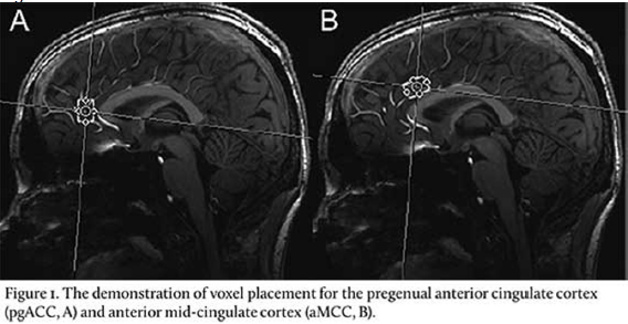
We used a glutamine sensitive magnetic resonance spectroscopy protocol at 7 Tesla to compare the longitudinal changes of glutamine/glutamate after 1 hour and 24 hours in pregenual ACC (pgACC, Figure 1A) and anterior mid-cingulate cortex (aMCC, Figure 1B) in 59 healthy controls which underwent a double-blind, placebo-controlled ketamine infusion.
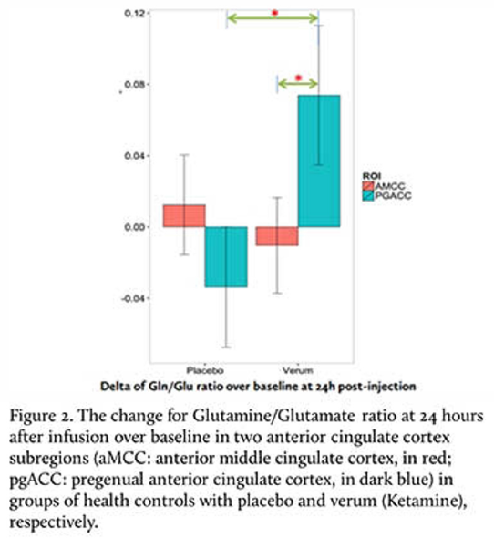
A significant interaction of time, region, and treatment was found (F = 3.881, p<0.028). Follow up analysis revealed that, only in pgACC and only after 24 hours, we found significantly increased glutamine/glutamate ratios in ketamine group compared with placebo (T = 2.331, p < 0.042) (Figure 2). We also found that the changes in pgACC over baseline are significant larger than changes in aMCC (T = 2.710, p<0.022) at 24 hours after ketamine infusion. Changes of glutamine/glutamate ratios were mainly driven by glutamine but not glutamate levels.
We found that elevated glutamine/glutamate after a single subanesthetic dose of ketamine infusion was specific to pgACC, a region previously reported glutamategic deficit in MDD. This change of glutamine/glutamate in the pgACC also showed a temporal specificity at 24 hours after infusion.


Comments
No Comments have been published for this article.