MicroFlow I Rust-Resistant Self-Contained Workstation
The MicroFlow I Workstation is a ductless carbon-filtered workstation with activated carbon filtration designed to collect small amounts of non-hazardous fumes and odors. It is self-contained with an integral recessed work surface to contain spills and can be moved from station to station. A clear hood surround with safety viewing sash can be conformed for use with a microscope. A variable-speed fan control provides the option of high or medium speeds and low flow for sensitive operations.
HEMCO Corporation

Optimizing Laser Welding
In many welding applications high powers of up to tens of KW are used, demanding precise shaping of the spot on the working plane to achieve optimized process parameters such as seam angle, seam strength, throughput, and HAZ (heat affected zones). HOLO/OR's
adjustable diffractive beam shapers offer many custom diffractive optical element (DOE) solutions for beam shaping that help improve these parameters. DOEs are flat, compact, passive components that offer accurate beam shaping solutions with no angular tolerance.
HOLO/OR
www.holoor.co.il/beam-shaping-in-laser-welding

Excelitas Technologies Celebrates a Decade of Innovation and Success
Excelitas Technologies, a global technology leader delivering innovative, customized photonic solutions, is celebrating the 10-year anniversary of its founding and a decade of innovation and success. Since being established as an independent company spun out of Perkin Elmer in December 2010, Excelitas Technologies has enjoyed greater than three-fold growth in both revenue and headcount.
Excelitas Technologies

Tomocube Microscope Quantifies Effectiveness of Experimental Nanodrugs against Biggest Killer in Westernized Societies
Treatment of atherosclerosis, the disease responsible for approximately half of all deaths in westernized societies, is a step closer following correlative studies using Tomocube's HT-2 microscope. A Korean research team used the HT-2's 3D refractive index (RI) tomography and high-resolution fluorescence imaging to measure the accumulation of lipid droplets (LD) in foam cells. They also exploited machine-learning-based image analysis to quantify the therapeutic effects of a targeted nanodrug on individual living cells.
Tomocube
https://pubs.acs.org/doi/abs/10.1021/acsnano.9b07993

New Advances from Keyence
Keyence has announced several new capabilities, including the fully automatic VHX-7000, which allows users to quickly view and capture 4K resolution images and to take 2D and 3D measurements. The VK-X1000 is capable of quickly gathering nanometer-level surface roughness data on any material. In addition, the easy-to-use and versatile IM Series Instant Measurement System allows dimensional inspection of equipment parts.
Keyence

Using NIR Fluorescent Targeted Probes to Visualize Exosomes
Using Trifoil Imaging's InSyTe FLECT/CT in vivo fluorescence microscope, images of exosome-delivered miR-199a-5p in a mouse model of liver steatosis were acquired and confirm its delivery to the liver. The in vivo images complement the extensive genetic, biochemical, cellular, and histopathology work performed to confirm the delivery of miR-199a-5p to the liver, as well as assess its effect on MST1 regulation. This use of the InSyTe FLECT/CT provides a significant advancement in the study of non-alcoholic fatty liver disease (NAFLD).
Trifoil Imaging
https://link.springer.com/article/10.1007/s12072-020-10096-0

UA-Zero Application – Fixation, Staining, and Processing of Tissue
In 2019 Agar Scientific launched a new direct replacement for uranyl acetate: UA-Zero. Their September blog “UA-Zero Application: Fixation, staining and processing of kidney tissue” (agarscientific.com) describes the protocol for using UA-Zero for the fixation, staining, and processing of kidney tissue. The results from both uranyl acetate and UA-Zero are directly compared using images from several sections.
Agar Scientific

Optimizing the Surface of Multiphase Al Alloys for EBSD Analysis
A recent experiment brief shows that the combined analytical power of EDAX and Gatan products can improve results. It highlights the ability of the Gatan PECS™ II broad beam ion mill to prepare specimens with multiple phases and varying polishing response to ensure clean damage-free sample surfaces. This optimizes the electron backscatter diffraction (EBSD) signal from all sample phases. The brief also details the analytical capability of the EDAX Velocity™ Super EBSD camera, providing high-speed accurate results.
EDAX and Gatan

Linkam Stages Used for Beamline Analysis at Diamond, the UK's National Synchrotron Facility
Beamline analysis is a vital technique used in the study of structural and crystallographic properties of materials. Based in Oxfordshire, the Diamond Light Source is the UK's national synchrotron and is one of the most advanced scientific facilities in the world. Its pioneering capabilities are helping to keep the UK at the forefront of scientific research, with over 14,000 researchers from across both academia and industry using Diamond to conduct experiments on samples varying from flexible electronics and jet engines to biological samples including unknown virus structures.
Linkam Scientific Instruments

Porotec Becomes Part of Verder Scientific
Porotec, located near Frankfurt, Germany, offers a broad range of innovative scientific instruments for the characterization of particles and porous materials, which satisfy the highest demands in science as well as industrial standards for quality control. The application laboratory has equipment for mercury porosimetry, physisorption, chemisorption, vapor sorption, density measurement, and particle size and shape. Porotec also operates as a dealer for Microtrac MRB, a company under the roof of the Verder Scientific Division that also specializes in particle characterization.
Porotec GMBH

Janelia's Optical Interest Group Seminar Series Now on HHMI's YouTube Channel
The Janelia Research Campus Optical Interest Group has launched a YouTube playlist (https://www.youtube.com/playlist?list=PLqwpOkZ9dxzKUjBx3dyaqjv6igKhGvAOG) with a mixture of optical imaging tool development (microscope and labeling technology) and image analysis videos. Some videos that are currently included are “How programmable microscopes can improve activity imaging” (Kaspar Podgorski); “Whole-organism segmentation: finding all cells in an EM-imaged Platynereis dumerilii”(Anna Kreshuk); “Turning nanobodies ‘on’ and ‘off’” (Helen Farrants); and “jGCaMP8, a new generation of ultrafast calcium indicators” (Yan Zhang).
Howard Hughes Medical Institute (HHMI), Janelia Research Campus

Graphene Markets: Orders Arrive, Consolidation Awaits, Reports IDTechEx
Following decades of development, 2021 and 2022 should be notable years for the graphene industry. Some in the graphene sector are now seeing their labors bear fruit in the form of commercial success. IDTechEx's “Graphene Market and 2D Materials Assessment 2021–2031” has released a market report on the subject and forecasts the market. The study gives detailed technical information about the graphene sector, including granular 10-year forecasts, comprehensive manufacturer analysis, and application analysis.
IDTechEx
www.idtechex.com/en/research-report/graphene-market-and-2d-materials-assessment-2021-2031/789

Identification of Microplastics in Surface Estuary Waters with Portable Raman
Microplastics are an environmental concern. The degrading plastic breaks down into small particles, even in tap water. It has become crucial to expand the capabilities of research laboratories to routinely analyze the chemical composition of candidate microplastics from environmental samples. Spectroscopic techniques are critical as they can confirm manual microplastic designation through polymer identification. The University of Delaware School of Marine Science uses a portable Raman microscope for the identification of microplastics in the environment.
B&W Tek
https://bwtek.com/learning-lab/application-notes

Winners of the 2020 Miltenyi Biotec Microscopy Award Competition
Researchers across North America using MACS Imaging and Microscopy systems were invited to submit entries for the 2020 North America Microscopy Award Competition. Entries were judged by an independent panel of microscopy experts based on biological complexity, sample quality, degree of labeling, and scientific relevance. The four prize winners can be found at www.miltenyibiotec.com/US-en/lp/north-america-microscopy-award.html?utm_source=email&utm_medium=local_singletopic.
Miltenyi Biotec

WITec Wins Wiley Analytical Science Award 2021
WITec GmbH, innovator of Raman and correlative imaging microscopes, has received a 2021 Wiley Analytical Science Award for its ParticleScout automated particle analysis tool. ParticleScout won second place in the category Spectroscopy and Microscopy. This award celebrates outstanding innovation in equipment used for scientific analysis.
WITec

Equipment Helps Science Teachers Offer Hands-On Laboratories and Live Demonstrations
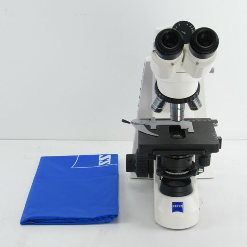
ZEISS recently donated a Primo Star Digital Classroom Microscope to the White Plains School Department, in White Plains, NY. The Primo Star features an integrated HD streaming camera and Labscope imaging software, an easy-to-use imaging app that enables teachers to connect several classrooms to a network. Also donated was an Axiocam 208 camera, ideal for helping science teachers presenting and sharing laboratory activities. Several White Plains High School science teachers are using the equipment in classes to enhance lab activities for students. The donation was made as part of ZEISS's Science Classroom Outreach Program for Educators (SCOPEs) Grant, established to help teachers face the new challenge of educating students through remote learning.
Zeiss

Crest V3 High-Speed Confocal System
The X-Light V3 is a cutting-edge confocal imaging system designed for the challenging microscopy applications. It is ideal for low light or high-speed imaging and has a software-controlled bypass mode for widefield or other imaging methods. The X-Light V3 comes with motorized excitation, dichroic, and emission filter wheels and dual simultaneous camera mounts with an automated mirror slider. The spinning disk box has a motorized bypass mode and is exchangeable for use with different pinhole patterns.
Crest Optics
https://crestoptics.com/x-light-v3

Easy Monochromator for Ultraviolet Light
McPherson launched an improved compact Model 234/302 monochromator. A monochromator separates light into constituent wavelengths, usually with narrow spectral bandwidth. The Model 234/302 does that well, with hundreds installed around the world. Internal surfaces have an optimized low-scatter finish, spectrograph accessories, and an improved turret with a range of masterpiece gratings.
McPherson

Aurion Gold Nanoparticles – Carboxyl-Functionalized
Electron Microscopy Sciences has introduced a series of carboxyl-functionalized gold nanoparticles. This product has been developed to facilitate conjugating gold nanoparticles to molecules that cannot be conjugated via the classic direct adsorption method. The product is especially suited for covalent conjugation of small ligands.
Electron Microscopy Sciences (EMS)
www.electronmicroscopysciences.com

Coxem Introduces the EM-30C
The EM-30C features a Cerium Hexaboride (CeB6) electron source providing 10× higher brightness and longer life compared to a tungsten filament, making it ideal for high-resolution imaging even at low accelerating voltages. The EM-30C is powered by Coxem's 4th Generation NanoStation software with automatic functions that simplify operation and speed analysis. Auto Focus, Brightness, and Contrast generate high-quality images fast and easy, while Panorama mode automatically stitches hundreds or even thousands of single images together to provide high-resolution mosaic images covering large areas.
Coxem

Gain Deeper Insights into Complex 3D Biology
The Molecular Devices ImageXpress® Micro Confocal High-Content Imaging System expands 3D imaging capabilities and generates greater phenotypic data without sacrificing throughput or quality. Key characteristics include acquisition of sharper, crisper images with minimal distortion of 3D samples with water immersion objectives; deeper penetration into thick samples with high-performance lasers and a deep tissue confocal disk; acquisition of data 10× faster; reduced data storage requirements with QuickID targeted acquisition; and 3D volumetric analysis within an existing workflow.
Molecular Devices

New DHM T100 Microscope from Lyncée Tec
Lyncée Tec has introduced the DHM T100 microscope system with a single and fixed objective (large choice of magnifications available); manual sample stage adjustment; a single wavelength; and acquisition, control, and analysis software. Upgrade packages include a turret with multiple objectives, a motorized stage for multisite measurements, a fluorescence module, and environmental control chamber. Analysis software modules include Multisite, End-points, Time-Lapse, Phase-Fluo Correlation, and 4D Tracking.
Lyncée Tec

Accelerate Discovery with Gatan K3 Base
Gatan has introduced the K3 Base, which is a small-form-factor, direct-detection camera that extends cryo-electron microscopy studies across more microscopes. The camera can turn screening microscopes into data-collection microscopes with a 14-megapixel sensor and >25 full fps transfer speed. Whether looking to add or distribute the workload to low-energy microscopes, the K3 Base camera provides a cost-effective solution to generate high-resolution structures.
Gatan

New Dragonfly Software Release
Dragonfly 2020.2 builds on the success of version 2020.1 by providing powerful new options, including advancements for the Segmentation Wizard that provide an easy-to-use, guided workflow for implementing powerful deep learning and machine learning segmentation of multi-dimensional images. New features include automated workflows, the option to extract an object's history as a macro, and options to find and preview the center of rotation for reconstructing cone beam and parallel beam projections with Dragonfly's CT Reconstruction module. More information can be found at: www.theobjects.com/assets/docs/dragonfly/dragonfly-release-notes-2020-2.pdf
Dragonfly
https://theobjects.com/dragonfly

Olympus LEXT™ OLS5100 Laser Microscope's Smart Features Empower Faster Experiment Workflows
The Olympus LEXT™ OLS5100 laser microscope offers guaranteed accuracy and precision with smart features, making materials science experiment workflows faster and more efficient. It provides high levels of accuracy and precision required for sub-micron 3D observation and surface roughness measurement. Some tasks, such as creating and managing experiment plans and choosing an objective lens, are time-consuming and sources of potential error. The microscope's smart features address these challenges.
Olympus

Fastec High-Speed Cameras: Better Lab Workflow in the Times of COVID-19
Fastec Imaging's new HS7 and HS5 high-speed cameras allow researchers and engineers to capture, display, analyze, transport, and store massive amounts of image data in much less time than with conventional cameras. The HS Series system architecture uses a fiber optic link to provide the fastest possible image transfers from camera to controller. This avoids waiting on the camera to download images before starting analysis or beginning the next test.
Fastec Imaging
www.fastecimaging.com/fastec-hs-series-cameras

Hitachi's SEM Heating Holder with Norcada MEMS Chips
The Hitachi Heating Holder combined with Norcada's MEMS in-situ Heating Chips enable visualization of thermal reactions when looking at materials and performing reactive experiments. For example, the holder and MEMS chips can be used for in-situ electron and X-ray microscopy work on micropores, nanopores, and single crystal silicon foils for radiation physics, in addition to use for a variety of other materials. Every MEMS chip is TEM-, SEM-, FIB-, and X-ray compatible.
Hitachi High Technologies
www.hitachi-hightech.com/us/product_list/?ld=sms2

Bruker's New e-Flash XS EBSD System
To significantly increase the number of labs able to acquire an integrated EDS and EBSD system, Bruker Nano Analytics developed e-Flash XS, a unique EBSD detector dedicated to the affordable part of the SEM market. It was purposely designed to be installed on low-footprint SEMs, for example, tabletop SEMs and standard SEMs with small chambers. It is integrated with a sixth-generation XFlash® EDS detector under the ESPRIT 2 software to provide a powerful combination of analytical techniques.
Bruker

ZEISS Enhances its Field Emission SEMs
The new ZEISS GeminiSEM family delivers more information from any sample, minimizes sample damage, and prevents sample artifacts. All three Gemini models come with a new chamber design, which allows researchers to bring in larger samples enabling core facilities to serve more analytical applications in a single instrument. The larger chamber enables configurability and flexibility to adapt to upcoming research tasks and optimizes analytical workflows.
ZEISS
www.zeiss.com/microscopy/us/products/scanning-electron-microscopes.html

Seiwa Optical has Introduced Virtual Booth
Seiwa Optical has created a virtual booth. Seiwa Optical Virtual Booth is web-based and used for conferences and shows to offer safe meetings during the times of COVID concerns. Seiwa Optical has been a provider of customizable optical solutions for machine vision, inspection, and industrial processing for over 50 years. They have offices established worldwide and are also a distributor for industrial vision products.
Seiwa Optical
www.seiwaamerica.com/virtual-booth

Vacuubrand Pumps
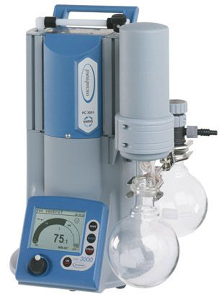
VACUUBRAND pumps are designed for the demands of a modern lab. These pumps are reliable, sustainable, and whisper-quiet. Most importantly, they provide the strong vacuum required for scientific research. With a choice of manually controlled pumps or automated, demand-responsive VARIO® models, networks can be tailored to meet a project's technical and budgetary requirements.
Vacuubrand

Micro CT System for Angstrom Scientific
The inCiTe™ micro-CT scanner from KA Imaging is the first commercial X-ray CT scanner that uses a patented high spatial resolution amorphous selenium (a-Se) detector technology exclusively developed by KA Imaging. The high detection efficiency of a-Se enables high-speed sampling at low X-ray exposure, allowing unprecedented volumetric scan speed at full spatial resolution. In addition, the inCiTe™ micro-CT scanner is designed with propagation-based phase-contrast imaging. Phase-contrast, low-density materials that are X-ray transparent in conventional X-ray imaging can be visualized.
Angstrom Scientific
www.angstrom.us/micro-ct-system


