The rodent population expansion in major cities is a growing public health concern. Mites rely on rodents as primary hosts. They have the potential to cause human disease including dermatitis and the transmission of zoonotic infection. Mites are difficult to identify, and infestations with these ectoparasites can be challenging to confirm and contain, especially in healthcare settings. Examining the affected host (humans or animals) often reveals nonspecific findings.Reference Engel, Welzel, Maass, Schramm and Wolff1
Ornithonyssus bacoti, also known as the tropical rat mite, is commonly found in temperate climates around the world.Reference Engel, Welzel, Maass, Schramm and Wolff1 Whereas Liponyssoides sanguineus (formerly Allodermanyssus sanguineus or house mouse mite) has an unclear geographic distribution. However, riskettsialpox, a zoonotic infection carried by L. sanguineus, has been reported in the Americas, Asia, Africa and Europe.Reference Paddock, Koss and Eremeeva2 We report an outbreak of dermatitis due to infestation of O. bacoti and L. sanguineus in a hospital clinic causing healthcare worker exposure and dermatitis.
Case presentation
A clinic staff member developed a pruritic, papular rash involving upper and lower extremities as well as the neck. Within a day of this incident, 8 additional clinic staff members developed similar symptoms. None had systemic symptoms. One staff member reported insects rising to the surface of the water during a bath. Infection prevention staff, with assistance from clinic leadership, occupational health, environmental services, environmental health and safety, laboratory leadership and hospital leadership, initiated an investigation.
The clinic is located at Tufts Medical Center, an urban hospital in Boston, Massachusetts. In the weeks prior to the initial report, clinic staff had noted sounds in the walls consistent with possible rodent activity. Concurrent with the cases of dermatitis, staff began to notice small insects in the environment, particularly in the work area utilized by front desk staff.
Microbiologic identification
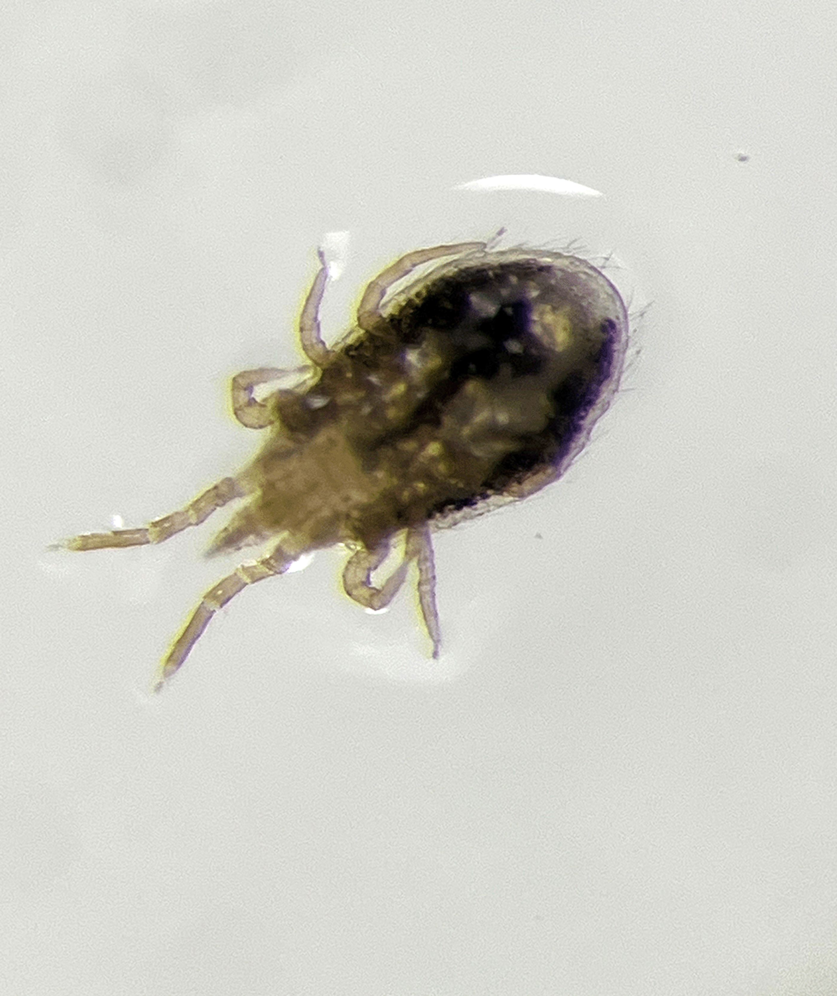
Mites were collected from the clinic environment in a specimen cup and sent to the Cummings School of Veterinary Medicine at Tufts University for further identification by a parasitologist. Two types of mites were morphologically identified: Ornithonyssus bacoti (Fig. 1) and Liponyssoides sanguineus.

Fig. 1. Image from specimen obtained from the environment during the infestation, representative of Ornithonysus bacoti.
Eradication
The mite infestation was linked to rodents that had infested the trash-compactor area located below the clinic. Rodent traps were set in the trash-compactor area. A juvenile hormone analog (Gentrol insect growth regulator; active ingredient, (S)-hydroprene) was sprayed in the clinic and compactor area. The agent mimics abnormally high levels of the juvenile hormone, interfering with the normal development of reproductive adults. The clinic was closed to patients and staff for 24 hours. No rodent or mite activity was noted for 2 weeks, when staff reported mite re-emergence. Additional extermination expertise was brought in.
Clinic operations were relocated to a different area. All items including printers, furniture, and papers were removed. The environmental health and safety team identified a clinic wall and an attached cabinet as a possible source of the mite infestation. There were several sightings of mites in that area. The cabinet and parts of the wall were removed, revealing a connection with the compactor area below and detection of live mites throughout. The wall was subsequently demolished for more extensive treatment of the area and to eliminate the connection with the compactor area. Over 2 weeks, additional wall demolition was conducted. In addition to juvenile hormone analog, a miticide, abamectin (AVID .15 EC miticide, Syngenta, Basel, Switzerland), which controls mites by stimulating the release of γ amino butyric acid leading to mite paralysis, was sprayed to kill existing mites.
The clinic was monitored for a week following the extermination with no signs of rodent or mite activity. Clinic repairs were then completed. Continued surveillance for 6 months of clinic and trash compactor areas did not show signs of mite recurrence.
Discussion
To our knowledge, this is the first report of an outbreak of dermatitis due to mite infestation in an acute-care hospital. A few such infestations in other settings have been reported, including animal research facilities, houses, laboratory personnel, and congregate settings.Reference Engel, Welzel, Maass, Schramm and Wolff1,Reference Fox3,Reference Baumstark, Beck and Hofmann4 O. bacoti and L. sanguineus are both ectoparasites that require hosts for feeding but can survive in the environment, often in nesting materials, when not feeding. The preferred primary hosts are rodents; however, humans can be secondary hosts in cases of heavy mite infestation or after extermination of the primary host.
Dermatitis and potential for zoonotic infections
The bite of O. bacoti can be painful and is typically followed by the eruption of a pruritic erythematous macular rash. The rash can be concentrated in body parts that are accessible by the mite or at parts around tightly fitting clothes.Reference Engel, Welzel, Maass, Schramm and Wolff1 Vesicular rash and nodules have also been described. Treatment is directed toward symptom management with antihistamines and glucocorticoids since the mite is easily washed with a shower without the requirement of mite directed therapies.
O. bacoti and L. sanguineus have the potential for the transmission of zoonotic infections. L. sanguineus is the main known carrier of Rickettsia akari. Reference Paddock, Koss and Eremeeva2 A few reports in the literature regarding zoonotic infections directly linked to O. bacoti have described endemic typhus transmission and Bartonella henselae infection. Experimental studies have shown the potential for vector transmission of the agents of Q fever, tularemia, and epidemic hemorrhagic fever, coxsackievirus, Yersinia pestis, and rickettsialpox.Reference Watson5
Rickettsialpox cases have been reported across the United States and it remains endemic in New York City. Some reports have revealed exoskeletons of L. sanguineus in buildings and others have described isolated cases without clear identification of a mite.Reference Koss, Carter and Grossman6
Eradication
Complete eradication of mite infestations can be challenging and requires coordination to eliminate both the mites and the rodents. Following rodent extermination, mites can travel hundreds of feet and can survive for weeks without feeding. Thus, reinfestation is common but can be prevented by employing multiple treatments. The early identification of outbreaks could facilitate eradication, thus minimizing the impact on human health. In addition to juvenile hormone analog and abamectin, several other pesticides, including benzyl benzoate and gamma benzene hexachloride 1% (Lindane) or fumigated silica aerogel dust into walls, have been used to eradicate tropical rat mites.Reference Fishman7
Rodent activity in urban centers
Climate change leading to warmer, wet winters and drought in the summers is conductive to the expansion of rodent populations. Furthermore, urbanization can affect the behavioral traits of rodents making them more explorative and bolder and increasing the likelihood of close contact with humans.Reference Dammhahn, Mazza, Schirmer, Göttsche and Eccard8 Boston reported its highest number of rodent complaints in 2021 compared to prior years, according to the city’s 311 data. The city of Chicago reported a 39% increase in public rat complaints from 2008 to 2017. Similar findings have been reported in California, Texas, and New York City.9 This growing rodent problem in urban centers will inevitably increase human exposure to vectorborne illnesses and zoonotic diseases.
In conclusion, successful and expeditious eradication of a clinic infestation by O. bacoti and L. sanguineus were achieved due to several important actions: (1) rapid identification of the mites utilizing the expertise of a veterinary parasitology consultant; (2) collaboration between the infection prevention team, clinic leadership, occupational health, environmental health and safety, environmental services, laboratory leadership, and hospital leadership; and (3) aggressive mitigation, which included necessary demolition of walls and other structures. Our experience could provide direction in future outbreaks.
Acknowledgments
We thank Stefan J. Gustafson for his assistance in communicating information related to the outbreak.
Financial support
No financial support was provided relevant to this article.
Competing interest
All authors report no conflicts of interest relevant to this article.


