Hostname: page-component-586b7cd67f-rcrh6 Total loading time: 0 Render date: 2024-11-29T13:44:46.978Z Has data issue: false hasContentIssue false

Whole-Cell Tomography Using a Conventional Scanning Electron Microscope

Published online by Cambridge University Press:  23 September 2015

Taiga Okumura
Affiliation:
Hitachi, Ltd., Central Research Laboratory, Tokyo, Japan
Minami Shoji
Affiliation:
Hitachi, Ltd., Central Research Laboratory, Tokyo, Japan
Akiko Hisada
Affiliation:
Hitachi, Ltd., Central Research Laboratory, Tokyo, Japan
Yusuke Ominami
Affiliation:
Hitachi High-Technologies Corporation, Ibaraki, Japan
Tatsuo Ushiki
Affiliation:
Graduate School of Medical and Dental Sciences, Niigata University, Niigata, Japan
Masato Nakajima
Affiliation:
Graduate School of Medical and Dental Sciences, Niigata University, Niigata, Japan
Takashi Ohshima
Affiliation:
Hitachi, Ltd., Central Research Laboratory, Tokyo, Japan

Abstract

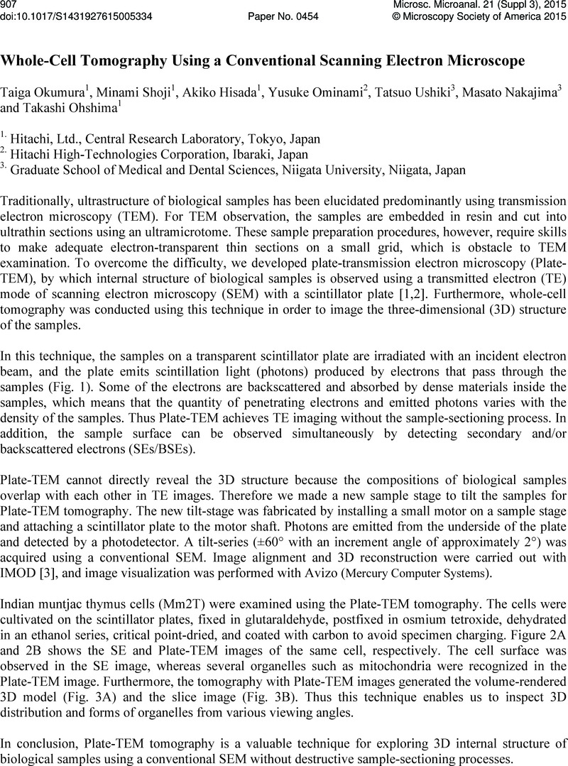
Image of the first page of this content. For PDF version, please use the ‘Save PDF’ preceeding this image.'
Type
Abstract
Copyright
Copyright © Microscopy Society of America 2015 

References

References:

[1] Ominami, Y, et al, Scanning Microscopies (2014) 923608.Google Scholar
[2] Ominami, Y, et al, Microscopy and Microanalysis (2014) 3233.Google Scholar
[3] Kremer, JR, Mastronarde, DN & McIntosh, JR, J. Struct. Biol. 116 (1996) 7176.Google Scholar