Hostname: page-component-cd9895bd7-fscjk Total loading time: 0 Render date: 2024-12-25T19:52:17.492Z Has data issue: false hasContentIssue false

Volume Imaging By Tracking Sparse Topological Features In Electron Micrograph Tilt Series

Published online by Cambridge University Press:  22 July 2022

T. C. Petersen*
Affiliation:
Monash Centre for Electron Microscopy, Monash University, Clayton, Victoria, Australia
C. Zhao
Affiliation:
Interdisciplinary Nanoscience Centre, Aarhus University, Aarhus, Midtjylland, Denmark
E. D. Bøjesen
Affiliation:
Monash Centre for Electron Microscopy, Monash University, Clayton, Victoria, Australia Interdisciplinary Nanoscience Centre, Aarhus University, Aarhus, Midtjylland, Denmark
N. L. N. Broge
Affiliation:
Interdisciplinary Nanoscience Centre, Aarhus University, Aarhus, Midtjylland, Denmark
S. Hata
Affiliation:
Department of Advanced Materials Science, Kyushu University, Fukuoka, Japan
Y. Liu
Affiliation:
Research School of Chemistry, The Australian National University, Canberra, Acton, Australia
J. Etheridge
Affiliation:
Monash Centre for Electron Microscopy, Monash University, Clayton, Victoria, Australia
*
*Corresponding author: [email protected]

Abstract

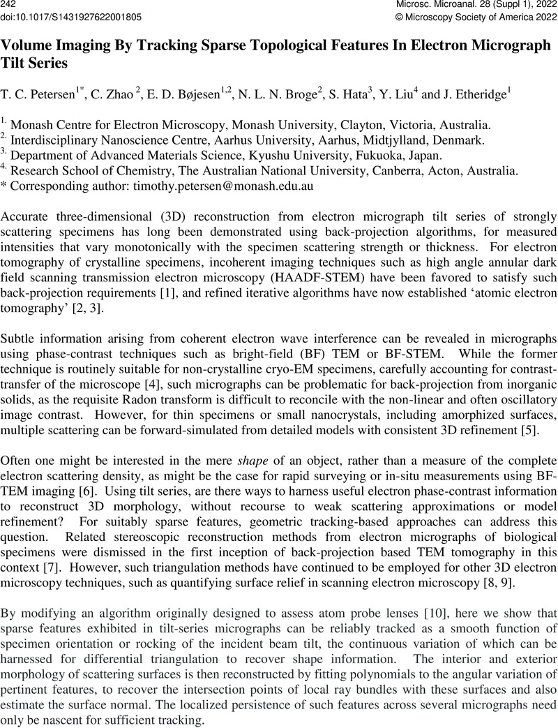
Image of the first page of this content. For PDF version, please use the ‘Save PDF’ preceeding this image.'
Type
Advanced 3D Imaging and Analysis Methods for New Opportunities in Material Science
Copyright
Copyright © Microscopy Society of America 2022

References

Weyland, M, Midgley, PA and Thomas, JM, J. Phys. Chem. B 105 (2001), p. 7882.CrossRefGoogle Scholar
Yang, Y et al. , Nature 542 (2017), p. 75.CrossRefGoogle Scholar
Zhou, J et al. , Nature 570 (2019), p. 500.CrossRefGoogle Scholar
Frank, J in “Electron tomography: three-dimensional imaging with the transmission electron microscope” (Plenum Press, New York, 1992).Google Scholar
Ren, D et al. , Ultramicroscopy 208 (2020), p. 112860.CrossRefGoogle Scholar
Hata, S et al. , Curr. Opin. Solid State Mater. Sci. 24 (2020), p. 100850.CrossRefGoogle Scholar
De Rosier, DJ and Klug, A, Nature 217 (1968), p. 130.CrossRefGoogle Scholar
Hilliard, JE, J. Microsc. 95 (1972), p. 45.CrossRefGoogle Scholar
Podsiadlo, P and Stachowiak, G, Wear 230 (1999), p. 184.CrossRefGoogle Scholar
Haley, D et al. , J. Microsc. 244 (2011), p. 170.CrossRefGoogle Scholar
Ortega, D et al. , PLoS ONE 14 (2019), p. e0215531.CrossRefGoogle Scholar
Petersen, TC et al. , Ultramicroscopy (2022) p. 113475, https://doi.org/10.1016/j.ultramic.2022.113475.CrossRefGoogle Scholar
Mitchell, DRG and Schaffer, B, Ultramicroscopy 103 (2005), p. 319.CrossRefGoogle Scholar
This work was supported by the Australian Research Council (ARC) via grants LE0454166 and LE170100118, as well as the Japan Society for the Promotion of Science (JSPS)/Ministry of Education, Culture, Sports, Science and Technology (MEXT), Japan KAKENHI (JP18H05479, JP20H02426); Japan Science and Technology Agency (JST) CREST (\#JPMJCR18J4). Bo Brummerstedt Iversen of Aarhus University is thanked for support, along with that of the Villum Foundation. J. Etheridge acknowledges funding from ARC grant DP150104483. Matt Weyland and Laure Bourgeois are thanked for their assistance at the Monash Centre for Electron Microscopy.Google Scholar