Hostname: page-component-586b7cd67f-rcrh6 Total loading time: 0 Render date: 2024-11-27T22:55:37.032Z Has data issue: false hasContentIssue false

Tracking the Mn Diffusion in the Carbon-Supported Nanoparticles Through the Collaborative Analysis of Atom Probe and Evaporation Simulation

Published online by Cambridge University Press:  17 October 2022

Chanwon Jung
Affiliation:
Department of Materials Science and Engineering, Korea Advanced Institute of Science and Technology (KAIST), 291 Daehak-ro, Yuseong-gu, Daejeon 34141, Republic of Korea Max-Planck-Institut für Eisenforschung GmbH, Düsseldorf 40237, Germany
Hosun Jun
Affiliation:
Department of Materials Science and Engineering, Korea Advanced Institute of Science and Technology (KAIST), 291 Daehak-ro, Yuseong-gu, Daejeon 34141, Republic of Korea
Kyuseon Jang
Affiliation:
Department of Materials Science and Engineering, Korea Advanced Institute of Science and Technology (KAIST), 291 Daehak-ro, Yuseong-gu, Daejeon 34141, Republic of Korea
Se-Ho Kim
Affiliation:
Max-Planck-Institut für Eisenforschung GmbH, Düsseldorf 40237, Germany
Pyuck-Pa Choi*
Affiliation:
Department of Materials Science and Engineering, Korea Advanced Institute of Science and Technology (KAIST), 291 Daehak-ro, Yuseong-gu, Daejeon 34141, Republic of Korea
*
*Corresponding author: Pyuck-Pa Choi, E-mail: [email protected]
Get access

Abstract

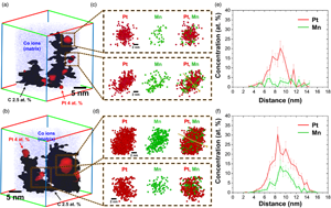
Carbon-supported nanoparticles have been used widely as efficient catalysts due to their enhanced surface-to-volume ratio. To investigate their structure–property relationships, acquiring 3D elemental distribution is required. Here, carbon-supported Pt, PtMn alloy, and ordered Pt3Mn nanoparticles are synthesized and analyzed with atom probe tomography as model systems. A significant difference of Mn distribution after the heat-treatment was found. Finally, the field evaporation behavior of the carbon support was discussed and each acquired reconstruction was compared with computational results from an evaporation simulation. This paper provides a guideline for studies using atom probe tomography on the heterogeneous carbon-supported nanoparticle system that leads to insights toward a wide variety of applications.

Type
Materials Science Applications
Copyright
Copyright © The Author(s), 2022. Published by Cambridge University Press on behalf of the Microscopy Society of America

Access options

Get access to the full version of this content by using one of the access options below. (Log in options will check for institutional or personal access. Content may require purchase if you do not have access.)

Footnotes

These authors contributed equally to this work.

References

Antolini, E (2009). Carbon supports for low-temperature fuel cell catalysts. Appl Catal B 88, 124.CrossRefGoogle Scholar
Barroo, C, Akey, AJ & Bell, DC (2019). Atom probe tomography for catalysis applications: A review. Appl Sci 9, 2721.CrossRefGoogle Scholar
Chi, M, Wang, C, Lei, Y, Wang, G, Li, D, More, KL, Lupini, A, Allard, LF, Markovic, NM & Stamenkovic, VR (2015). Surface faceting and elemental diffusion behaviour at atomic scale for alloy nanoparticles during in situ annealing. Nat Commun 6, 19.CrossRefGoogle ScholarPubMed
Exertier, F, Wang, J, Fu, J & Marceau, RKW (2021). Understanding the effects of graphene coating on the electrostatic field at the tip of an atom probe tomography specimen. Microsc Microanal 28, 112.Google Scholar
Felfer, P, Benndorf, P, Masters, A, Maschmeyer, T & Cairney, JM (2014). Revealing the distribution of the atoms within individual bimetallic catalyst nanoparticles. Angew Chem Int Ed 53, 1119011193.CrossRefGoogle ScholarPubMed
Felfer, P, Stevenson, L & Li, T (2018). Atom probe tomography. Praktische Metallogr/Practical Metallogr 55, 515526.CrossRefGoogle Scholar
Gault, B, Breen, AJ, Chang, Y, He, J, Jägle, EA, Kontis, P, Kürnsteiner, P, da Silva, AK, Makineni, SK & Mouton, I (2018). Interfaces and defect composition at the near-atomic scale through atom probe tomography investigations. J Mater Res 33, 40184030.CrossRefGoogle Scholar
Gault, B, Moody, MP, Cairney, JM & Ringer, SP (2012). Atom Probe Microscopy. New York, USA: Springer Science & Business Media.CrossRefGoogle Scholar
Gault, B, Yang, W, Ratinac, KR, Zheng, R, Braet, F & Ringer, SP (2010). Atom probe microscopy of self-assembled monolayers: Preliminary results. Langmuir 26, 52915294.CrossRefGoogle ScholarPubMed
Jang, K, Kim, S-H, Jun, H, Jung, C, Yu, J, Lee, S & Choi, P-P (2021). Three-dimensional atomic mapping of ligands on palladium nanoparticles by atom probe tomography. Nat Commun 12, 110.Google ScholarPubMed
Johansen, M & Liu, F (2021). Best practices for analysis of carbon fibers by atom probe tomography. Microsc Microanal 28, 110.Google Scholar
Johansen, M, Schlueter, C, Tam, PL, Asp, LE & Liu, F (2021). Mapping nitrogen heteroatoms in carbon fibres using atom probe tomography and photoelectron spectroscopy. Carbon 179, 2027.CrossRefGoogle Scholar
Jun, H, Jang, K, Jung, C & Choi, P-P (2021). Atom probe tomography investigations of Ag nanoparticles embedded in pulse-electrodeposited Ni films. Microsc Microanal 27, 110.CrossRefGoogle Scholar
Jung, C, Kang, K, Marshal, A, Pradeep, KG, Seol, JB, Lee, HM & Choi, PP (2019). Effects of phase composition and elemental partitioning on soft magnetic properties of AlFeCoCrMn high entropy alloys. Acta Mater 171, 3139. doi:10.1016/j.actamat.2019.04.007.CrossRefGoogle Scholar
Jung, C, Lee, C, Bang, K, Lim, J, Lee, H, Ryu, HJ, Cho, E & Lee, HM (2017). Synthesis of chemically ordered Pt3Fe/C intermetallic electrocatalysts for oxygen reduction reaction with enhanced activity and durability via a removable carbon coating. ACS Appl Mater Interfaces 9, 3180631815.CrossRefGoogle Scholar
Kelly, TF & Miller, MK (2007). Atom probe tomography. Rev Sci Instrum 78, 31101.CrossRefGoogle ScholarPubMed
Kim, K, Jeong, I, Cho, Y, Shin, D, Song, S, Ahn, SK, Eo, YJ, Cho, A, Jung, C, Jo, W, Kim, JH, Choi, PP, Gwak, J & Yun, JH (2019). Mechanisms of extrinsic alkali incorporation in CIGS solar cells on flexible polyimide elucidated by nanoscale and quantitative analyses. Nano Energy 67, 104201.CrossRefGoogle Scholar
Kim, SH, Lim, J, Sahu, R, Kasian, O, Stephenson, LT, Scheu, C & Gault, B (2020 b). Direct imaging of dopant and impurity distributions in 2D MoS2. Adv Mater 32, 1907235.CrossRefGoogle ScholarPubMed
Kim, S-H, Antonov, S, Zhou, X, Stephenson, LT, Jung, C, El-Zoka, AA, Schreiber, DK, Conroy, M & Gault, B (2022). Atom probe analysis of electrode materials for Li-ion batteries: Challenges and ways forward. J Mater Chem A 10, 49264935.CrossRefGoogle ScholarPubMed
Kim, S-H, Jang, K, Kang, PW, Ahn, J-P, Seol, J-B, Kwak, C-M, Hatzoglou, C, Vurpillot, F & Choi, P-P (2020 a). Characterization of Pd and Pd@Au core-shell nanoparticles using atom probe tomography and field evaporation simulation. J Alloys Compd 831, 154721.CrossRefGoogle Scholar
Kim, S-H, Kang, PW, Park, OO, Seol, J-B, Ahn, J-P, Lee, JY & Choi, P-P (2018). A new method for mapping the three-dimensional atomic distribution within nanoparticles by atom probe tomography (APT). Ultramicroscopy 190, 3038.CrossRefGoogle Scholar
Kürnsteiner, P, Wilms, MB, Weisheit, A, Barriobero-Vila, P, Jägle, EA & Raabe, D (2017). Massive nanoprecipitation in an Fe-19Ni-xAl maraging steel triggered by the intrinsic heat treatment during laser metal deposition. Acta Mater 129, 5260.CrossRefGoogle Scholar
Larson, DJ, Prosa, TJ, Ulfig, RM, Geiser, BP & Kelly, TF (2013). Local Electrode Atom Probe Tomography.CrossRefGoogle Scholar
Lee, C, Shin, K, Jung, C, Choi, P-P, Henkelman, G & Lee, HM (2019 a). Atomically embedded Ag via electrodiffusion boosts oxygen evolution of CoOOH nanosheet arrays. ACS Catal 10, 562569.CrossRefGoogle Scholar
Lee, H, Jang, Y, Nam, S-W, Jung, C, Choi, P-P, Gwak, J, Yun, JH, Kim, K & Shin, B (2019 b). Passivation of deep-level defects by cesium fluoride post-deposition treatment for improved device performance of Cu(In,Ga)Se 2 solar cells. ACS Appl Mater Interfaces 11, 3565335660.CrossRefGoogle Scholar
Li, T, Bagot, PAJ, Christian, E, Theobald, BRC, Sharman, JDB, Ozkaya, D, Moody, MP, Tsang, SCE & Smith, GDW (2014). Atomic imaging of carbon-supported Pt, Pt/Co, and Ir@Pt nanocatalysts by atom-probe tomography. ACS Catal 4, 695702.CrossRefGoogle Scholar
Lim, J, Jung, C, Hong, D, Bak, J, Shin, J, Kim, M, Song, D, Lee, C, Lim, J & Lee, H (2022). Atomically ordered Pt3Mn intermetallic electrocatalysts for the oxygen reduction reaction in fuel cells. J Mater Chem A 10, 7399–7408.CrossRefGoogle Scholar
Lim, J, Kim, S, Aymerich Armengol, R, Kasian, O, Choi, P, Stephenson, LT, Gault, B & Scheu, C (2020). Atomic-scale mapping of impurities in partially reduced hollow TiO2 nanowires. Angew Chem 132, 57005704.CrossRefGoogle Scholar
Madaan, N, Bao, J, Nandasiri, M, Xu, Z, Thevuthasan, S & Devaraj, A (2015). Impact of dynamic specimen shape evolution on the atom probe tomography results of doped epitaxial oxide multilayers: Comparison of experiment and simulation. Appl Phys Lett 107, 91601.CrossRefGoogle Scholar
Miller, MK (2012). Atom Probe Tomography: Analysis at the Atomic Level. New York, USA: Springer Science & Business Media.Google Scholar
Miller, MK & Forbes, RG (2014). Atom-Probe Tomography. New York, USA: Springer.CrossRefGoogle Scholar
Oberdorfer, C, Eich, SM & Schmitz, G (2013). A full-scale simulation approach for atom probe tomography. Ultramicroscopy 128, 5567. doi:10.1016/j.ultramic.2013.01.005.CrossRefGoogle ScholarPubMed
Peng, L, Zhou, L, Kang, W, Li, R, Qu, K, Wang, L & Li, H (2020). Electrospinning synthesis of carbon-supported Pt3Mn intermetallic nanocrystals and electrocatalytic performance towards oxygen reduction reaction. Nanomaterials 10, 1893.CrossRefGoogle ScholarPubMed
Qiu, S, Zheng, C, Zhou, Q, Dong, D, Shi, Q, Garg, V, Cheng, W, Marceau, RKW, Sha, G & Fu, J (2020). Direct imaging of liquid–nanoparticle interfaces with atom probe tomography. J Phys Chem C 124, 1938919395.CrossRefGoogle Scholar
Raghuwanshi, M, Cojocaru-Mirédin, O & Wuttig, M (2020). Investigating bond rupture in resonantly bonded solids by field evaporation of carbon nanotubes. Nano Lett 20, 116121.CrossRefGoogle ScholarPubMed
Schirhagl, R, Raatz, N, Meijer, J, Markham, M, Gerstl, SSA & Degen, CL (2015). Nanometer-scale isotope analysis of bulk diamond by atom probe tomography. Diamond Relat Mater 60, 6065.CrossRefGoogle Scholar
Solodenko, H, Stender, P & Schmitz, G (2021). Atom probe study of 1-octadecanethiol self-assembled monolayers on platinum (111) and (200) surfaces. Microsc Microanal 28, 110.Google Scholar
Stoffers, A, Oberdorfer, C & Schmitz, G (2012). Controlled field evaporation of fluorinated self-assembled monolayers. Langmuir 28, 5659.CrossRefGoogle ScholarPubMed
Tedsree, K, Li, T, Jones, S, Chan, CWA, Yu, KMK, Bagot, PAJ, Marquis, EA, Smith, GDW & Tsang, SCE (2011). Hydrogen production from formic acid decomposition at room temperature using a Ag–Pd core–shell nanocatalyst. Nat Nanotechnol 6, 302307.CrossRefGoogle ScholarPubMed
Thompson, K, Lawrence, D, Larson, DJ, Olson, JD, Kelly, TF & Gorman, B (2007). In situ site-specific specimen preparation for atom probe tomography. Ultramicroscopy 107, 131139.CrossRefGoogle ScholarPubMed
Uchida, M, Aoyama, Y, Tanabe, M, Yanagihara, N, Eda, N & Ohta, A (1995). Influences of both carbon supports and heat-treatment of supported catalyst on electrochemical oxidation of methanol. J Electrochem Soc 142, 2572.CrossRefGoogle Scholar
Ungar, T, Gubicza, J, Ribarik, G, Pantea, C & Zerda, TW (2002). Microstructure of carbon blacks determined by X-ray diffraction profile analysis. Carbon 40, 929937.CrossRefGoogle Scholar
Supplementary material: File

Jung et al. supplementary material

Jung et al. supplementary material

Download Jung et al. supplementary material(File)
File 12.2 MB