Hostname: page-component-586b7cd67f-vdxz6 Total loading time: 0 Render date: 2024-11-25T13:18:33.806Z Has data issue: false hasContentIssue false

Towards Artifact-free FIB-SEM Datasets: Strategies Before, During, and After Image Acquisition

Published online by Cambridge University Press:  30 July 2020

Kedar Narayan
Affiliation:
National Cancer Institute, NIH & Frederick National Laboratory, Frederick, Maryland, United States
Alexandre Laquerre
Affiliation:
Fibics Inc, Ottawa, Ontario, Canada
Michael Phaneuf
Affiliation:
Fibics Inc, Ottawa, Ontario, Canada

Abstract

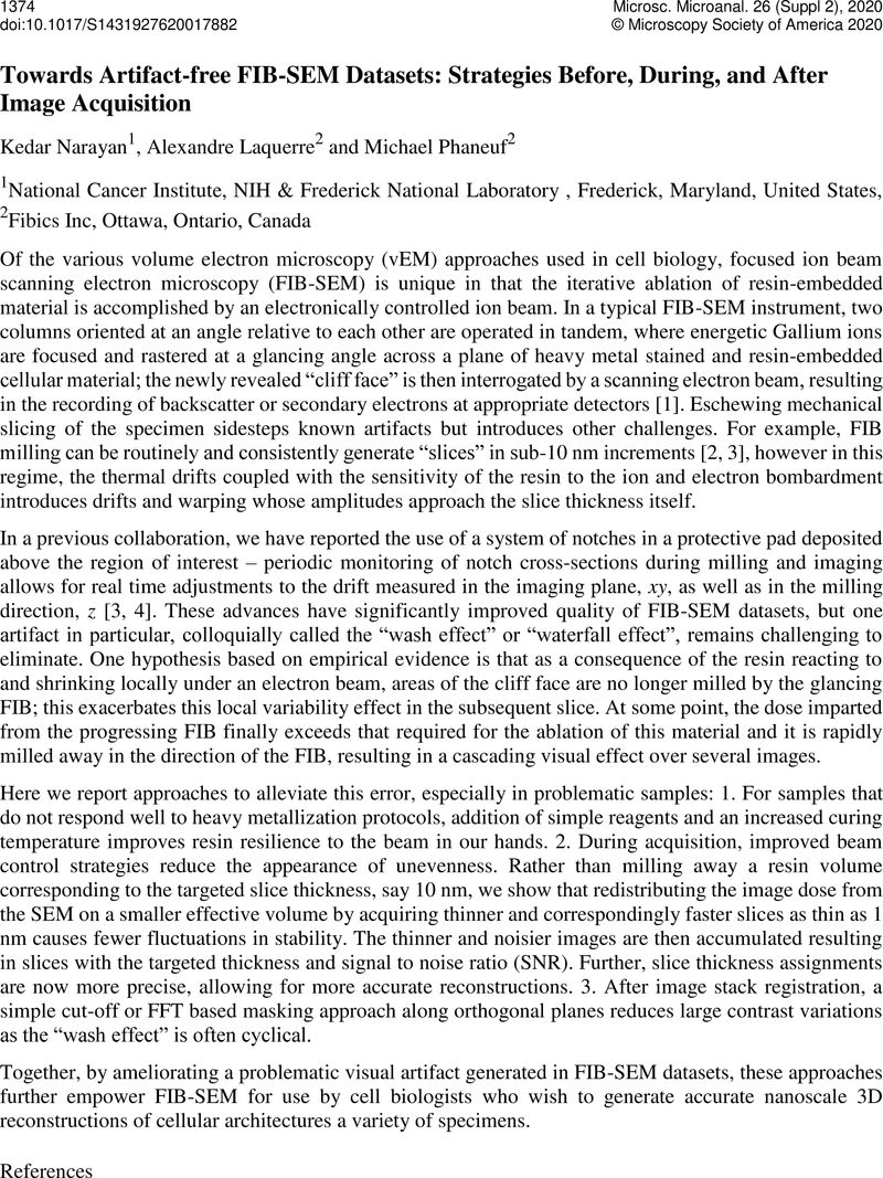
Image of the first page of this content. For PDF version, please use the ‘Save PDF’ preceeding this image.'
Type
3D Scanning Electron Microscopy Imaging of Biological Samples
Copyright
Copyright © Microscopy Society of America 2020

References

Narayan, K. and Subramaniam, S. (2015). Focused Ion Beams in Biology. Nature Methods Nov;12(11):1021-3110.1038/nmeth.3623CrossRefGoogle ScholarPubMed
Xu, C. S. et al. (2017). Enhanced FIB-SEM systems for large-volume 3D imaging. Elife May 13;6Google Scholar
Narayan, K. et al. (2014). Multi-resolution correlative focused ion beam scanning electron microscopy: applications to cell biology. Journal of Structural Biology. Mar 185(3):278-8410.1016/j.jsb.2013.11.008CrossRefGoogle ScholarPubMed
Phaneuf, M. W. and Lagarec, K. G. (2017). Microscopy imaging method and system US9812290B2 filed 31 Jan 2017 & issued 07 Nov 2017.Google Scholar