Hostname: page-component-78c5997874-94fs2 Total loading time: 0 Render date: 2024-11-04T20:27:55.801Z Has data issue: false hasContentIssue false

Structural and Ultrastructural Changes in the Tongue of mdx Mice

Published online by Cambridge University Press:  24 January 2022

André N. Tomiate
Affiliation:
Laboratório de Morfologia e Atividade Física (LAMAF), Instituto de Biociências, Universidade Estadual Paulista (UNESP), Rio Claro, São Paulo, Brasil
Gabriela K. Barbosa
Affiliation:
Laboratório de Morfologia e Atividade Física (LAMAF), Instituto de Biociências, Universidade Estadual Paulista (UNESP), Rio Claro, São Paulo, Brasil
Rose E. G. Rici
Affiliation:
Departamento de Cirurgia, Faculdade de Medicina Veterinária e Zootecnia, Universidade de São Paulo, São Paulo, Brasil
Sonia Regina Y. de Almeida
Affiliation:
Departamento de Anatomia, Instituto de Ciências Biomédicas - ICB III, Universidade de São Paulo, São Paulo, Brasil
Ii-sei Watanabe
Affiliation:
Departamento de Cirurgia, Faculdade de Medicina Veterinária e Zootecnia, Universidade de São Paulo, São Paulo, Brasil Departamento de Anatomia, Instituto de Ciências Biomédicas - ICB III, Universidade de São Paulo, São Paulo, Brasil
Adriano P. Ciena*
Affiliation:
Laboratório de Morfologia e Atividade Física (LAMAF), Instituto de Biociências, Universidade Estadual Paulista (UNESP), Rio Claro, São Paulo, Brasil
*
*Corresponding author: Adriano P. Ciena, E-mail: [email protected]
Get access

Abstract

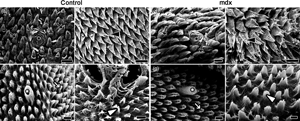
The mdx mouse is an experimental model of Duchenne muscular dystrophy, a genetic disorder characterized by progressive muscular degeneration which affects the oral cavity musculature, and promotes difficulty in swallowing. This study aimed to describe morphological, structural, and ultrastructural changes in the tongue mucosa and musculature of mdx mice. Forty six-month-old mice were divided into two groups: Control C57bl/10 (n = 20) and mdx C57bl/10mdx (n = 20). The tongue was dissected and analyzed with light microscopy, scanning electron microscopy, and transmission electron microscopy techniques. Our results showed conical and triangular filiform, fungiform, foliate, and vallate papillae, and their connective tissue cores. The epithelium layers identified were corneum, granulosum, spinosum, and basale. The mdx group had a thicker epithelium. Lamina propria was composed of reddish and greenish collagen. In mdx, collagen was present in the musculature of the tongue's body and in the muscular tissue between mucous and serous glands of the caudal region. Musculature was also characterized by a shorter length of sarcoplasmic invaginations, myocytolysis in mitochondrial groupings, and inflammatory focus. In conclusion, the tongue of 6-month-old mdx mice had morphology, structure, and ultrastructure revealed, showing higher wear of filiform papillae indirect reflex from the muscular degeneration process.

Type
Micrographia
Copyright
Copyright © The Author(s), 2022. Published by Cambridge University Press on behalf of the Microscopy Society of America

Access options

Get access to the full version of this content by using one of the access options below. (Log in options will check for institutional or personal access. Content may require purchase if you do not have access.)

References

Allen, DG, Whitehead, NP & Froehner, SC (2016). Absence of dystrophin disrupts skeletal muscle signaling: Roles of Ca2+, reactive oxygen Species, and nitric oxide in the development of muscular dystrophy. Physiol Rev 96(1), 253305. doi:10.1152/physrev.00007.2015CrossRefGoogle ScholarPubMed
Aloysius, A, Born, P, Kinali, M, Davis, T, Pane, M & Mercuri, E (2008). Swallowing difficulties in duchenne muscular dystrophy: Indications for feeding assessment and outcome of videofluoroscopic swallow studies. Eur J Paediatr Neurol 12(3), 239245. doi:10.1016/j.ejpn.2007.08.009CrossRefGoogle Scholar
Barbosa, GK, Jacob, CS, Pimentel Neto, J, de Oliveira, MF, Rici, REG, Watanabe, I & Ciena, AP (2020). Structural and ultrastructural characteristics of the tongue of the collared peccary (Pecari tajacu, Linnaeus, 1758). Anat Histol Embryol 49(4), 532540. doi:10.1111/ahe.12557CrossRefGoogle Scholar
Bertoldo, BB, Etchebehere, RM, Furtado, TCDS, Faria, JBD, Silva, CB, Araújo, MFD, Rodrigues, DBR & Pereira, SADL (2019). Lingual salivary gland hypertrophy and decreased acinar density in chagasic patients without megaesophagus. Rev Inst Med Trop Sao Paulo 61. doi:10.1590/S1678-9946201961067CrossRefGoogle ScholarPubMed
Bulfield, G, Siller, WG, Wight, PAL & Moore, KJ (1984). X chromosome-linked muscular dystrophy (mdx) in the mouse. Proc Natl Acad Sci U S A 81, 11891192. doi:10.1073/pnas.81.4.1189CrossRefGoogle Scholar
Chamberlain, JS, Metzger, J, Reyes, M, Townsend, D & Faulkner, JA (2007). Dystrophin-deficient mdx mice display a reduced life span and are susceptible to spontaneous rhabdomyosarcoma. FASEB J 21, 21952204. doi:10.1096/fj.06-7353comCrossRefGoogle ScholarPubMed
Ciena, AP, Bolina, CDS, de Almeida, SRY, Rici, REG, de Oliveira, MF, Silva, MCPD, Miglino, MA & Watanabe, I (2013). Structural and ultrastructural features of the agouti tongue (Dasyprocta aguti Linnaeus, 1766). J Anat 223(2), 152158. doi:10.1111/joa.12065CrossRefGoogle Scholar
Ciena, AP, dos Santos, AC, Vasconcelos, BG, Rici, REG, Assis-Neto, AC, Almeida, SRY, Miglino, MA & Watanabe, I (2019). Morphological characteristics of the papillae and lingual epithelium of guinea pig (Cavia porcellus). Acta Zool 100, 5360. doi:10.1111/azo.12230CrossRefGoogle Scholar
Cros, D, Harnden, P, Pellissier, JF & Serratrice, G (1989). Muscle hypertrophy in duchenne muscular dystrophy. J Neurol 236, 4347. doi:10.1007/BF00314217CrossRefGoogle ScholarPubMed
de Aro, MM, dos Santos, AC, da Silveira, EE, Lisboa Neto, AFS, de Oliveira, MF & de Assis Neto, AC (2019). Morphological tools to evaluate the digestory apparatus in rocky cavy (Kerodon rupestris). Microsc Res Tech 82, 696708. doi:10.1002/jemt.23216CrossRefGoogle Scholar
Deconinck, N & Dan, B (2007). Pathophysiology of duchenne muscular dystrophy: Current hypotheses. J Pediatr Neurol 36(1), 17. doi:10.1016/j.pediatrneurol.2006.09.016CrossRefGoogle ScholarPubMed
Emura, S, Okumura, T & Chen, H (2011). Morphology of the lingual papillae in the patagonian cavy. Okajimas Folia Anat Jpn 88(3), 121125. doi:10.2535/ofaj.88.121CrossRefGoogle ScholarPubMed
Hoffman, EP, Brown, RH & Kunkel, LM (1987). Dystrophin: The protein product of the duchenne muscular dystrophy locus. Cell 51, 919928. doi:10.1016/0092-8674(87)90579-4CrossRefGoogle ScholarPubMed
Igbokwe, CO & Mbajiorgu, FE (2019). Anatomical and scanning electron microscopic study of the tongue in the African giant pouched rats (Cricetomys gambianus, Waterhouse). Anat Histol Embryol 48, 455465. doi:10.1111/ahe.12467CrossRefGoogle Scholar
Kitajima, K & Kobayashi, K (1992). Light and electron microscopic studies on the lingual papillae and their connective tissue cores in hamster. Jpn J Oral Biol 34, 503530. doi:10.2330/joralbiosci1965.34.503CrossRefGoogle Scholar
Kobayashi, K & Iwasaki, S (1989). Comparative studies on the stereo architecture of the connective tissue papillae in some mammalian tongues. Prog Clin Biol Res 295, 303308.Google ScholarPubMed
Kobayashi, K, Miyata, K, Takahashi, K & Iwasaki, S (1989). Three-dimensional architecture of the connective tissue papillae of the mouse tongue as viewed by scanning electron microscopy. Kaibogaku Zasshi 64, 523538.Google ScholarPubMed
Martinez, GZ, Grillo, B, Rocha, LC, Jacob, C, Pimentel Neto, J, Tomiate, AN, Barbosa, GK, Watanabe, I & Ciena, AP (2021). Morphological changes in the myotendinous junction of mdx mice. Microsc Microanal 27(5), 12901294. doi:10.1017/S1431927621012496CrossRefGoogle Scholar
Muller, J, Vayssiere, N, Royuela, M, Leger, ME, Muller, A, Bacou, F, Pons, F, Hugon, G & Mornet, D (2001). Comparative evolution of muscular dystrophy in diaphragm, gastrocnemius and masseter muscles from old mdx mice. J Muscle Res Cell Motil 22, 133139. doi:10.1023/a:1010305801236CrossRefGoogle Scholar
Pane, M, Vasta, I, Messina, S, Sorleti, D, Aloysius, A, Sciarra, F, Mangiola, F, Kinali, M, Ricci, E & Mercuri, E (2006). Feeding problems and weight gain in duchenne muscular dystrophy. Eur J Paediatr Neurol 10, 231236. doi:10.1016/j.ejpn.2006.08.008CrossRefGoogle ScholarPubMed
Pastoret, C & Sebille, A (1995). Age-related differences in regeneration of dystrophic (mdx) and normal muscle in the mouse. Muscle Nerve 18(10), 11471154. doi:10.1002/mus.880181011CrossRefGoogle ScholarPubMed
Porter, JD, Khanna, S, Kaminski, HJ, Rao, JS, Merriam, AP, Richmonds, CR, Leahy, P, Li, J, Guo, W & Andrade, FH (2002). A chronic inflammatory response dominates the skeletal muscle molecular signature in dystrophin-deficient mdx mice. Hum Mol Genet 11(3), 263272. doi:10.1093/hmg/11.3.263CrossRefGoogle ScholarPubMed
Reginato, GDS, Bolina, CDS, Watanabe, I & Ciena, AP (2014). Three-dimensional aspects of the lingual papillae and their connective tissue cores in the tongue of rats: A scanning electron microscope study. Sci World J 2014. doi:10.1155/2014/841879CrossRefGoogle Scholar
Spassov, A, Gredes, T, Gedrange, T, Lucke, S, Palvolic, D & Kunert-Keil, C (2010). Histological changes in masticatory muscles of mdx mice. Arch Oral Biol 55, 318324. doi:10.1016/j.archoralbio.2010.02.005CrossRefGoogle ScholarPubMed
Tomiate, A, Barbosa, G, Rocha, L, de Almeida, S, de Oliveira, M, Watanabe, I & Ciena, A (2021b). Structural and ultrastructural characteristics of the Red-rumped agouti (Dasyprocta leporina—Linnaeus, 1758) palatine epithelium. Microsc Microanal 27(3), 645649. doi:10.1017/S1431927621000350CrossRefGoogle Scholar
Tomiate, AN, Barbosa, GK, Rocha, LC, Vasconcelos, BG, de Almeida, SRY, Miglino, MA, Watanabe, I & Ciena, AP (2021a). Structural and ultrastructural characterization of the palatine epithelium of the Guinea pig: a new record of telocytes in the oral cavity. Microsc Res Tech 84, 16211627. doi:10.1002/jemt.23722CrossRefGoogle Scholar
Vainzof, M, Ayub-Guerrieri, D, Onofre, PCG, Martins, PCM, Lopes, VF, Zilberztajn, D, Maia, LS, Sell, K & Yamamoto, LU (2008). Animal models for genetic neuromuscular diseases. J Mol Neurosci 34, 241248. doi:10.1007/s12031-007-9023-9CrossRefGoogle ScholarPubMed
van den Engel-Hoek, L, de Groot, IJM, Sie, LT, van Bruggen, HW, de Groot, SAF, Erasmus, CE & van Alfen, N (2016). Dystrophic changes in masticatory muscles related chewing problems and malocclusions in duchenne muscular dystrophy. Neuromuscul Disord 26, 354360. doi:10.1016/j.nmd.2016.03.008CrossRefGoogle ScholarPubMed
van den Engel-Hoek, L, Erasmus, CE, Hendriks, JCM, Geurts, ACH, Klein, WM, Pillen, S, Sie, LT, Swart, BJM & de Groot, IJM (2013). Oral muscles are progressively affected in duchenne muscular dystrophy: Implication for dysphagia treatment. J Neurol 260, 12951303. doi:10.1007/s00415-012-6793-yCrossRefGoogle Scholar
Watanabe, I, Haemmerle, CAS, Dias, FJ, Cury, DP, da Silva, MCP, Sosthines, MCK, dos Santos, TC, Guimarães, JP & Miglino, MA (2013). Structural characterization of the capybara (Hydrochaeris hydrochaeris) tongue by light, scanning, and transmission electron microscopy. Microsc Res Tech 76, 141155. doi:10.1002/jemt.22145CrossRefGoogle ScholarPubMed