Hostname: page-component-5f745c7db-nzk4m Total loading time: 0 Render date: 2025-01-06T07:32:58.911Z Has data issue: true hasContentIssue false

Real-time 3D Tomographic Imaging of Biological Samples with Zooming to Features of Interest

Published online by Cambridge University Press:  22 July 2022

Viktor Nikitin*
Affiliation:
Advanced Photon Source, Argonne National Laboratory, Lemont, IL, United States
Pavel Shevchenko
Affiliation:
Advanced Photon Source, Argonne National Laboratory, Lemont, IL, United States
Francesco De Carlo
Affiliation:
Advanced Photon Source, Argonne National Laboratory, Lemont, IL, United States
*
*Corresponding author: [email protected]

Abstract

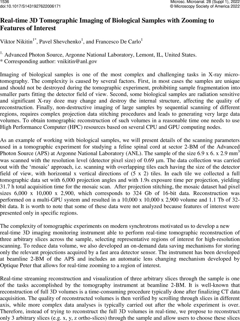
Image of the first page of this content. For PDF version, please use the ‘Save PDF’ preceeding this image.'
Type
From Images to Insights: Working With Large Multi-modal Data in Cell Biological Imaging
Copyright
Copyright © Microscopy Society of America 2022

References

Rivers, M, AIP Conference Proceedings 1234 (2010), p. 51-54. doi: 10.1063/1.3463256CrossRefGoogle Scholar
Tomopy, CLI, https://tomopycli.readthedocs.io/en/latest/ (accessed February 12, 2022).Google Scholar
Gursoy, D, Journal of Synchrotron Radiation, 21 (2014), p. 1188-1193. doi: 10.1107/S1600577514013939.CrossRefGoogle Scholar
Tomocupy, CLI, https://tomocupycli.readthedocs.io/en/latest/ (accessed February 12, 2022).Google Scholar
This research used resources of the Advanced Photon Source, a U.S. Department of Energy (DOE) Office of Science User Facility at Argonne National Laboratory and is based on research supported by the U.S. DOE Office of Science-Basic Energy Sciences, under Contract No. DE-AC02-06CH11357. The authors also acknowledge the group led by Dr. Chardon from the Department of Neuroscience at Northwestern University for collaborative work on the feline spinal cord imaging.Google Scholar