Hostname: page-component-78c5997874-ndw9j Total loading time: 0 Render date: 2024-11-17T03:23:07.246Z Has data issue: false hasContentIssue false

Rabbit Endothelial Progenitor Cells Derived From Peripheral Blood and Bone Marrow: An Ultrastructural Comparative Study

Published online by Cambridge University Press:  17 March 2022
Hana Duranova*
Affiliation:
AgroBioTech Research Centre, Slovak University of Agriculture, Tr. A. Hlinku 2, Nitra 94976, Slovak Republic
Veronika Valkova
Affiliation:
AgroBioTech Research Centre, Slovak University of Agriculture, Tr. A. Hlinku 2, Nitra 94976, Slovak Republic
Lucia Olexikova
Affiliation:
NPPC, Research Institute for Animal Production Nitra, Institute of Farm Animal Genetics and Reproduction, Hlohovecká 2, Lužianky 951 41, Slovak Republic
Barbora Radochova
Affiliation:
Laboratory of Biomathematics, Institute of Physiology, The Czech Academy of Sciences, Vídeňská 1083, Prague 4 CZ-14220, Czech Republic
Andrej Balazi
Affiliation:
NPPC, Research Institute for Animal Production Nitra, Institute of Farm Animal Genetics and Reproduction, Hlohovecká 2, Lužianky 951 41, Slovak Republic
Peter Chrenek
Affiliation:
NPPC, Research Institute for Animal Production Nitra, Institute of Farm Animal Genetics and Reproduction, Hlohovecká 2, Lužianky 951 41, Slovak Republic Faculty of Biotechnology and Food Science, Institute of Biotechnology, Slovak University of Agriculture, Tr. A. Hlinku 2, Nitra 94976, Slovak Republic
Jaromir Vasicek
Affiliation:
NPPC, Research Institute for Animal Production Nitra, Institute of Farm Animal Genetics and Reproduction, Hlohovecká 2, Lužianky 951 41, Slovak Republic Faculty of Biotechnology and Food Science, Institute of Biotechnology, Slovak University of Agriculture, Tr. A. Hlinku 2, Nitra 94976, Slovak Republic
*
*Corresponding author: Hana Duranova, E-mail: [email protected]
Get access

Abstract

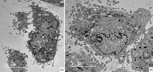
The present study was designed to compare the ultrastructure of early endothelial progenitor cells (EPCs) derived from rabbit peripheral blood (PB-EPCs) and bone marrow (BM-EPCs). After the cells had been isolated and cultivated up to passage 3, microphotographs obtained from transmission electron microscope were evaluated from qualitative and quantitative (unbiased stereological approaches) points of view. Our results revealed that both cell populations displayed almost identical ultrastructural characteristics represented by abundant cellular organelles dispersed in the cytoplasm. Moreover, the presence of very occasionally occurring mature endothelial-specific Weibel–Palade bodies (WPBs) confirmed their endothelial lineage origin. The more advanced stage of their differentiation was also demonstrated by the relatively low nucleus/cytoplasm (N/C) ratios (0.41 ± 0.19 in PB-EPCs; 0.37 ± 0.25 in BM-EPCs). Between PB-EPCs and BM-EPCs, no differences in proportions of cells occupied by nucleus (28.13 ± 8.97 versus 25.10 ± 11.48%), mitochondria (3.71 ± 1.33 versus 4.23 ± 1.00%), and lipid droplets (0.65 ± 1.01 versus 0.36 ± 0.40%), as well as in estimations of the organelles surface densities were found. The data provide the first quantitative evaluation of the organelles of interest in PB-EPCs and BM-EPCs, and they can serve as a research framework for understanding cellular function.

Type
Biological Applications
Copyright
Copyright © The Author(s), 2022. Published by Cambridge University Press on behalf of the Microscopy Society of America

Access options

Get access to the full version of this content by using one of the access options below. (Log in options will check for institutional or personal access. Content may require purchase if you do not have access.)

References

Alberts, B, Johnson, A, Lewis, J, Raff, M, Roberts, K & Walter, P (2002). Molecular Biology of the Cell, 4th ed. New York: Garland Science.Google Scholar
Amini, AR, Laurencin, CT & Nukavarapu, SP (2012). Differential analysis of peripheral blood-and bone marrow-derived endothelial progenitor cells for enhanced vascularization in bone tissue engineering. J Orthop Res 30, 15071515.CrossRefGoogle ScholarPubMed
Aon, MA, Bhatt, N & Cortassa, SC (2014). Mitochondrial and cellular mechanisms for managing lipid excess. Front Physiol 5, 282.CrossRefGoogle ScholarPubMed
Bai, C, Hou, L, Zhang, M, Pu, Y, Guan, W & Ma, Y (2012). Characterization of vascular endothelial progenitor cells from chicken bone marrow. BMC Vet Res 8, 54.CrossRefGoogle ScholarPubMed
Balbarini, A & Di Stefano, R (2008). Circulating endothelial progenitor cells – characterisation, function and relationship with cardiovascular risk factors. Eur Cardiol 4, 1619.CrossRefGoogle Scholar
Barber, CL & Iruela-Arispe, ML (2006). The ever-elusive endothelial progenitor cell: Identities, functions and clinical implications. Pediatr Res 59, 26R32R.CrossRefGoogle ScholarPubMed
Berezin, AE (2017). Preconditioned endothelial progenitor cells as biomarker of vascular reparation. Insights Biomed 2, 1.CrossRefGoogle Scholar
Bozzola, JJ & Russell, LD (1999). Electron Microscopy: Principles and Techniques for Biologists. Sudbury, MA: Jones & Bartlett Learning.Google Scholar
Broskey, NT, Daraspe, J, Humbel, BM & Amati, F (2013). Skeletal muscle mitochondrial and lipid droplet content assessed with standardized grid sizes for stereology. J Appl Physiol 115, 765770.CrossRefGoogle ScholarPubMed
Cabello, PDP, Fernandez, M, Chamorro, CA, Fernandez, JG & Villar, JM (1988). Stereological study of the early ultrastructural differentiation of chick embryo neuroepithelial cells during neurulation. Acta Anat 132, 1216.CrossRefGoogle Scholar
Čakić-Milošević, M, Koko, V, Davidović, VS & Radovanović, J (2004). Ultrastructural alterations of rat brown adipocytes after short-term corticosterone treatment. Acta Vet 54, 95104.Google Scholar
Chopra, HHMK, Hung, MK, Kwong, DL, Zhang, CF & Pow, EHN (2018). Insights into endothelial progenitor cells: Origin, classification, potentials, and prospects. Stem Cells Int 2018, 9847015.CrossRefGoogle ScholarPubMed
Cónsole, GM, Jurado, SB, Oyhenart, E, Ferese, C, Pucciarelli, H & Gómez Dumm, CLA (2001). Morphometric and ultrastructural analysis of different pituitary cell populations in undernourished monkeys. Braz J Med Biol Res 34, 6574.CrossRefGoogle ScholarPubMed
Cortassa, S, Sollott, SJ & Aon, MA (2017). Mitochondrial respiration and ROS emission during β-oxidation in the heart: An experimental-computational study. PLoS Comput Biol 13, e1005588.CrossRefGoogle Scholar
Crosier, AE, Farin, PW, Dykstra, MJ, Alexander, JE & Farin, CE (2000). Ultrastructural morphometry of bovine compact morulae produced in vivo or in vitro. Biol Reprod 62, 14591465.CrossRefGoogle ScholarPubMed
Dadarwal, D, Adams, GP, Hyttel, P, Brogliatti, GM, Caldwell, S & Singh, J (2015). Organelle reorganization in bovine oocytes during dominant follicle growth and regression. Reprod Biol Endocrinol 13, 124.CrossRefGoogle ScholarPubMed
de Assis, GF, Ribeiro, DA, Campos, PD, Cestari, TM & Taga, R (2003). Comparative stereologic study between secretory and maturation ameloblasts in rat incisors. J Appl Oral Sci 11, 144149.CrossRefGoogle Scholar
de Souza, DB, Costa, WS, Cardoso, LE, Benchimol, M, Pereira-Sampaio, MA & Sampaio, FJ (2013). Does prolonged pneumoperitoneum affect the kidney? Oxidative stress, stereological and electron microscopy study in a rat model. Int Braz J Urol 39, 3036.CrossRefGoogle Scholar
Donois, E, Surlève-Bazeille, , del Marmol, V & Ghanem, G (1998). Comparison of HPLC and stereologic image analysis for the quantitation of eu- and pheomelanins in nevus cells and stimulated melanoma cells. J Invest Dermatol 111, 422428.CrossRefGoogle ScholarPubMed
Duranova, H, Valkova, V, Knazicka, Z, Olexikova, L & Vasicek, J (2020). Mitochondria: A worthwhile object for ultrastructural qualitative characterization and quantification of cells at physiological and pathophysiological states using conventional transmission electron microscopy. Acta Histochem 122, 151646.CrossRefGoogle ScholarPubMed
Fang, X, Zhao, R, Wang, K, Li, Z, Yang, P, Huang, Q, Xu, Y, Hong, B & Liu, J (2012). Bone marrow-derived endothelial progenitor cells are involved in aneurysm repair in rabbits. J Clin Neurosci 19, 12831286.CrossRefGoogle ScholarPubMed
Fedorovich, NE, Haverslag, RT, Dhert, WJ & Alblas, J (2010). The role of endothelial progenitor cells in prevascularized bone tissue engineering: Development of heterogeneous constructs. Tissue Eng Part A 16, 23552367.CrossRefGoogle ScholarPubMed
Fujimoto, T, Ohsaki, Y, Suzuki, M & Cheng, J (2013). Imaging lipid droplets by electron microscopy. Methods Cell Biol 116, 227251.CrossRefGoogle ScholarPubMed
Gao, Q & Goodman, JM (2015). The lipid droplet – a well-connected organelle. Front Cell Dev Biol 3, 49.CrossRefGoogle ScholarPubMed
Garcia, Y, Breen, A, Burugapalli, K, Dockery, P & Pandit, A (2007). Stereological methods to assess tissue response for tissue-engineered scaffolds. Biomaterials 28, 175186.CrossRefGoogle ScholarPubMed
Gong, X, Li, B, Yang, Y, Huang, Y, Sun, Y, Liu, M, Jia, X & Fan, Y (2019). Bone marrow derived endothelial progenitor cells retain their phenotype and functions after a limited number of culture passages and cryopreservation. Cytotechnology 71, 114.CrossRefGoogle ScholarPubMed
Guan, XM, Cheng, M, Li, H, Cui, XD, Li, X, Wang, YL, Sun, JL & Zhang, XY (2013). Biological properties of bone marrow-derived early and late endothelial progenitor cells in different culture media. Mol Med Rep 8, 17221728.CrossRefGoogle ScholarPubMed
He, H, Shirota, T, Yasui, H & Matsuda, T (2003). Canine endothelial progenitor cell-lined hybrid vascular graft with nonthrombogenic potential. J Thorac Cardiovasc Surg 126, 455464.CrossRefGoogle ScholarPubMed
He, T, Smith, LA, Harrington, S, Nath, KA, Caplice, NM & Katusic, ZS (2004). Transplantation of circulating endothelial progenitor cells restores endothelial function of denuded rabbit carotid arteries. Stroke 35, 23782384.CrossRefGoogle ScholarPubMed
Howard, V & Reed, M (2005). Unbiased Stereology: Three-Dimensional Measurement in Microscopy, 2nd ed. Oxon: Garland Science/Bios Scientific Publishers.Google Scholar
Hur, J, Yoon, CH, Kim, HS, Choi, JH, Kang, HJ, Hwang, KK, Oh, BH, Lee, MM & Park, YB (2004). Characterization of two types of endothelial progenitor cells and their different contributions to neovasculogenesis. Arterioscler Thromb Vasc Biol 24, 288293.CrossRefGoogle ScholarPubMed
Kubínová, L & Kutík, J (2007). Surface density and volume density measurements of chloroplast thylakoids in maize (Zea mays L.) under chilling conditions. Photosynthetica 45, 481488.CrossRefGoogle Scholar
Kumar, S, Ciraolo, G, Hinge, A & Filippi, MD (2014). An efficient and reproducible process for transmission electron microscopy (TEM) of rare cell populations. J Immunol Methods 404, 8790.CrossRefGoogle ScholarPubMed
Lica, JJ, Wieczór, M, Grabe, GJ, Heldt, M, Jancz, M, Misiak, M, Gucwa, K, Brankiewicz, W, Maciejewska, N, Stupak, A, Bagiński, M, Rolka, K, Hellmann, A & Składanowski, A (2021). Effective drug concentration and selectivity depends on fraction of primitive cells. Int J Mol Sci 22, 4931.CrossRefGoogle ScholarPubMed
Lindal, S, Gunnes, S, Lund, I, Straume, BK, Jørgensen, L & Sørlie, D (1990). Ultrastructural changes in rat hearts following cold cardioplegic ischemia of differing duration and differing modes of reperfusion. Scand J Thorac Cardiovasc Surg 24, 213222.CrossRefGoogle ScholarPubMed
Loomans, CJM, Wan, H, de Crom, R, van Haperen, R, de Boer, HC, Leenen, PJM, Drexhage, HA, Rabelink, TJ, van Zonneveld, AJ & Staal, FJT (2006). Angiogenic murine endothelial progenitor cells are derived from a myeloid bone marrow fraction and can be identified by endothelial NO synthase expression. Arterioscler Thromb Vasc Biol 26, 17601767.CrossRefGoogle ScholarPubMed
Lopes-Coelho, F, Silva, F, Gouveia-Fernandes, S, Martins, C, Lopes, N, Domingues, G, Brito, C, Almeida, AM, Pereira, SA & Serpa, J (2020). Monocytes as endothelial progenitor cells (EPCs), another brick in the wall to disentangle tumor angiogenesis. Cells 9, 107.CrossRefGoogle Scholar
Luo, T, Shu, J, Lu, Z, Han, T, Fang, G & Xue, X (2019). Potential role of caveolin-1 in regulating the function of endothelial progenitor cells from experimental MODS model. Mediators Inflamm 2019, 8297391.CrossRefGoogle ScholarPubMed
Luo, TH, Wang, Y, Lu, ZM, Zhou, H, Xue, XC, Bi, JW, Ma, LY & Fang, GE (2009). The change and effect of endothelial progenitor cells in pig with multiple organ dysfunction syndromes. Crit Care 13, R118.CrossRefGoogle ScholarPubMed
Ma, ZL, Mai, XL, Sun, JH, Ju, SH, Yang, X, Ni, Y & Teng, GJ (2009). Inhibited atherosclerotic plaque formation by local administration of magnetically labeled endothelial progenitor cells (EPCs) in a rabbit model. Atherosclerosis 205, 8086.CrossRefGoogle Scholar
Maeshima, K, Iino, H, Hihara, S & Imamoto, N (2011). Nuclear size, nuclear pore number and cell cycle. Nucleus 2, 113118.CrossRefGoogle ScholarPubMed
Magwere, T, Goodall, S, Skepper, J, Mair, W, Brand, MD & Partridge, L (2006). The effect of dietary restriction on mitochondrial protein density and flight muscle mitochondrial morphology in Drosophila. J Gerontol A Biol Sci Med Sci 61, 3647.CrossRefGoogle ScholarPubMed
Mai, XL, Ma, ZL, Sun, JH, Ju, SH, Ma, M & Teng, GJ (2009). Assessments of proliferation capacity and viability of New Zealand rabbit peripheral blood endothelial progenitor cells labeled with superparamagnetic particles. Cell Transplant 18, 171181.CrossRefGoogle ScholarPubMed
Malik, D & Kaul, D (2018). Human cellular mitochondrial remodelling is governed by miR-2909 RNomics. PLoS One 13, e0203614.CrossRefGoogle ScholarPubMed
Mandarim-de-Lacerda, CA (2003). Stereological tools in biomedical research. An Acad Bras Cienc 75, 469486.CrossRefGoogle ScholarPubMed
Maul, GG, Deaven, LL, Freed, JJ, Campbell, LM & Becak, W (1980). Investigation of the determinants of nuclear pore number. Cytogenet Cell Genet 26, 175190.CrossRefGoogle ScholarPubMed
Medina, RJ, O'Neill, CL, Sweeney, M, Guduric-Fuchs, J, Gardiner, TA, Simpson, DA & Stitt, AW (2010). Molecular analysis of endothelial progenitor cell (EPC) subtypes reveals two distinct cell populations with different identities. BMC Med Genomics 3, 18.CrossRefGoogle ScholarPubMed
Meinild Lundby, AK, Jacobs, RA, Gehrig, S, de Leur, J, Hauser, M, Bonne, TC, Flück, D, Dandanell, S, Kirk, N, Kaech, A, Ziegler, U, Larsen, S & Lundby, C (2018). Exercise training increases skeletal muscle mitochondrial volume density by enlargement of existing mitochondria and not de novo biogenesis. Acta Physiol 222, e12905.CrossRefGoogle Scholar
Neumüller, J, Neumüller-Guber, SE, Lipovac, M, Mosgoeller, W, Vetterlein, M, Pavelka, M & Huber, J (2006). Immunological and ultrastructural characterization of endothelial cell cultures differentiated from human cord blood derived endothelial progenitor cells. Histochem Cell Biol 126, 649664.CrossRefGoogle ScholarPubMed
Nova-Lamperti, E, Zúñiga, F, Ormazábal, V, Escudero, C & Aguayo, C (2016). Vascular regeneration by endothelial progenitor cells in health and diseases. In Microcirculation Revisited: From Molecules to Clinical Practice, Lenasi, H (Ed.), pp. 231258. Rijeka, Croatia: IntechOpen.Google Scholar
Novotová, M, Pavlovicová, M, Veksler, VI, Ventura-Clapier, R & Zahradník, I (2006). Ultrastructural remodeling of fast skeletal muscle fibers induced by invalidation of creatine kinase. Am J Physiol Cell Physiol 291, C1279C1285.CrossRefGoogle ScholarPubMed
Novotová, M, Tarabová, B, Tylková, L, Ventura-Clapier, R & Zahradník, I (2016). Ultrastructural remodelling of slow skeletal muscle fibres in creatine kinase deficient mice: A quantitative study. Gen Physiol Biophys 35, 477486.CrossRefGoogle ScholarPubMed
Olexikova, L, Makarevich, AV, Pivko, J & Chrenek, P (2013). Ultrastructure of rabbit embryos exposed to hyperthermia and anti-Hsp 70. Anat Histol Embryol 42, 285291.CrossRefGoogle ScholarPubMed
Ørtenblad, N (2018). Mitochondrial increase in volume density with exercise training: More, larger or better? Acta Physiol 222, e12976.CrossRefGoogle ScholarPubMed
Ozkok, A & Yildiz, A (2018). Endothelial progenitor cells and kidney diseases. Kidney Blood Press Res 43, 701718.CrossRefGoogle ScholarPubMed
Paine, PL & Horowitz, SB (1980). The movement of material between nucleus and cytoplasm. In Cell Biology: A Comprehensive Treatise V4: Gene Expression: Translation and the Behavior of Proteins, Prescott, DM & Goldstein, L (Eds.), pp. 299339. New York: Academic Press.CrossRefGoogle Scholar
Pasquinelli, G, Vinci, MC, Gamberini, C, Orrico, C, Foroni, L, Guarnieri, C, Parenti, A, Gargiulo, M, Ledda, F, Caldarera, CM & Muscari, C (2009). Architectural organization and functional features of early endothelial progenitor cells cultured in a hyaluronan-based polymer scaffold. Tissue Eng Part A 15, 27512762.CrossRefGoogle Scholar
Pavelka, M & Roth, J (2015). Functional Ultrastructure: Atlas of Tissue Biology and Pathology. Wien: Springer.Google Scholar
Pires-Luís, AS, Rocha, E, Bartosch, C, Oliveira, E, Silva, J, Barros, A, , R & Sousa, M (2016). A stereological study on organelle distribution in human oocytes at prophase I. Zygote 24, 346354.CrossRefGoogle Scholar
Romek, M & Krzysztofowicz, E (2005). Stereological analysis of mitochondria in embryos of Rana temporaria and Bufo bufo during cleavage. Folia Histochem Cytobiol 43, 5763.Google ScholarPubMed
Rozen, N, Bick, T, Bajayo, A, Shamian, B, Schrift-Tzadok, M, Gabet, Y, Yayon, A, Bab, I, Soudry, M & Lewinson, D (2009). Transplanted blood-derived endothelial progenitor cells (EPC) enhance bridging of sheep tibia critical size defects. Bone 45, 918924.CrossRefGoogle ScholarPubMed
Seemann, I, te Poele, JA, Hoving, S & Stewart, FA (2014). Mouse bone marrow-derived endothelial progenitor cells do not restore radiation-induced microvascular damage. ISRN Cardiol 2014, 506348.Google Scholar
Tomaiuolo, M, Litvinov, RI, Weisel, JW & Stalker, TJ (2020). Use of electron microscopy to study platelets and thrombi. Platelets 31, 580588.CrossRefGoogle ScholarPubMed
Turgeon, ML (2005). Clinical Hematology: Theory and Procedures, 4th ed. Philadelphia, PA: Lippincott Williams & Wilkins.Google Scholar
Vaickus, LJ & Tambouret, RH (2015). Young investigator challenge: The accuracy of the nuclear-to-cytoplasmic ratio estimation among trained morphologists. Cancer Cytopathol 123, 524530.CrossRefGoogle ScholarPubMed
Vašíček, J, Baláži, A, Bauer, M, Svoradová, A, Tirpáková, M, Tomka, M & Chrenek, P (2021). Molecular profiling and gene banking of rabbit EPCs derived from two biological sources. Genes 12, 366.CrossRefGoogle ScholarPubMed
Vašíček, J, Kováč, M, Baláži, A, Kulíková, B, Tomková, M, Olexiková, L, Čurlej, J, Bauer, M, Schnabl, S, Hilgarth, M, Hubmann, R, Shehata, M, Makarevich, AV & Chrenek, P (2020). Combined approach for characterization and quality assessment of rabbit bone marrow-derived mesenchymal stem cells intended for gene banking. N Biotechnol 54, 112.CrossRefGoogle ScholarPubMed
Vašíček, J, Shehata, M, Schnabl, S, Hilgarth, M, Hubmann, R, Jäger, U, Bauer, M & Chrenek, P (2018). Critical assessment of the efficiency of CD34 and CD133 antibodies for enrichment of rabbit hematopoietic stem cells. Biotechnol Prog 34, 12781289.CrossRefGoogle ScholarPubMed
Wang, HH, Wu, YJ, Tseng, YM, Su, CH, Hsieh, CL & Yeh, HI (2019). Mitochondrial fission protein 1 up-regulation ameliorates senescence-related endothelial dysfunction of human endothelial progenitor cells. Angiogenesis 22, 569582.CrossRefGoogle ScholarPubMed
Wang, Q, Zhang, W, He, G, Sha, H & Quan, Z (2016). Method for in vitro differentiation of bone marrow mesenchymal stem cells into endothelial progenitor cells and vascular endothelial cells. Mol Med Rep 14, 55515555.CrossRefGoogle ScholarPubMed
Wei, H, Zhao, X, Yuan, R, Dai, X, Li, Y & Liu, L (2015 b). Effects of PB-EPCs on homing ability of rabbit BMSCs via endogenous SDF-1 and MCP-1. PLoS One 10, e0145044.CrossRefGoogle ScholarPubMed
Wei, MQ, Wen, DD, Wang, XY, Huan, Y, Yang, Y, Xu, J, Cheng, K & Zheng, MW (2015 a). Experimental study of endothelial progenitor cells labeled with superparamagnetic iron oxide in vitro. Mol Med Rep 11, 38143819.CrossRefGoogle ScholarPubMed
White, FH & Gohari, K (1981). Variations in the nuclear-cytoplasmic ratio during epithelial differentiation in experimental oral carcinogenesis. J Oral Pathol 10, 164172.CrossRefGoogle ScholarPubMed
White, FH, Jin, Y & Yang, L (1997). An evaluation of the role of nuclear cytoplasmic ratios and nuclear volume densities as diagnostic indicators in metaplastic, dysplastic and neoplastic lesions of the human cheek. Histol Histopathol 12, 6977.Google ScholarPubMed
Yang, N, Li, D, Jiao, P, Chen, B, Yao, S, Sang, H, Yang, M, Han, J, Zhang, Y & Qin, S (2011). The characteristics of endothelial progenitor cells derived from mononuclear cells of rat bone marrow in different culture conditions. Cytotechnology 63, 217226.CrossRefGoogle ScholarPubMed
Zhao, H, Perkins, G, Yao, H, Callacondo, D, Appenzeller, O, Ellisman, M, la Spada, AR & Haddad, GG (2018). Mitochondrial dysfunction in IPSC-derived neurons of subjects with chronic mountain sickness. J Appl Physiol 125, 832840.CrossRefGoogle ScholarPubMed
Zhu, C (2014). Endothelial progenitor cell (EPC)-seeded intravascular stents. In Cardiac Regeneration and Repair: Biomaterials and Tissue Engineering, Li, RK & Weisel, RD (Eds.), pp. 110124. Cambridge: Woodhead Publishing.CrossRefGoogle Scholar
Zielińska, KA, Grealy, M & Dockery, P (2020). A stereological study of developmental changes in hepatocyte ultrastructure of zebrafish (Danio rerio). J Anat 236, 9961003.CrossRefGoogle Scholar