Hostname: page-component-586b7cd67f-vdxz6 Total loading time: 0 Render date: 2024-11-28T07:53:48.348Z Has data issue: false hasContentIssue false

Nondestructive Materials Characterization in 3D by Laboratory Diffraction Contrast Tomography – Applications and Future Directions

Published online by Cambridge University Press:  25 July 2016

C. Holzner
Affiliation:
Carl Zeiss X-ray Microscopy, Inc., Pleasanton, CAUSA
L. Lavery
Affiliation:
Carl Zeiss X-ray Microscopy, Inc., Pleasanton, CAUSA
H. Bale
Affiliation:
Carl Zeiss X-ray Microscopy, Inc., Pleasanton, CAUSA
A. Merkle
Affiliation:
Carl Zeiss X-ray Microscopy, Inc., Pleasanton, CAUSA
S. McDonald
Affiliation:
Manchester X-ray Imaging Facility, School of Materials, University of Manchester, Manchester M13 9PL, UK
P. Withers
Affiliation:
Manchester X-ray Imaging Facility, School of Materials, University of Manchester, Manchester M13 9PL, UK
Y. Zhang
Affiliation:
Department of Wind Energy, Technical University of Denmark, Risø Campus, 4000 Roskilde, Denmark
D. Juul Jensen
Affiliation:
Department of Wind Energy, Technical University of Denmark, Risø Campus, 4000 Roskilde, Denmark
P. Reischig
Affiliation:
Xnovo Technology ApS, 4600, Køge, Denmark
E. Lauridsen
Affiliation:
Xnovo Technology ApS, 4600, Køge, Denmark

Abstract

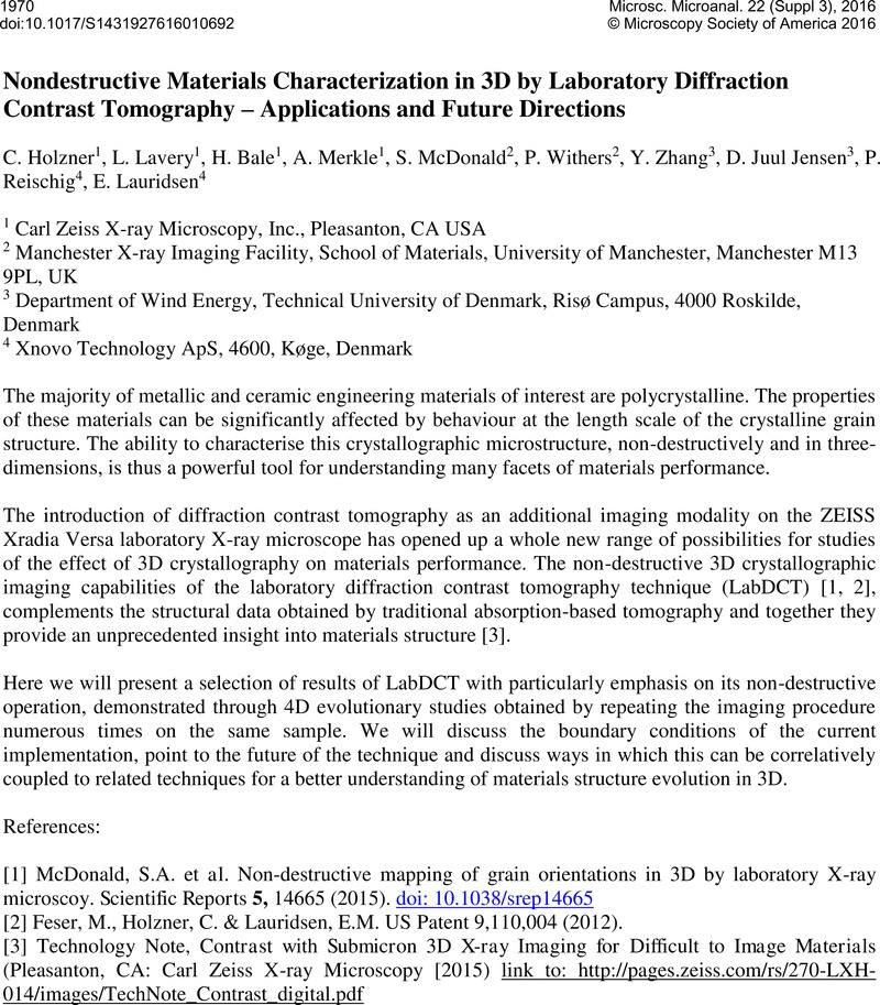
Image of the first page of this content. For PDF version, please use the ‘Save PDF’ preceeding this image.'
Type
Abstract
Copyright
© Microscopy Society of America 2016 

References

References:

[1] McDonald, S.A., et al., Non-destructive mapping of grain orientations in 3D by laboratory X-ray microscoy. Scientific Reports 5, 14665 (2015) doi: 10.1038/srep14665.Google Scholar
[2] Feser, M., Holzner, C. & Lauridsen, E.M. US Patent 9,110,004 (2012).Google Scholar
[3] Technology Note, Contrast with Submicron 3D X-ray Imaging for Difficult to Image Materials (Pleasanton, CA: Carl Zeiss X-ray Microscopy [2015) link to: http://pages.zeiss.com/rs/270-LXH-014/images/TechNote_Contrast_digital.pdf.Google Scholar