Hostname: page-component-745bb68f8f-b6zl4 Total loading time: 0 Render date: 2025-01-07T18:36:52.871Z Has data issue: false hasContentIssue false

Microwave Brain Tissue Processing: Making Life Easier for Ultrastructural Immunohistochemical Analysis of Synapses in Rodent Models of Neurodegeneration

Published online by Cambridge University Press:  22 July 2022

Cynthia Moore
Affiliation:
Neurocytology Lab, VA Medical Center/Portland, Oregon, USA
Charles K. Meshul*
Affiliation:
Neurocytology Lab, VA Medical Center/Portland, Oregon, USA Department of Behavioral Neuroscience and Pathology, Oregon Health & Science University, Portland, OR, USA
*
*Corresponding author: [email protected]

Abstract

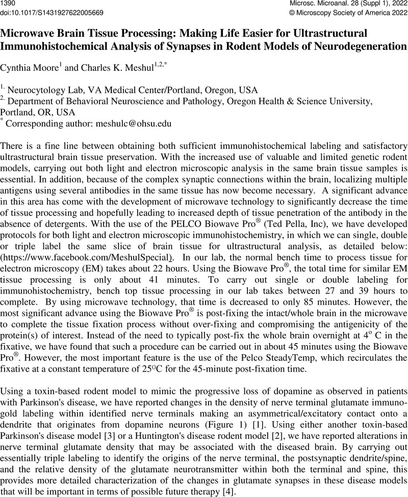
Image of the first page of this content. For PDF version, please use the ‘Save PDF’ preceeding this image.'
Type
Challenges and Advances in Electron Microscopy Research and Diagnosis of Diseases in Humans, Plants and Animals
Copyright
Copyright © Microscopy Society of America 2022

References

Moore, C, Xu, M, Bohlen, JK, and Meshul, CK. European Journal of Neuroscience, 53, (2021), 20612077. doi: 10.1111/ejn.14894CrossRefGoogle Scholar
Parievsky, A, Moore, C, Kamdjou, T, Cepeda, C, Meshul, CK, and Levine, MS. Neurobiology of Disease, 108, (2017), 29-44. doi: 10.1016/j.nbd.2017.07.020.CrossRefGoogle Scholar
R Walker, H, Moore, C, Davies, G, Dirling, LB, Koch, RJ, and Meshul, CK. PLoS One, 7(3):e32919 (2012). doi: 10.1371/journal.pone.0032919.Google Scholar
The authors acknowledge funding from the Department of Veterans Affairs Merit Review Program. A very personal thanks to Ted Pella, a true gentleman, and his company for their continued support of not only my lab but of our local Pacific Northwest Microscopy Society. He is truly missed but his legacy continues.Google Scholar