No CrossRef data available.
Article contents
Imaging Sub-cellular 3D Structures Using Soft X-ray Microscopy
Published online by Cambridge University Press: 30 July 2020
Abstract
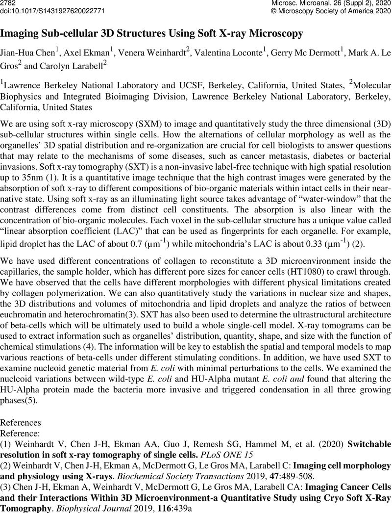
An abstract is not available for this content so a preview has been provided. As you have access to this content, a full PDF is available via the ‘Save PDF’ action button.

- Type
- Biological Soft X-ray Tomography
- Information
- Copyright
- Copyright © Microscopy Society of America 2020
References
Weinhardt, V, Chen, J-H, Ekman, AA, Guo, J, Remesh, SG, Hammel, M, et al. (2020) Switchable resolution in soft x-ray tomography of single cells . PLoS ONE 15 10.1371/journal.pone.0227601CrossRefGoogle ScholarPubMed
Weinhardt, V, Chen, J-H, Ekman, A, McDermott, G, Le Gros, MA, Larabell, C: Imaging cell morphology and physiology using X-rays. Biochemical Society Transactions 2019, 47:489–508.10.1042/BST20180036CrossRefGoogle ScholarPubMed
Chen, J-H, Ekman, A, Weinhardt, V, McDermott, G, Le Gros, MA, Larabell, CA: Imaging Cancer Cells and their Interactions Within 3D Microenvironment-a Quantitative Study using Cryo Soft X-Ray Tomography. Biophysical Journal 2019, 116:439a10.1016/j.bpj.2018.11.2364CrossRefGoogle Scholar
Singla, J, McClary, KM, White, KL, Alber, F, Sali, A, Stevens RC: Opportunities and challenges in building a spatiotemporal multi-scale model of the human pancreatic beta-cell. Cell (2018) 173(1):11–19.10.1016/j.cell.2018.03.014CrossRefGoogle ScholarPubMed
Hammel, M, Amlanjyoti, D, Reyes, FE, Chen, J-H, Parpana, R, Tang, HY, Larabell, CA, Tainer, JA, Adhya, S: HU multimerization shift controls nucleoid compaction. Science advances 2016, 2:e1600650.10.1126/sciadv.1600650CrossRefGoogle ScholarPubMed


