Hostname: page-component-cd9895bd7-dk4vv Total loading time: 0 Render date: 2024-12-31T23:51:30.577Z Has data issue: false hasContentIssue false

FE-SEM Characterization of α-Mannose Density and Surface Mapping Changes in Human Sperm Head During In Vitro Capacitation

Published online by Cambridge University Press:  30 October 2020
María José Gómez-Torres*
Affiliation:
Departamento de Biotecnología, Facultad de Ciencias, Universidad de Alicante, Ap. C. 99, Alicante 03080, Spain Cátedra Human Fertility, Universidad de Alicante, Alicante, Spain
Laura Robles-Gómez
Affiliation:
Departamento de Biotecnología, Facultad de Ciencias, Universidad de Alicante, Ap. C. 99, Alicante 03080, Spain
Natalia Huerta-Retamal
Affiliation:
Departamento de Biotecnología, Facultad de Ciencias, Universidad de Alicante, Ap. C. 99, Alicante 03080, Spain
Paula Sáez-Espinosa
Affiliation:
Departamento de Biotecnología, Facultad de Ciencias, Universidad de Alicante, Ap. C. 99, Alicante 03080, Spain
Manuel Avilés
Affiliation:
Departamento de Biología Celular e Histología, Facultad de Medicina, Universidad de Murcia e IMIB-Arrixaca, Murcia, Spain
Jon Aizpurua
Affiliation:
IVF Spain, Reproductive Medicine, Alicante, Spain
Alejandro Romero
Affiliation:
Departamento de Biotecnología, Facultad de Ciencias, Universidad de Alicante, Ap. C. 99, Alicante 03080, Spain
*
*Author for correspondence: María José Gómez-Torres, E-mail: [email protected]
Get access

Abstract

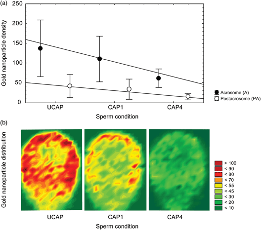
Sperm capacitation includes the reorganization of plasma membrane components and the outstanding modification of the glycocalyx. The α-mannose presence and location during in vitro capacitation have been commonly described in human spermatozoa using Concanavalin A (Con A) lectin. However, it is still unclear to date how in vitro capacitation time affects the α-mannose residues and their topographic spatial distribution on sperm membrane. Here, we characterized the α-mannose density and specific membrane domain locations before and after in vitro capacitation (1–4 h) using high-resolution field emission scanning electron microscopy (FE-SEM). Results showed that α-mannose residues were present preferably on the acrosome domains for all physiological conditions. Uncapacitated sperm comparatively exhibits significant highest labeling densities of α-mannose residues. In addition, as in vitro capacitation takes place, significant and progressive decreasing of sugar residues was combined with their relocation mostly affecting acrosomal domain apical areas. Our findings reveal that combined approach using FE-SEM and gold nanoparticle topographical mapping offers new human sperm biomolecular and structural details during capacitation events.

Type
Biological Applications
Copyright
Copyright © The Author(s), 2020. Published by Cambridge University Press

Access options

Get access to the full version of this content by using one of the access options below. (Log in options will check for institutional or personal access. Content may require purchase if you do not have access.)

Footnotes

a

M. J. Gómez-Torres and L. Robles-Gómez contributed equally as co-first authors.

References

Baibakov, B, Boggs, NA, Yauger, B, Baibakov, G & Dean, J (2012). Human sperm bind to the N-terminal domain of ZP2 in humanized zonae pellucidae in transgenic mice. J Cell Biol 197, 897905.CrossRefGoogle Scholar
Bains, H, Sehgal, S & Bawa, SR (1992). Human sperm surface mapping with lectins. Acta Anat (Basel) 145, 207211.CrossRefGoogle ScholarPubMed
Baker, SS, Thomas, M & Thaler, CD (2004). Sperm membrane dynamics assessed by changes in lectin fluorescence before and after capacitation. J Androl 25, 744751.CrossRefGoogle ScholarPubMed
Bernabó, N, Ramal-Sánchez, M, Valbonetti, L, Greco, L, Capacchietti, G, Mattioli, M & Barboni, B (2018). Membrane dynamics of spermatozoa during capacitation: New insight in germ cells signalling. In Germ Cell, pp. pp. 73100. London, UK: IntechOpen.Google Scholar
Boerke, A, Tsai, PS, García-Gil, N, Brewis, IA & Gadella, BM (2008). Capacitation-dependent reorganization of microdomains in the apical sperm head plasma membrane: Functional relationship with zona binding and the zona-induced acrosome reaction. Theriogenology 70, 11881196.CrossRefGoogle ScholarPubMed
Brewer, F, Bhattacharyya, L, Brown, RD 3rd & Koenig, SH (1985). Interactions of Concanavalin A with a trimannosyl oligosaccharide fragment of complex and high mannose type glycopeptides. Biochem Biophys Res Commun 127, 10661071.CrossRefGoogle ScholarPubMed
Clark, GF (2013). The role of carbohydrate recognition during human sperm-egg binding. Hum Reprod 28, 566577.CrossRefGoogle ScholarPubMed
Cross, NL (2004). Reorganization of lipid rafts during capacitation of human sperm. Biol Reprod 71, 13671373.CrossRefGoogle ScholarPubMed
Cross, NL & Overstreet, JW (1987). Glycoconjugates of the human sperm surface: Distribution and alterations that accompany capacitation in vitro. Gamete Res 16, 2325.CrossRefGoogle ScholarPubMed
De Jonge, C (2017). Biological basis for human capacitation revisited. Hum Reprod Update 23, 289299.Google ScholarPubMed
De Souza, W, Campanati, L & Attias, M (2008). Strategies and results of field emission scanning electron microscopy (FE-SEM) in the study of parasitic protozoa. Micron 39, 7787.CrossRefGoogle Scholar
Diekman, AB (2003). Glycoconjugates in sperm function and gamete interactions: How much sugar does it take to sweet-talk the egg? Cell Mol Life Sci 60, 298308.CrossRefGoogle ScholarPubMed
Gadella, BM & Boerke, A (2016). An update on post-ejaculatory remodeling of the sperm surface before mammalian fertilization. Theriogenology 85, 113124.CrossRefGoogle ScholarPubMed
Gadella, BM & Van Gestel, RA (2004). Bicarbonate and its role in mammalian sperm function. Anim Reprod Sci 82–83, 307319.CrossRefGoogle ScholarPubMed
Goldberg, MW (2008). Immunolabelling for scanning electron microscopy (SEM) and field emission SEM. Method Cell Biol 88, 109130.CrossRefGoogle ScholarPubMed
Goldberg, MW & Fiserová, J (2016). Immunogold labeling for scanning electron microscopy. In High-Resolution Imaging of Cellular Proteins. Methods in Molecular Biology, vol. 1474, Schwartzbach, S, Skalli, O & Schikorski, T (Eds.), pp. 309325. New York, NY: Humana Press.Google Scholar
Gómez-Torres, MJ, Avilés, M, Girela, JL, Murcia, V, Fernández-Colom, PJ, Romeu, A & De Juan, J (2012). Characterization of the lectin binding pattern in human spermatozoa after swim-up selection. Histol Histopathol 27, 16211628.Google ScholarPubMed
Gordon, M & Dandekar, PV (1976). Electron microscope assessment of fertilization of rabbit ova treated with Concanavalin A and Wheat germ agglutinin. J Exp Zool 198, 437442.CrossRefGoogle ScholarPubMed
Gupta, SK (2015). Role of zona pellucida glycoproteins during fertilization in humans. J Reprod Immunol 108, 9097.CrossRefGoogle ScholarPubMed
Gupta, SK, Bhandari, B, Shrestha, A, Biswal, BK, Palaniappan, C, Malhotra, SS & Gupta, N (2012). Mammalian zona pellucida glycoproteins: Structure and function during fertilization. Cell Tissue Res 349, 665678.CrossRefGoogle ScholarPubMed
Jiménez, I, González-Márquez, H, Ortiz, R, Herrera, JA, García, A, Betancourt, M & Fierro, R (2003). Changes in the distribution of lectin receptors during capacitation and acrosome reaction in boar spermatozoa. Theriogenology 59, 11711180.CrossRefGoogle ScholarPubMed
Kinsey, WH & Koehler, JK (1978). Cell surface changes associated with in vitro capacitation of hamster sperm. J Ultrastruct Res 64, 113.CrossRefGoogle ScholarPubMed
Lewin, LM, Weissenberg, R, Sobel, JS, Marcus, Z & Nebel, L (1979). Differences in Concanavalin A-FITC binding to rat spermatozoa during epididymal maturation and capacitation. Syst Biol Reprod Med 2, 279281.Google ScholarPubMed
Liu, M (2016). Capacitation-associated glycocomponents of mammalian sperm. Reprod Sci 23, 572594.CrossRefGoogle ScholarPubMed
Monné, M & Jovine, L (2011). A structural view of egg coat architecture and function in fertilization. Biol Reprod 85, 661669.CrossRefGoogle ScholarPubMed
Pang, PC, Tissot, B, Drobnis, EZ, Sutovsky, P, Morris, HR, Clark, GF & Dell, A (2007). Expression of bisecting type and Lewis x/Lewis y terminated N-glycans on human sperm. J Biol Chem 282, 3659336602.CrossRefGoogle Scholar
Runnebaum, IB, Schill, WB & Topfer-Petersen, E (1995). ConA-binding proteins of the sperm surface are conserved through evolution and in sperm maturation. Andrologia 27, 8190.CrossRefGoogle ScholarPubMed
Sáez-Espinosa, P, Huerta-Retamal, N, Robles-Gómez, L, Avilés, M, Aizpurua, J, Velasco, I, Romero, A & Gómez-Torres, MJ (2019). Influence of in vitro capacitation time on structural and functional human sperm parameters. Asian J Androl 21, 17.Google Scholar
Schwarz, MA & Koehler, JK (1979). Alterations in lectin binding to guinea pig spermatozoa accompanying in vitro capacitation and the acrosome reaction. Biol Reprod 21, 12951307.CrossRefGoogle ScholarPubMed
Sosa, CM, Pavarotti, MA, Zanetti, MN, Zoppino, FCM, De Blas, GA & Mayorga, LS (2015). Kinetics of human sperm acrosomal exocytosis. Mol Hum Reprod 21, 244254.CrossRefGoogle ScholarPubMed
Stoffel, MH, Busato, A & Friess, AE (2002). Density and distribution of anionic sites on boar ejaculated and epididymal spermatozoa. Histochem Cell Biol 117, 441445.CrossRefGoogle ScholarPubMed
Suárez, SS (2008). Regulation of sperm storage and movement in the mammalian oviduct. Int J Dev Biol 52, 455462.CrossRefGoogle ScholarPubMed
Tecle, E & Gagneux, P (2015). Sugar-coated sperm: Unraveling the functions of the mammalian sperm glycocalyx. Mol Reprod Dev 82, 635650.CrossRefGoogle ScholarPubMed
Vázquez, JM, Martínez, E, Pastor, LM, Roca, J, Matas, C & Calvo, A (1996). Lectin histochemistry during in vitro capacitation and acrosome reaction in boar spermatozoa: New lectins for evaluating acrosomal status of boar spermatozoa. Acta Histochem 98, 93100.CrossRefGoogle ScholarPubMed
World Health Organization (2010). WHO Laboratory Manual for the Examination and Processing of Human Semen. Geneva: World Health Organization.Google Scholar
Supplementary material: File

Gómez-Torres et al. supplementary material

Gómez-Torres et al. supplementary material 1

Download Gómez-Torres et al. supplementary material(File)
File 14.3 KB
Supplementary material: Image

Gómez-Torres et al. supplementary material

Gómez-Torres et al. supplementary material 2

Download Gómez-Torres et al. supplementary material(Image)
Image 4 MB
Supplementary material: Image

Gómez-Torres et al. supplementary material

Gómez-Torres et al. supplementary material 3

Download Gómez-Torres et al. supplementary material(Image)
Image 311.3 KB