Hostname: page-component-586b7cd67f-gb8f7 Total loading time: 0 Render date: 2024-11-28T15:47:05.471Z Has data issue: false hasContentIssue false

Electron- microscopy characterization of cells infected with a new phlebovirus isolated in sandflies from South Portugal

Published online by Cambridge University Press:  10 September 2015

F. Amaro*
Affiliation:
Centro de Estudos de Vectores e Doenças Infecciosas/Instituto Nacional de Saúde Dr. Ricardo Jorge, Avenida da Liberdade n.º 5, 2965-575 Águas de Moura, PORTUGAL
L. Zé-Zé
Affiliation:
Centro de Estudos de Vectores e Doenças Infecciosas/Instituto Nacional de Saúde Dr. Ricardo Jorge, Avenida da Liberdade n.º 5, 2965-575 Águas de Moura, PORTUGAL
M. J. Alves
Affiliation:
Centro de Estudos de Vectores e Doenças Infecciosas/Instituto Nacional de Saúde Dr. Ricardo Jorge, Avenida da Liberdade n.º 5, 2965-575 Águas de Moura, PORTUGAL
A. P. Alves de Matos
Affiliation:
Cooperativa de Ensino Superior - Egas Moniz, Campus Universitário, Quinta da Granja, 2829- 511 Monte de Caparica, Almada, PORTUGAL

Abstract

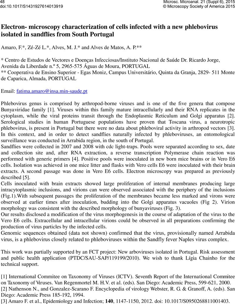
Image of the first page of this content. For PDF version, please use the ‘Save PDF’ preceeding this image.'
Type
Life Sciences
Copyright
Copyright © Microscopy Society of America 2015 

References

[1]International Commitee on Taxonomy of Viruses (ICTV). Seventh Report of the International Commitee on Taxonomy of Viruses, Van Regenmortel, M. H.V.et al. (eds). San Diego: Academic Press, 599621, 2000.Google Scholar
[2]Nathenson, N. & Gonzalez-Scarano, F.Encyclopedia of virology, Webster, R. G. & Granoff, A. (eds) San Diego: Academic Press 185192, 1994.Google Scholar
[3]Amaro, F., et al, Epidemiology and Infection 140, 11471150, 2012. doi: 10.1017/S0950268811001403.CrossRefGoogle Scholar
[4]Sánchez-Seco, M. P., et al, Jounal of 21Medical Virology 71, 140149, 2003.Google Scholar
[5]Alves de Matos, A.P., et al, Microscopy and Microanalysis 17, 101108, 2011.CrossRefGoogle Scholar