Hostname: page-component-cd9895bd7-gxg78 Total loading time: 0 Render date: 2024-12-25T06:53:04.123Z Has data issue: false hasContentIssue false

Automatic Diffraction Analysis and Lattice Fitting for Convergent-Beam Electron Diffraction Patterns in 4D-STEM

Published online by Cambridge University Press:  30 July 2021

Sihan Wang
Affiliation:
North Carolina State University, North Carolina, United States
Tim Eldred
Affiliation:
North Carolina State University, United States
Jacob Smith
Affiliation:
North Carolina State University, United States
Wenpei Gao
Affiliation:
North Carolina State University, United States

Abstract

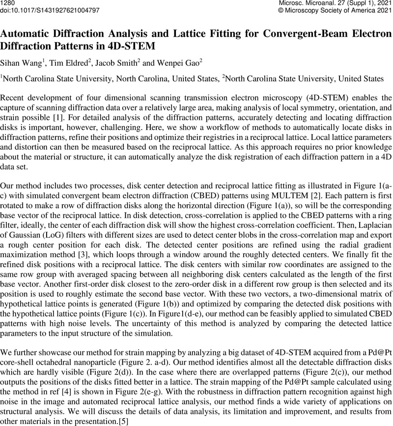
Image of the first page of this content. For PDF version, please use the ‘Save PDF’ preceeding this image.'
Type
Diffraction Imaging Across Disciplines
Copyright
Copyright © The Author(s), 2021. Published by Cambridge University Press on behalf of the Microscopy Society of America

References

Ophus, C., Four-Dimensional Scanning Transmission Electron Microscopy (4D-STEM): From Scanning Nanodiffraction to Ptychography and Beyond, Microsc Microanal. 25 (2019) 563582. https://doi.org/10.1017/S1431927619000497.CrossRefGoogle ScholarPubMed
Lobato, I., Van Dyck, D., MULTEM: A new multislice program to perform accurate and fast electron diffraction and imaging simulations using Graphics Processing Units with CUDA, Ultramicroscopy. 156 (2015) 917. https://doi.org/10.1016/j.ultramic.2015.04.016.CrossRefGoogle ScholarPubMed
Müller, K., Rosenauer, A., Schowalter, M., Zweck, J., Fritz, R., Volz, K., Strain Measurement in Semiconductor Heterostructures by Scanning Transmission Electron Microscopy, Microsc Microanal. 18 (2012) 9951009. https://doi.org/10.1017/S1431927612001274.CrossRefGoogle ScholarPubMed
Rouvière, J.L., Sarigiannidou, E., Theoretical discussions on the geometrical phase analysis, Ultramicroscopy. 106 (2005) 117. https://doi.org/10.1016/j.ultramic.2005.06.001.CrossRefGoogle ScholarPubMed
This work was supported by start-up fund from the College of Engineering and the Department of Materials Science and Engineering at North Carolina State University. Electron microscopy was performed at the Analytical Instrumentation Facility (AIF) at North Carolina State University, which is supported by the State of North Carolina and the National Science Foundation (award number ECCS-2025064). The AIF is a member of the North Carolina Research Triangle Nanotechnology Network (RTNN), a site in the National Nanotechnology Coordinated Infrastructure (NNCI).Google Scholar