Hostname: page-component-586b7cd67f-dsjbd Total loading time: 0 Render date: 2024-11-24T17:50:57.073Z Has data issue: false hasContentIssue false

Automated ExEm-spFRET Microscope

Published online by Cambridge University Press:  21 February 2022
Han Sun
Affiliation:
Key Laboratory of Laser Life Science & Institute of Laser Life Science, College of Biophotonics, South China Normal University, Guangzhou 510631, China Guangdong Provincial Key Laboratory of Laser Life Science, College of Biophotonics, South China Normal University, Guangzhou 510631, China
Chenshuang Zhang
Affiliation:
Key Laboratory of Laser Life Science & Institute of Laser Life Science, College of Biophotonics, South China Normal University, Guangzhou 510631, China Guangdong Provincial Key Laboratory of Laser Life Science, College of Biophotonics, South China Normal University, Guangzhou 510631, China
Ye Yuan
Affiliation:
Key Laboratory of Laser Life Science & Institute of Laser Life Science, College of Biophotonics, South China Normal University, Guangzhou 510631, China Guangdong Provincial Key Laboratory of Laser Life Science, College of Biophotonics, South China Normal University, Guangzhou 510631, China
Lu Gao
Affiliation:
Key Laboratory of Laser Life Science & Institute of Laser Life Science, College of Biophotonics, South China Normal University, Guangzhou 510631, China Guangdong Provincial Key Laboratory of Laser Life Science, College of Biophotonics, South China Normal University, Guangzhou 510631, China
Shixian Zhai
Affiliation:
Key Laboratory of Laser Life Science & Institute of Laser Life Science, College of Biophotonics, South China Normal University, Guangzhou 510631, China Guangdong Provincial Key Laboratory of Laser Life Science, College of Biophotonics, South China Normal University, Guangzhou 510631, China
Hongce Chen
Affiliation:
Key Laboratory of Laser Life Science & Institute of Laser Life Science, College of Biophotonics, South China Normal University, Guangzhou 510631, China Guangdong Provincial Key Laboratory of Laser Life Science, College of Biophotonics, South China Normal University, Guangzhou 510631, China
Qilin Tang
Affiliation:
Key Laboratory of Laser Life Science & Institute of Laser Life Science, College of Biophotonics, South China Normal University, Guangzhou 510631, China Guangdong Provincial Key Laboratory of Laser Life Science, College of Biophotonics, South China Normal University, Guangzhou 510631, China
Zhengfei Zhuang*
Affiliation:
Key Laboratory of Laser Life Science & Institute of Laser Life Science, College of Biophotonics, South China Normal University, Guangzhou 510631, China Guangdong Provincial Key Laboratory of Laser Life Science, College of Biophotonics, South China Normal University, Guangzhou 510631, China SCNU Qingyuan Institutes of Science and Technology Innovation Co., Ltd., Qingyuan 511517, China
Tongsheng Chen*
Affiliation:
Key Laboratory of Laser Life Science & Institute of Laser Life Science, College of Biophotonics, South China Normal University, Guangzhou 510631, China Guangdong Provincial Key Laboratory of Laser Life Science, College of Biophotonics, South China Normal University, Guangzhou 510631, China SCNU Qingyuan Institutes of Science and Technology Innovation Co., Ltd., Qingyuan 511517, China
*
*Corresponding author: Zhengfei Zhuang, E-mail: [email protected]; Tongsheng Chen, E-mail: [email protected]; [email protected]
Get access

Abstract

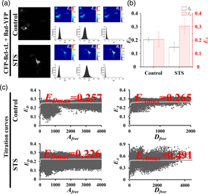
Excitation–emission-spectral unmixing-based fluorescence resonance energy transfer (ExEm-spFRET) microscopy exhibits excellent robustness in living cells. We here develop an automatic ExEm-spFRET microscope with 3.04 s of time resolution for a quantitative FRET imaging. The user-friendly interface software has been designed to operate in two modes: administrator and user. Automatic background recognition, subtraction, and cell segmentation were integrated into the software, which enables FRET calibration or measurement in a one-click operation manner. In administrator mode, both correction factors and spectral fingerprints are only calibrated periodically for a stable system. In user mode, quantitative ExEm-spFRET imaging is directly implemented for FRET samples. We implemented quantitative ExEm-spFRET imaging for living cells expressing different tandem constructs (C80Y, C40Y, C10Y, and C4Y, respectively) and obtained consistent results for at least 3 months, demonstrating the stability of our microscope. Next, we investigated Bcl-xL-Bad interaction by using ExEm-spFRET imaging and FRET two-hybrid assay and found that the Bcl-xL-Bad complexes exist mainly in Bad-Bcl-xL trimers in healthy cells and Bad-Bcl-xL2 trimers in apoptotic cells. We also performed time-lapse FRET imaging on our system for living cells expressing Yellow Cameleon 3.6 (YC3.6) to monitor ionomycin-induced rapid extracellular Ca2+ influx with a time interval of 5 s for total 250 s.

Type
Software and Instrumentation
Copyright
Copyright © The Author(s), 2022. Published by Cambridge University Press on behalf of the Microscopy Society of America

Access options

Get access to the full version of this content by using one of the access options below. (Log in options will check for institutional or personal access. Content may require purchase if you do not have access.)

References

Behera, S, Wang, N, Zhang, C, Schmitz-Thom, I, Strohkamp, S, Schültke, S, Hashimoto, K, Xiong, L & Kudla, J (2015). Analyses of Ca2+ dynamics using a ubiquitin-10 promoter-driven yellow cameleon 3.6 indicator reveal reliable transgene expression and differences in cytoplasmic Ca2+ responses in Arabidopsis and rice (Oryza sativa) roots. New Phytol 206, 751760. doi:10.1111/nph.13250CrossRefGoogle ScholarPubMed
Belosludtsev, KN, Dubinin, MV, Belosludtseva, NV & Mironova, GD (2019). Mitochondrial Ca2+ transport: Mechanisms, molecular structures, and role in cells. Biochemistry 84, 593607. doi:10.1134/S0006297919060026Google ScholarPubMed
Butz, ES, Ben-Johny, M, Shen, M, Yang, PS, Sang, L, Biel, M, Yue, DT & Wahl-Schott, C (2016). Quantifying macromolecular interactions in living cells using FRET two-hybrid assays. Nat Protoc 11, 24702498. doi:10.1038/nprot.2016.128CrossRefGoogle ScholarPubMed
Chai, L, Zhang, J, Zhang, L & Chen, T (2015). Miniature fiber optic spectrometer-based quantitative fluorescence resonance energy transfer measurement in single living cells. J Biomed Opt 20, 037008. doi:10.1117/1.JBO.20.3.037008CrossRefGoogle ScholarPubMed
Clegg, RM (1992). Fluorescence resonance energy transfer and nucleic acids. Meth Enzymol 211, 353388. doi:10.1016/0076-6879(92)11020-jCrossRefGoogle ScholarPubMed
De Los Santos, C, Chang, CW, Mycek, MA & Cardullo, RA (2015). FRAP, FLIM, and FRET: Detection and analysis of cellular dynamics on a molecular scale using fluorescence microscopy. Mol Reprod Dev 82, 587604. doi:10.1002/mrd.22501CrossRefGoogle ScholarPubMed
Du, M, Zhang, L, Xie, S & Chen, T (2016). Wide-field microscopic FRET imaging using simultaneous spectral unmixing of excitation and emission spectra. Opt Express 24, 1603716051. doi:10.1364/OE.24.016037CrossRefGoogle ScholarPubMed
Elangovan, M, Wallrabe, H, Chen, Y, Day, RN, Barroso, M & Periasamy, A (2003). Characterization of one- and two-photon excitation fluorescence resonance energy transfer microscopy. Methods 29, 5873. doi:10.1016/s1046-2023(02)00283-9CrossRefGoogle ScholarPubMed
Hoppe, AD, Scott, BL, Welliver, TP, Straight, SW & Swanson, JA (2013). N-way FRET microscopy of multiple protein-protein interactions in live cells. PLoS One 8, e64760. doi:10.1371/journal.pone.0064760CrossRefGoogle ScholarPubMed
Koushik, SV, Blank, PS & Vogel, SS (2009). Anomalous surplus energy transfer observed with multiple FRET acceptors. PLoS One 4, e8031. doi:10.1371/journal.pone.0008031CrossRefGoogle ScholarPubMed
Krebs, M, Held, K, Binder, A, Hashimoto, K, Den Herder, G, Parniske, M, Kudla, J & Schumacher, K (2012). FRET-based genetically encoded sensors allow high-resolution live cell imaging of Ca2+ dynamics. Plant J 69, 181192. doi:10.1111/j.1365-313X.2011.04780.xCrossRefGoogle Scholar
Levy, S, Wilms, CD, Brumer, E, Kahn, J, Pnueli, L, Arava, Y, Eilers, J & Gitler, D (2011). SpRET: Highly sensitive and reliable spectral measurement of absolute FRET efficiency. Microsc Microanal 17, 176190. doi:10.1017/S1431927610094493CrossRefGoogle ScholarPubMed
Li, H, Yu, H & Chen, T (2012). Partial acceptor photobleaching-based quantitative FRET method completely overcoming emission spectral crosstalks. Microsc Microanal 18, 10211029. doi:10.1017/S1431927612001110CrossRefGoogle ScholarPubMed
Lin, F, Du, M, Yang, F, Wei, L & Chen, T (2018). Improved spectrometer-microscope for quantitative fluorescence resonance energy transfer measurement based on simultaneous spectral unmixing of excitation and emission spectra. J Biomed Opt 23, 110. doi:10.1117/1.JBO.23.1.016006Google ScholarPubMed
Mustafa, S, Hannagan, J, Rigby, P, Pfleger, K & Corry, B (2013). Quantitative Förster resonance energy transfer efficiency measurements using simultaneous spectral unmixing of excitation and emission spectra. J Biomed Opt 18, 26024. doi:10.1117/1.JBO.18.2.026024CrossRefGoogle ScholarPubMed
Petros, AM, Nettesheim, DG, Wang, Y, Olejniczak, ET, Meadows, RP, Mack, J, Swift, K, Matayoshi, ED, Zhang, H, Thompson, CB & Fesik, SW (2000). Rationale for Bcl-xL/Bad peptide complex formation from structure, mutagenesis, and biophysical studies. Protein Sci 9, 25282534. doi:10.1110/ps.9.12.2528CrossRefGoogle ScholarPubMed
Su, W, Du, M, Lin, F, Zhang, C & Chen, T (2019). Quantitative FRET measurement based on spectral unmixing of donor, acceptor and spontaneous excitation-emission spectra. J Biophotonics 12, e201800314. doi:10.1002/jbio.201800314CrossRefGoogle ScholarPubMed
Sun, H, Zhang, C, Ma, Y, Du, M & Chen, T (2019). Controlling and online measurement of automatic dual-channel E-FRET microscope. Biomed Signal Process Control 53, 101585. doi:10.1016/j.bspc.2019.101585CrossRefGoogle Scholar
Thaler, C, Koushik, SV, Blank, PS & Vogel, SS (2005). Quantitative multiphoton spectral imaging and its use for measuring resonance energy transfer. Biophys J 89, 27362749. doi:10.1529/biophysj.105.061853CrossRefGoogle ScholarPubMed
Valentijn, AJ, Metcalfe, AD, Kott, J, Streuli, CH & Gilmore, AP (2003). Spatial and temporal changes in Bax subcellular localization during anoikis. J Cell Biol 162, 599612. doi:10.1083/jcb.200302154CrossRefGoogle ScholarPubMed
Vanderhoof, B, Nelson, R, Beiner, G, Raicu, V & Oliver, J (2020). Tissue factor oligomerization in living cells using förster resonance energy transfer. Microsc Microanal 26(S2), 828829. doi:10.1017/S1431927620015986CrossRefGoogle Scholar
Yu, H, Zhang, J, Li, H & Chen, T (2013). Ma-PbFRET: Multiple acceptors FRET measurement based on partial acceptor photobleaching. Microsc Microanal 19, 171179. doi:10.1017/S1431927612014079CrossRefGoogle ScholarPubMed
Zha, J, Harada, H, Osipov, K, Jockel, J, Waksman, G & Korsmeyer, SJ (1997). BH3 domain of BAD is required for heterodimerization with BCL-XL and pro-apoptotic activity. J Biol Chem 272, 2410124104. doi:10.1074/jbc.272.39.24101CrossRefGoogle ScholarPubMed
Zhang, C, Liu, Y, Qu, W, Su, W, Du, M, Yang, F & Chen, T (2019). ExEm-FRET two-hybrid assay: FRET two-hybrid assay based on linear unmixing of excitation-emission spectra. Opt Express 27, 1828218295. doi:10.1364/OE.27.018282CrossRefGoogle ScholarPubMed
Zhang, M, Cao, HZ, Hou, L, Song, SQ, Zeng, JY & Pei, Y (2017). In vivo imaging of Ca2+ accumulation during cotton fiber initiation using fluorescent indicator YC3.60. Plant Cell Rep 36, 911918. doi:10.1007/s00299-017-2122-3CrossRefGoogle ScholarPubMed
Supplementary material: File

Sun et al. supplementary material

Sun et al. supplementary material

Download Sun et al. supplementary material(File)
File 41.3 MB