Hostname: page-component-586b7cd67f-vdxz6 Total loading time: 0 Render date: 2024-11-24T09:10:24.308Z Has data issue: false hasContentIssue false

Application of Blind Deconvolution Based on the New Weighted L1-norm Regularization with Alternating Direction Method of Multipliers in Light Microscopy Images

Published online by Cambridge University Press:  11 September 2020
Ji-Youn Kim
Affiliation:
Department of Dental Hygiene, College of Health Science, Gachon University, 191, Hambakmoero, Yeonsu-gu, Incheon, Republic of Korea
Kyuseok Kim
Affiliation:
Department of Radiation Convergence Engineering, Yonsei University, 1, Yonseidae-gil, Wonju-si, Gangwon-do, Republic of Korea
Youngjin Lee*
Affiliation:
Department of Radiological Science, College of Health Science, Gachon University, 191, Hambakmoero, Yeonsu-gu, Incheon, Republic of Korea
*
*Author for correspondence: Youngjin Lee, E-mail: [email protected]
Get access

Abstract

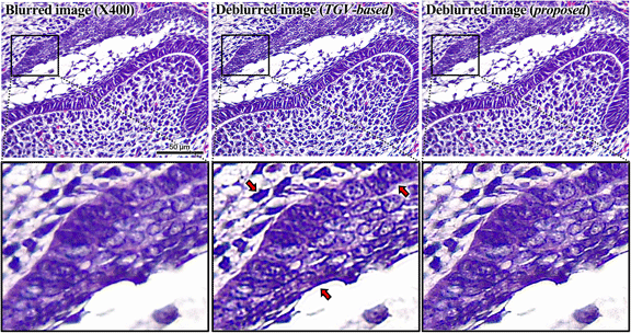
This study aimed to develop and evaluate a blind-deconvolution framework using the alternating direction method of multipliers (ADMMs) incorporated with weighted L1-norm regularization for light microscopy (LM) images. A presimulation study was performed using the Siemens star phantom prior to conducting the actual experiments. Subsequently, the proposed algorithm and a total generalized variation-based (TGV-based) method were applied to cross-sectional images of a mouse molar captured at 40× and 400× on-microscope magnifications and the results compared, and the resulting images were compared. Both simulation and experimental results confirmed that the proposed deblurring algorithm effectively restored the LM images, as evidenced by the quantitative evaluation metrics. In conclusion, this study demonstrated that the proposed deblurring algorithm can efficiently improve the quality of LM images.

Type
Software and Instrumentation
Copyright
Copyright © Microscopy Society of America 2020

Access options

Get access to the full version of this content by using one of the access options below. (Log in options will check for institutional or personal access. Content may require purchase if you do not have access.)

References

Almeida, MSC & Figueiredo, MAT (2013). Deconvolving images with unknown boundaries using the alternating direction method of multipliers. IEEE Trans Image Process 22, 30743086.CrossRefGoogle ScholarPubMed
Banham, M & Katsaggelos, A (1997). Digital image restoration. IEEE Signal Process Mag 14, 2441.CrossRefGoogle Scholar
Becker, K, Saghafi, S, Pende, M, Sabdyusheva-Litschauer, I, Hahn, CM, Foroughipour, M, Jahrling, N & Hans-Ulrich, D (2019). Deconvolution of light sheet microscopy recording. Sci Rep 9. doi:10.1038/s41598-019-53875-y.CrossRefGoogle Scholar
Bradley, CW, Jeffery, LS & Carey, EP (1997). A method for detecting microcalcifications in digital mammograms. J Digit Imaging 10, 136139.Google Scholar
Campisi, P & Egiazarian, K (2007). Blind Image Deconvolution: Theory and Applications. Boca Raton, FL: CRC. doi:10.1201/9781420007299.Google Scholar
Chan, SH, Khoshabeh, R, Gibson, KB, Gill, PE & Nquyen, TQ (2011). An augmented Lagrangian method for total variation video restoration. IEEE Trans Image Process 20, 30973111.CrossRefGoogle ScholarPubMed
Chan, SH, Wang, X & Elgendy, OA (2017). Plug-and-play ADMM for image restoration: Fixed-point convergence and applications. IEEE Trans Comput Imaging 3, 8498.CrossRefGoogle Scholar
Chang, H & Marchesini, S (2016). A general framework for denoising phaseless diffraction measurements. arXiv preprint arXiv:1611.01417.Google Scholar
Conte, F, Germani, A & Iannello, G (2013). A Kalman filter approach for denoising and deblurring 3D microscopy images. IEEE Trans Image Process 22, 53065321.CrossRefGoogle Scholar
Dapson, RW & Horobin, RW (2009). Dyes from a twenty-first century perspective. Biotech Histochem 84, 135137.CrossRefGoogle ScholarPubMed
Dolui, S & Michailovich, O (2011). Blind deconvolution of medical ultrasound images using variable splitting and proximal point methods. In 2011 IEEE International Symposium on Biomedical Imaging: From Nano to Macro (ISBI 2011), April, Chicago, IL, USA.Google Scholar
Fergus, R, Singh, B, Hertzmann, A, Roweis, S & Freeman, W (2003). Removing camera shake from a single photograph. ACM Trans Graphics 25, 787794.CrossRefGoogle Scholar
Figueiredo, M & Nowak, R (2003). An EM algorithm for wavelet-based image restoration. IEEE Trans Image Process 12, 906916.CrossRefGoogle Scholar
Kim, K, Park, S, Kim, G, Cho, H, Je, U, Park, C, Lim, H, Lee, D, Park, Y & Woo, T (2017). Industrial x-ray inspection system with improved image characterization using blind deblurring based on compressed-sensing scheme. Instrum Sci Technol 45, 248258.CrossRefGoogle Scholar
Kindur, D & Hatzinakos, D (1996). Blind image deconvolution. IEEE Signal Process Mag 13, 4364.CrossRefGoogle Scholar
Kirsch, R (1971). Computer determination of the constituent structure of biological images. Comput Biomed Res 4, 315328.CrossRefGoogle ScholarPubMed
Li, G, Luo, S, Yan, Y & Gu, N (2015). A method of extending the depth of focus of the high-resolution X-ray imaging system employing optical lens and scintillator: A phantom study. Biomed Eng Online 14. doi:10.1186/1475-925X-14-S1-S15.CrossRefGoogle ScholarPubMed
Louchet, C & Moisan, L (2011). Total variation as a local filter. SIAM J Imaging Sci 4, 651694.CrossRefGoogle Scholar
Nasonov, A & Krylov, A (2018). An improvement of BM3D image denoising and deblurring algorithm by generalized total variation. In 2018 7th European Workshop on Visual Information Processing (EUVIP), November, Tampere, Finland.CrossRefGoogle Scholar
Parikh, N & Boyd, S (2013). Proximal algorithms. Found Trends Optim 1, 123231.Google Scholar
Reeves, S & Mersereau, R (1992). Blur identification by the method of generalized cross-validation. IEEE Trans Image Process 1, 301311.CrossRefGoogle ScholarPubMed
Ren, D (2018). Blur kernel estimation and image restoration methods for blind deconvolution. PhD Dissertations. Hong Kong Polytechnic University.Google Scholar
Ren, D, Zuo, W, Zhang, D, Xu, J & Zhang, L (2018). Partial deconvolution with inaccurate blur kernel. IEEE Trans Image Process 27, 511524.CrossRefGoogle ScholarPubMed
Sattar, F, Floreby, L, Salomonsson, G & Lovstrom, B (1997). Image enhancement based on a nonlinear multiscale method. IEEE Trans Image Process 6, 888895.CrossRefGoogle ScholarPubMed
Stockham, T, Cannon, T & Ingebretsen, R (1975). Blind deconvolution through digital signal processing. Proc IEEE 63, 678692.CrossRefGoogle Scholar
Swedlow, J (2003). Quantitative fluorescence microscopy and image deconvolution. Methods Cell Biol 72, 349367.CrossRefGoogle ScholarPubMed
Takeda, H, Farsiu, S & Milanfar, P (2008). Deblurring using regularized locally adaptive kernel regression. IEEE Trans Image Process 17, 550563.CrossRefGoogle ScholarPubMed
Taruttis, A & Ntziachristos, V (2015). Advances in real-time multispectral optoacoustic imaging and its applications. Nature Photonics 9, 219227.CrossRefGoogle Scholar
Vardi, Y & Lee, D (1993). From image deblurring to optimal investments: Maximum likelihood solutions for positive linear inverse problems. J R Stat Soc Series B 55, 569612.Google Scholar
Wang, Z & Bovik, AC (2004). Image quality assessment: From error visibility to structural similarity. IEEE Trans Image Process 13, 600612.CrossRefGoogle ScholarPubMed
Xu, H, Huang, T, Lv, X & Liu, J (2014 a). The implementation of LSMR in image deblurring. Appl Math Inf Sci 8, 30413048.CrossRefGoogle Scholar
Xu, L, Ren, JS, Liu, C & Jia, J (2014 b). Deep convolutional neural network for image deconvolution. Proc Adv Neural Inf Process Syst 1, 17901798.Google Scholar
Yu, C, Zhang, C & Xie, L (2012). A blind deconvolution approach to ultrasound imaging. IEEE Trans Ultrason Ferr 59, 271280.Google ScholarPubMed
Zhang, H, Ouyang, L, Huang, J, Ma, J, Chen, W & Wang, J (2014). Few-view cone-beam CT reconstruction with deformed prior image. Med Phys 41, 121905.CrossRefGoogle ScholarPubMed
Supplementary material: File

Kim et al. supplementary material

Kim et al. supplementary material 1

Download Kim et al. supplementary material(File)
File 59.7 KB
Supplementary material: Image

Kim et al. supplementary material

Kim et al. supplementary material 2

Download Kim et al. supplementary material(Image)
Image 394.2 KB