Hostname: page-component-586b7cd67f-2plfb Total loading time: 0 Render date: 2024-11-30T20:37:44.818Z Has data issue: false hasContentIssue false

Plasma Focused Ion Beam Serial Sectioning as a Technique to Characterize Nonmetallic Inclusions in Superelastic Nitinol Fine Wires

Published online by Cambridge University Press:  08 December 2020

Janet L. Gbur*
Affiliation:
Department of Materials Science and Engineering, Case Western Reserve University, Cleveland, OH 44106, USA
Ronald Kelley
Affiliation:
Materials & Structural Analysis, Thermo Fisher Scientific, Hillsboro, OR 97124, USA
John J. Lewandowski
Affiliation:
Department of Materials Science and Engineering, Case Western Reserve University, Cleveland, OH 44106, USA
*
*Author for correspondence: Janet L. Gbur, E-mail: [email protected]
Get access

Abstract

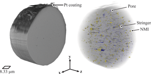
Nonmetallic inclusion (NMI) populations in superelastic (SE) Nitinol fine wires (<140 μm in diameter) were investigated by combining plasma focused ion beam (PFIB) serial sectioning with scanning electron microscopy (SEM). High purity (HP)—lower oxygen content and standard purity (SP)—higher oxygen content Nitinol wires were sectioned and imaged. The three-dimensional (3D) reconstructions provided more complete connectivity of NMIs and pores as well as information about the distribution of the features within the wire volume that is not possible with traditional two-dimensional (2D) imaging techniques. NMIs were present alone and with pores in the leading and/or trailing edges of the inclusions, in addition to stringers (i.e., fractured, elongated NMI, and intermixed with pores adjacent to each other), all of which were parallel to the wire drawing axis. The area percentages for the NMIs were 0.01% (HP Nitinol) and 0.04% (SP Nitinol), while the volume percentages measured 0.09% (HP Nitinol) and 0.47% (SP Nitinol). The combined PFIB-SEM serial sectioning approach provided the requisite resolution necessary to distinguish between NMIs and pores at micron and submicron sizes. Information gathered from this technique can be used to better inform models and predictions for fatigue lifetimes based on statistical analyses of these feature populations.

Type
Materials Science Applications
Copyright
Copyright © The Author(s), 2020. Published by Cambridge University Press on behalf of the Microscopy Society of America

Access options

Get access to the full version of this content by using one of the access options below. (Log in options will check for institutional or personal access. Content may require purchase if you do not have access.)

References

ASTM (2008). Standard practice for determining the inclusion or second-phase constituent content of metals by automatic image analysis. In E1245-03 (Reapproved 2008), pp. 18. West Conshohocken, PA, USA: ASTM International.Google Scholar
ASTM (2012). Standard specification for wrought nickel-titanium shape memory alloys for medical devices and surgical implants. In F2063-12, pp. 16. West Conshohocken, PA, USA: ASTM International.Google Scholar
ASTM (2013 a). Standard specification for wrought seamless nickel-titanium shape memory alloy tube for medical devices and surgical implants. In F2633-13, pp. 15. West Conshohocken, PA: ASTM International.Google Scholar
ASTM (2013 b). Standard test methods for determining the inclusion content of steel. In E45-13, pp. 119. West Conshohocken, PA, USA: ASTM International.Google Scholar
ASTM (2015). Standard test methods for rating and classifying inclusions in steel using the scanning electron microscope. In E2142-08 (Reapproved 2015), pp. 114. West Conshohocken, PA: ASTM International.Google Scholar
Bonsignore, C (2017). Present and future approaches to lifetime prediction of superelastic nitinol. Theor Appl Fract Mec 92, 298305. doi:10.1016/j.tafmec.2017.04.001.CrossRefGoogle Scholar
Burnett, TL, Kelley, R, Winiarski, B, Contreras, L, Daly, M, Gholinia, A, Burke, MG & Withers, PJ (2016). Large volume serial section tomography by Xe plasma FIB dual beam microscopy. Ultramicroscopy 161, 119129. doi:10.1016/j.ultramic.2015.11.001.CrossRefGoogle ScholarPubMed
Coda, A, Zilio, S, Norwich, D & Sczerzenie, F (2012). Characterization of inclusions in VIM/VAR niti alloys. J Mater Eng Perform 21(12), 25722577. doi:10.1007/s11665-012-0366-1.CrossRefGoogle Scholar
Daly, M, Burnett, TL, Pickering, EJ, Tuck, OCG, Léonard, F, Kelley, R, Withers, PJ & Sherry, AH (2017). A multi-scale correlative investigation of ductile fracture. Acta Mater 130, 5668. doi:10.1016/j.actamat.2017.03.028.CrossRefGoogle Scholar
DeHoff, RT (1983). Quantitative serial sectioning analysis: Preview. J Microsc 131(3), 259263.CrossRefGoogle Scholar
Gbur, JL (2018). Inclusion effects on the lifetime performance of superelastic nitinol wires. PhD/Case. Western Reserve University.Google Scholar
Gbur, JL & Lewandowski, JJ (2016). Fatigue and fracture of wires and cables for biomedical applications. Int Mater Rev 61(4), 231314. doi:10.1080/09506608.2016.1152347.CrossRefGoogle Scholar
Gbur, JL & Lewandowski, JJ (2021). Effects of nitinol purity and inclusions on the fracture and fatigue behavior of fine NiTi wire. in preparation.Google Scholar
Graham, R, Van Doren, B, Henson, R & DiCello, J (2004). Characteristics of high purity Nitinol. Paper Presented at the International Conference on Shape Memory and Superelastic Technologies, Pacific Grove, California, USA.Google Scholar
Kramer, GM (2009). A comparison of chemistry and inclusion distribution and morphology versus melting method of NiTi alloys. J Mater Eng Perform 18(5–6), 479483. doi:10.1007/s11665-009-9438-2.CrossRefGoogle Scholar
Launey, M, Robertson, SW, Vien, L, Senthilnathan, K, Chintapalli, P & Pelton, AR (2014). Influence of microstructural purity on the bending fatigue behavior of VAR-melted superelastic Nitinol. J Mech Behav Biomed Mater 34, 181186. doi:10.1016/j.jmbbm.2014.02.008.CrossRefGoogle ScholarPubMed
Morgan, N, Wick, A, DiCello, J & Graham, R (2006). Carbon and oxygen levels in Nitinol alloys and the implications for medical device manufacture and durability. Paper Presented at the Proceedings of the International Conference on Shape Memory and Superelastic Technologies, Pacific Grove, California, USA.Google Scholar
Patel, MM (2007). Characterizing fatigue response of nickel-titanium alloys by rotary beam testing. J ASTM Int 4(6), 111. doi:10.1520/JAI100390.CrossRefGoogle Scholar
Rahim, M, Frenzel, J, Frotscher, M, Pfetzing-Micklich, J, Steegmüller, R, Wohlschlögel, M, Mughrabi, H & Eggeler, G (2013). Impurity levels and fatigue lives of pseudoelastic NiTi shape memory alloys. Acta Mater 61(10), 36673686. doi:10.1016/j.actamat.2013.02.054.CrossRefGoogle Scholar
Robertson, SW, Launey, M, Shelley, O, Ong, I, Vien, L, Senthilnathan, K, Saffari, P, Schlegel, S & Pelton, AR (2015). A statistical approach to understand the role of inclusions on the fatigue resistance of superelastic Nitinol wire and tubing. J Mech Behav Biomed Mater 51, 119131. doi:10.1016/j.jmbbm.2015.07.003.CrossRefGoogle ScholarPubMed
Salvo, L, Cloetens, P, Maire, E, Zabler, S, Blandin, JJ, Buffière, JY & Josserond, C (2003). X-ray micro-tomography an attractive characterisation technique in materials science. Nucl Instrum Methods Phys Res B 200, 273286. doi:10.1016/S0168-583X(02)01689-0.CrossRefGoogle Scholar
Sczerzenie, F, Vergani, G & Belden, C (2012). The measurement of total inclusion content in nickel-titanium alloys. J Mater Eng Perform 21(12), 25782586. doi:10.1007/s11665-012-0377-y.CrossRefGoogle Scholar
Toro, A, Zhou, F, Wu, MH, Van Geertruyden, W & Misiolek, WZ (2009). Characterization of Non-metallic inclusions in superelastic NiTi tubes. J Mater Eng Perform 18(5–6), 448458. doi:10.1007/s11665-009-9410-1.CrossRefGoogle Scholar
Urbano, MF, Cadelli, A, Sczerzenie, F, Luccarelli, P, Beretta, S & Coda, A (2015). Inclusions size-based fatigue life prediction model of NiTi alloy for biomedical applications. Shap Mem Superelasticity 1(2), 240251. doi:10.1007/s40830-015-0016-1.CrossRefGoogle Scholar
Vander Voort, G (2015). Light microscopy. In Metallography and Microstructures, Vander Voort, G (Ed.), Vol. 9, pp. 332354. Materials Park, OH: ASM International.Google Scholar