Hostname: page-component-586b7cd67f-r5fsc Total loading time: 0 Render date: 2024-11-24T11:32:28.336Z Has data issue: false hasContentIssue false

Optimizing Nonrigid Registration for Scanning Transmission Electron Microscopy Image Series

Published online by Cambridge University Press:  23 November 2020

Chenyu Zhang
Affiliation:
Department of Materials Science and Engineering, University of Wisconsin – Madison, 1509 University Avenue, Madison, WI53706, USA
Jie Feng
Affiliation:
Department of Materials Science and Engineering, University of Wisconsin – Madison, 1509 University Avenue, Madison, WI53706, USA
Andrew B. Yankovich
Affiliation:
Department of Materials Science and Engineering, University of Wisconsin – Madison, 1509 University Avenue, Madison, WI53706, USA
Alexander Kvit
Affiliation:
Department of Materials Science and Engineering, University of Wisconsin – Madison, 1509 University Avenue, Madison, WI53706, USA
Benjamin Berkels
Affiliation:
Aachen Institute for Advanced Study in Computational Engineering Science, RWTH Aachen University, Schinkelstr. 2, 52056Aachen, Germany
Paul M. Voyles*
Affiliation:
Department of Materials Science and Engineering, University of Wisconsin – Madison, 1509 University Avenue, Madison, WI53706, USA
*
*Author for correspondence: Paul M. Voyles, E-mail: [email protected]
Get access

Abstract

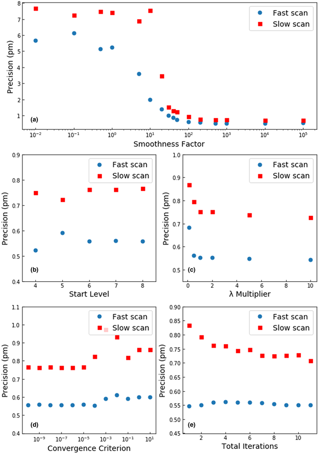
Achieving sub-picometer precision measurements of atomic column positions in high-resolution scanning transmission electron microscope images using nonrigid registration (NRR) and averaging of image series requires careful optimization of experimental conditions and the parameters of the registration algorithm. On experimental data from SrTiO3 [100], sub-pm precision requires alignment of the sample to the zone axis to within 1 mrad tilt and sample drift of less than 1 nm/min. At fixed total electron dose for the series, precision in the fast scan direction improves with shorter pixel dwell time to the limit of our microscope hardware, but the best precision along the slow scan direction occurs at 6 μs/px dwell time. Within the NRR algorithm, the “smoothness factor” that penalizes large estimated shifts is the most important parameter for sub-pm precision, but in general, the precision of NRR images is robust over a wide range of parameters.

Type
Software and Instrumentation
Copyright
Copyright © The Author(s), 2020. Published by Cambridge University Press

Access options

Get access to the full version of this content by using one of the access options below. (Log in options will check for institutional or personal access. Content may require purchase if you do not have access.)

References

Bals, S, Van Aert, S, Van Tendeloo, G & Ávila-Brande, D (2006). Statistical estimation of atomic positions from exit wave reconstruction with a precision in the picometer range. Phys Rev Lett 96, 096106.CrossRefGoogle ScholarPubMed
Berkels, B, Binev, P, Blom, DA, Dahmen, W, Sharpley, RC & Vogt, T (2014). Optimized imaging using non-rigid registration. Ultramicroscopy 138, 4656.CrossRefGoogle ScholarPubMed
Berkels, B & Liebscher, CH (2019). Joint non-rigid image registration and reconstruction for quantitative atomic resolution scanning transmission electron microscopy. Ultramicroscopy 198, 4957. doi:10.1016/j.ultramic.2018.12.016.CrossRefGoogle ScholarPubMed
Borisevich, AY, Ovchinnikov, OS, Chang, HJ, Oxley, MP, Yu, P, Seidel, J, Eliseev, EA, Morozovska, AN, Ramesh, R, Pennycook, SJ & Kalinin, SV (2010). Mapping octahedral tilts and polarization across a domain wall in BiFeO3 from Z-contrast scanning transmission electron microscopy image atomic column shape analysis. ACS Nano 4, 60716079.CrossRefGoogle ScholarPubMed
Braidy, N, Le Bouar, Y, Lazar, S & Ricolleau, C (2012). Correcting scanning instabilities from images of periodic structures. Ultramicroscopy 118, 6776. doi:10.1016/j.ultramic.2012.04.001.CrossRefGoogle ScholarPubMed
Briggs, WL, Henson, VE & McCormick, SF (2000). A multigrid tutorial, 2nd ed. Society for Industrial and Applied Mathematics. Available at https://www.researchgate.net/publication/220690328 (accessed August 29, 2020).CrossRefGoogle Scholar
Buban, JP, Ramasse, Q, Gipson, B, Browning, ND & Stahlberg, H (2010). High-resolution low-dose scanning transmission electron microscopy. J Electron Microsc 59, 103112.CrossRefGoogle ScholarPubMed
De Backer, A, Martinez, GT, MacArthur, KE, Jones, L, Béché, A, Nellist, PD & Van Aert, S (2014). Dose limited reliability of quantitative annular dark field scanning transmission electron microscopy for nano-particle atom-counting. Ultramicroscopy, 151, 5661.CrossRefGoogle ScholarPubMed
De Backer, A, Martinez, GT, Rosenauer, A & Van Aert, S (2013). Atom counting in HAADF STEM using a statistical model-based approach: Methodology, possibilities, and inherent limitations. Ultramicroscopy, 134, 2333.CrossRefGoogle ScholarPubMed
Feng, J, Kvit, AV, Zhang, C, Hoffman, J, Bhattacharya, A, Morgan, D & Voyles, PM (2017). Imaging of single La vacancies in LaMnO3. Microsc Microanal 22, 902903. Available at http://arxiv.org/abs/1711.06308.CrossRefGoogle Scholar
Han, J, Berkels, B, Droske, M, Hornegger, J, Rumpf, M, Schaller, C, Scorzin, J & Urbach, H (2007). Mumford–Shah model for one-to-one edge matching. IEEE Trans Image Process 16, 27202732.CrossRefGoogle ScholarPubMed
Hwang, J, Zhang, JY, D'Alfonso, AJ, Allen, LJ & Stemmer, S (2013). Three-dimensional imaging of individual dopant atoms in SrTiO3. Phys Rev Lett 111, 266101.CrossRefGoogle ScholarPubMed
Jones, L (2016). Quantitative ADF STEM: Acquisition, analysis and interpretation. In IOP Conference Series: Materials Science and Engineering 109, 012008. Available at http://stacks.iop.org/1757-899X/109/i=1/a=012008?key=crossref.9fd1cf232cf77eee645f9e71cc9d799c.Google Scholar
Jones, L & Nellist, PD (2013). Identifying and correcting scan noise and drift in the scanning transmission electron microscope. Microsc Microanal 19, 10501060.CrossRefGoogle ScholarPubMed
Jones, L, Varambhia, A, Beanland, R, Kepaptsoglou, D, Griffiths, I, Ishizuka, A, Azough, F, Freer, R, Ishizuka, K, Cherns, D, Ramasse, QM, Lozano-Perez, S & Nellist, PD (2018). Managing dose-, damage- and data-rates in multi-frame spectrum-imaging. Microscopy 67, i98i113.CrossRefGoogle ScholarPubMed
Jones, L, Wenner, S, Nord, M, Ninive, PH, Løvvik, OM, Holmestad, R & Nellist, PD (2017). Optimising multi-frame ADF-STEM for high-precision atomic-resolution strain mapping. Ultramicroscopy 179, 5762.CrossRefGoogle ScholarPubMed
Jones, L, Yang, H, Pennycook, TJ, Marshall, MSJ, Van Aert, S, Browning, ND, Castell, MR & Nellist, PD (2015). Smart align—A new tool for robust non-rigid registration of scanning microscope data. Adv Struct Chem Imaging 1, 8.CrossRefGoogle Scholar
Kimoto, K, Asaka, T, Yu, X, Nagai, T, Matsui, Y & Ishizuka, K (2010). Local crystal structure analysis with several picometer precision using scanning transmission electron microscopy. Ultramicroscopy 110, 778782.CrossRefGoogle ScholarPubMed
Kotakoski, J, Jin, CH, Lehtinen, O, Suenaga, K & Krasheninnikov, AV (2010). Electron knock-on damage in hexagonal boron nitride monolayers. Phys Rev B 82, 113404. doi:10.1103/PhysRevB.82.113404.CrossRefGoogle Scholar
Krause, FF, Schowalter, M, Grieb, T, Müller-Caspary, K, Mehrtens, T & Rosenauer, A (2016). Effects of instrument imperfections on quantitative scanning transmission electron microscopy. Ultramicroscopy 161, 146160. doi:10.1016/j.ultramic.2015.10.026.CrossRefGoogle ScholarPubMed
Lee, D, Lu, H, Gu, Y, Choi, S-Y, Li, S-D, Ryu, S, Paudel, TR, Song, K, Mikheev, E, Lee, S, Stemmer, S, Tenne, DA, Oh, SH, Tsymbal, EY, Wu, X, Chen, LQ, Gruverman, A & Eom, CB (2015). Emergence of room-temperature ferroelectricity at reduced dimensions. Science 349, 13141317.CrossRefGoogle ScholarPubMed
Muller, DA, Kirkland, EJ, Thomas, MG, Grazul, JL, Fitting, L & Weyland, M (2006). Room design for high-performance electron microscopy. Ultramicroscopy 106, 10331040.CrossRefGoogle ScholarPubMed
Nilsson Pingel, T, Jørgensen, M, Yankovich, AB, Grönbeck, H & Olsson, E (2018). Influence of atomic site-specific strain on catalytic activity of supported nanoparticles. Nat Commun 9, 2722.CrossRefGoogle ScholarPubMed
Ning, S, Fujita, T, Nie, A, Wang, Z, Xu, X, Chen, J, Chen, M, Yao, S & Zhang, TY (2018). Scanning distortion correction in STEM images. Ultramicroscopy 184, 274283.CrossRefGoogle ScholarPubMed
Oni, AA, Sang, X, Raju, SV, Dumpala, S, Broderick, S, Kumar, A, Sinnott, S, Saxena, S, Rajan, K & LeBeau, JM (2015). Large area strain analysis using scanning transmission electron microscopy across multiple images. Appl Phys Lett 106. doi:10.1063/1.4905368.CrossRefGoogle Scholar
Ophus, C, Ciston, J & Nelson, CT (2016). Correcting nonlinear drift distortion of scanning probe and scanning transmission electron microscopies from image pairs with orthogonal scan directions. Ultramicroscopy 162, 19.CrossRefGoogle ScholarPubMed
Rojac, T, Bencan, A, Drazic, G, Sakamoto, N, Ursic, H, Jancar, B, Tavcar, G, Makarovic, M, Walker, J, Malic, B & Damjanovic, D (2016). Domain-wall conduction in ferroelectric BiFeO3 controlled by accumulation of charged defects. Nat Mater 16, 322327.CrossRefGoogle ScholarPubMed
Sang, X & LeBeau, JM (2014). Revolving scanning transmission electron microscopy: Correcting sample drift distortion without prior knowledge. Ultramicroscopy 138, 2835.CrossRefGoogle ScholarPubMed
Sang, X & LeBeau, JM (2015). Characterizing the response of a scintillator-based detector to single electrons. Ultramicroscopy 161, 39.CrossRefGoogle ScholarPubMed
Savitzky, BH, El Baggari, I, Clement, CB, Waite, E, Goodge, BH, Baek, DJ, Sheckelton, JP, Pasco, C, Nair, H, Schreiber, NJ, Hoffman, J, Admasu, AS, Kim, J, Cheong, SW, Bhattacharya, A, Schlom, DG, McQueen, TM, Hovden, R & Kourkoutis, LF (2018). Image registration of low signal-to-noise cryo-STEM data. Ultramicroscopy 191, 5665. doi:10.1016/j.ultramic.2018.04.008.CrossRefGoogle ScholarPubMed
Sundaramoorthi, G, Yezzi, A & Mennucci, AC (2007). Sobolev active contours. Int J Comput Vis 73, 345366.CrossRefGoogle Scholar
Tang, YL, Zhu, YL & Ma, XL (2015). On the benefit of aberration-corrected HAADF-STEM for strain determination and its application to tailoring ferroelectric domain patterns. Ultramicroscopy 160, 5763.CrossRefGoogle ScholarPubMed
Van Aert, S, den Dekker, AJ, Van Dyck, D & van den Bos, A (2002). Optimal experimental design of STEM measurement of atom column positions. Ultramicroscopy 90, 273289.CrossRefGoogle ScholarPubMed
Wang, Y, Salzberger, U, Sigle, W, Eren Suyolcu, Y & van Aken, PA (2016). Oxygen octahedra picker: A software tool to extract quantitative information from STEM images. Ultramicroscopy 168, 4652.CrossRefGoogle ScholarPubMed
Yankovich, AB, Berkels, B, Dahmen, W, Binev, P, Sanchez, SI, Bradley, SA, Li, A, Szlufarska, I & Voyles, PM (2014). Picometre-precision analysis of scanning transmission electron microscopy images of platinum nanocatalysts. Nat Commun 5, 4155.CrossRefGoogle ScholarPubMed
Yankovich, AB, Berkels, B, Dahmen, W, Binev, P & Voyles, PM (2015). High-precision scanning transmission electron microscopy at coarse pixel sampling for reduced electron dose. Adv Struct Chem Imaging 1, 5.CrossRefGoogle Scholar
Zhang, D, Zhu, Y, Liu, L, Ying, X, Hsiung, C, Sougrat, R, Li, K & Han, Y (2018). Atomic-resolution transmission electron microscopy of electron beam-sensitive crystalline materials. Science 359, 675679.CrossRefGoogle ScholarPubMed