Hostname: page-component-586b7cd67f-2brh9 Total loading time: 0 Render date: 2024-11-24T14:55:46.477Z Has data issue: false hasContentIssue false

Morphological Changes in the Myotendinous Junction of mdx Mice

Published online by Cambridge University Press:  11 August 2021

Giovana Zerbo Martinez
Affiliation:
Laboratory of Morphology and Physical Activity (LAMAF), São Paulo State University (UNESP), Institute of Biosciences (IB), Avenue 24A, n. 1515, Rio Claro, SP 13506-900, Brazil
Bruna Aléxia Cristofoletti Grillo
Affiliation:
Laboratory of Morphology and Physical Activity (LAMAF), São Paulo State University (UNESP), Institute of Biosciences (IB), Avenue 24A, n. 1515, Rio Claro, SP 13506-900, Brazil
Lara Caetano Rocha
Affiliation:
Laboratory of Morphology and Physical Activity (LAMAF), São Paulo State University (UNESP), Institute of Biosciences (IB), Avenue 24A, n. 1515, Rio Claro, SP 13506-900, Brazil
Carolina dos Santos Jacob
Affiliation:
Laboratory of Morphology and Physical Activity (LAMAF), São Paulo State University (UNESP), Institute of Biosciences (IB), Avenue 24A, n. 1515, Rio Claro, SP 13506-900, Brazil
Jurandyr Pimentel Neto
Affiliation:
Laboratory of Morphology and Physical Activity (LAMAF), São Paulo State University (UNESP), Institute of Biosciences (IB), Avenue 24A, n. 1515, Rio Claro, SP 13506-900, Brazil
André Neri Tomiate
Affiliation:
Laboratory of Morphology and Physical Activity (LAMAF), São Paulo State University (UNESP), Institute of Biosciences (IB), Avenue 24A, n. 1515, Rio Claro, SP 13506-900, Brazil
Gabriela Klein Barbosa
Affiliation:
Laboratory of Morphology and Physical Activity (LAMAF), São Paulo State University (UNESP), Institute of Biosciences (IB), Avenue 24A, n. 1515, Rio Claro, SP 13506-900, Brazil
Ii-sei Watanabe
Affiliation:
Department of Anatomy, Institute of Biomedical Sciences III, University of São Paulo (USP), São Paulo, SP, Brazil
Adriano Polican Ciena*
Affiliation:
Laboratory of Morphology and Physical Activity (LAMAF), São Paulo State University (UNESP), Institute of Biosciences (IB), Avenue 24A, n. 1515, Rio Claro, SP 13506-900, Brazil
*
*Correspondent author: Adriano Polican Ciena, E-mail: [email protected]
Get access

Abstract

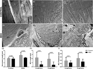
The myotendinous junction (MTJ) is the interface between muscle and tendon, and it is the main area of force transmission of the locomotor apparatus. Dystrophic processes promote pathological injury which affects the skeletal muscle and can influence the morphology of the MTJ. This study aimed to investigate the adaptations in MTJ morphology of mdx mice in the tibialis anterior muscle. Male mice (n = 24) were divided into Control—C57bl/10 and mdx—C57bl/10mdx (Duchenne muscular dystrophy experimental model). In the mdx group, centralized nuclei with a large area and greater deposition of type III collagen (fibrosis) were observed. Also, shorter sarcomeres and sarcoplasmatic projections of MTJ were observed. We concluded that the adaptations in mdx mice demonstrated extensive impairment in the MTJ region with reduced ultrastructures.

Type
Micrographia
Copyright
Copyright © The Author(s), 2021. Published by Cambridge University Press on behalf of the Microscopy Society of America

Access options

Get access to the full version of this content by using one of the access options below. (Log in options will check for institutional or personal access. Content may require purchase if you do not have access.)

References

Allen, DG, Whitehead, NP & Froehner, SC (2015). Absence of dystrophin disrupts skeletal muscle signaling: Roles of Ca2+, reactive oxygen species, and nitric oxide in the development of muscular dystrophy. Physiol Rev 96, 253305.CrossRefGoogle Scholar
Barker, RG, Wyckelsma, VL, Xu, H & Murphy, RM (2018). Mitochondrial content is preserved throughout disease progression in the mdx mouse model of Duchenne muscular dystrophy, regardless of taurine supplementation. Am J Physiol Cell Physiol 314, C483C491.CrossRefGoogle ScholarPubMed
Bhutda, S, Surve, MV, Anil, A, Kamath, K, Singh, N, Modi, D & Banerjee, A (2017). Histochemical staining of collagen and identification of its subtypes by picrosirius red dye in mouse reproductive tissues. Bio-Protocol 7, e2592.CrossRefGoogle ScholarPubMed
Bulfield, G, Siller, WG, Wight, PAL & Moore, KJ (1984). X chromosome-linked muscular dystrophy (mdx) in the mouse. Proc Natl Acad Sci 81, 11891192.CrossRefGoogle Scholar
Cardiff, RD, Miller, CH & Munn, RJ (2014). Manual hematoxylin and eosin staining of mouse tissue sections. Cold Spring Harbor Protoc 2, 655658.Google Scholar
Ciena, AP, Almeida, SRY, Bolina, CS, Bolina-Matos, RS, Rici, REG, Silva, MCP, Miglino, MA & Watanabe, I (2012). Ultrastructural features of the myotendinous junction of the sternomastoid muscle in Wistar rats: From newborn to aging. Microsc Res Tech 75, 12921296.CrossRefGoogle Scholar
Curzi, D, Lattanzi, D, Burattini, S, Tidball, JG & Falcieri, E (2013). Morphological changes of myotendinous junction generated by muscle disuse atrophy. Microscopie 19, 4652.Google Scholar
Curzi, D, Sartini, S, Guescini, M, Lattanzi, D, Di Palma, M, Ambrogini, P, Savelli, D, Stocchi, V, Cuppini, R & Falcieri, E (2016). Effect of different exercise intensities on the myotendinous junction plasticity. PLoS One 11, 112.CrossRefGoogle ScholarPubMed
Dellorusso, C, Crawford, RW, Chamberlain, JS & Brooks, SV (2001). Tibialis anterior muscles in mdx mice are highly susceptible to contraction-induced injury. J Muscle Res Cell Motil 22, 467475.CrossRefGoogle ScholarPubMed
Fenwick, AJ, Leighton, SR & Tanner, BCW (2016). Myosin MgADP release rate decreases as sarcomere length increases in skinned rat soleus muscle fibers. Biophys J 111, 20112023.CrossRefGoogle ScholarPubMed
Folker, ES & Baylies, MK (2013). Nuclear positioning in muscle development and disease. Front Physiol 4, 363.CrossRefGoogle ScholarPubMed
Gomes, ALO, Pinto, AN, Góes, ER, Hirosue, LN, Peixoto, BO, Caromano, FA & Blascovi-Assis, S (2011). Desempenho motor e funcional na Distrofia muscular de Duchenne: Estudo de um caso. J Health Sci Inst 29, 131135.Google Scholar
Gutpell, KM, Hrinivich, WT & Hoffman, LM (2015). Skeletal muscle fibrosis in the mdx/utrn+/− mouse validates its suitability as a murine model of Duchenne muscular dystrophy. PLoS One 10, 113.CrossRefGoogle Scholar
Hoffman, EP, Brown, RH & Kunkel, LM (1987). Dystrophin: The protein product of the Duchenne muscular dystrophy locus. Cell 51, 919928.CrossRefGoogle ScholarPubMed
Jacob, CS, Rocha, LC, Pimentel Neto, J, Watanabe, IS & Ciena, AP (2019). Effects of physical training on sarcomere lengths and muscle-tendon interface of the cervical region in an experimental model of menopause. Eur J Histochem 63, 131135.CrossRefGoogle Scholar
Knudsen, AB, Larsen, M, Mackey, AL, Hjort, M, Hansen, KK, Qvortrup, K, Kjaer, M & Krogsgaard, MR (2015). The human myotendinous junction: An ultrastructural and 3D analysis study. Scand J Med Sci Sports 25, e116e123.CrossRefGoogle ScholarPubMed
Kunz, RI, Coradini, JG, Soares, CLR, Brancalhão, RMC, Bertolini, GRF & Ribeiro, LFC (2013). Efeitos da imobilização e remobilização pela combinação natação e salto em meio aquático, sobre a morfologia do músculo tibial anterior de ratos. Publi UEPG Ci Biol Saúde 19, 123129.CrossRefGoogle Scholar
Lima, AAR & Cordeiro, L (2020). Fisioterapia aquática em indivíduos com distrofia muscular: uma revisão sistemática do tipo escopo. Fisioter Pesqui 27, 100111.CrossRefGoogle Scholar
Lynch, GS, Hinkle, RT, Chamberlain, JS, Brooks, SV & Faulkner, JA (2001). Force and power output of fast and slow skeletal muscles from mdx mice 6–28 months old. J Physiol 535, 591600.CrossRefGoogle ScholarPubMed
Manetti, M, Tani, A, Rosa, I, Chellini, F, Squecco, R, Idrizaj, E, Zecchi-Orlandini, S, Ibba-Manneschi, L & Sassoli, C (2019). Morphological evidence for telocytes as stromal cells supporting satellite cell activation in eccentric contraction-induced skeletal muscle injury. Sci Rep 9, 14515.CrossRefGoogle ScholarPubMed
Pimentel Neto, J, Rocha, LC, Barbosa, GK, Jacob, CS, Krause Neto, W, Watanabe, I & Ciena, AP (2020). Myotendinous junction adaptations to ladder-based resistance training: Identification of a new telocyte niche. Sci Rep 10, 14124.CrossRefGoogle ScholarPubMed
Ramaswamy, KS, Palmer, ML, Meulen, JHVD, Renoux, A, Kostrominova, TY, Michele, DE & Faulkner, JA (2011). Lateral transmission of force is impaired in skeletal muscles of dystrophic mice and very old rats. J Physiol 589, 11951208.CrossRefGoogle ScholarPubMed
Ribeiro, FAQ, Carvalho, MFP, Pereira, CSB & Tateno, DA (2015). Análise da concentração de colágeno tipo I e III presente no reparo de feridas tratadas com Mitomicina C em ratos. Arq Med Hosp Fac Cienc Med Santa Casa São Paulo 60, 2226.Google Scholar
Rocha, LC, Barbosa, GK, Pimentel Neto, J, Jacob, CS, Knudsen, AB, Watanabe, I & Ciena, AP (2021). Aquatic training after joint immobilization in rats promotes adaptations in myotendinous junctions. Int J Morphol Sci 22, 111.Google ScholarPubMed
Roman, W & Gomes, ER (2018). Nuclear positioning in skeletal muscle. Semin Cell Dev Biol 82, 5156.CrossRefGoogle ScholarPubMed
Sierra, LR, Fávaro, G, Cerri, BR, Rocha, LC, Almeida, SRY, Watanabe, I & Ciena, AP (2018). Myotendinous junction plasticity in aged ovariectomized rats submitted to aquatic training. Microsc Res Tech 81, 816822.CrossRefGoogle Scholar
Sudevan, S, Takiura, M, Kubota, Y, Higashitani, N, Cooke, M, Ellwood, RA, Etheridge, T, Szewczyk, NJ & Higashitani, A (2019). Mitochondrial dysfunction causes Ca2+ overload and ECM degradation-mediated muscle damage in C. elegans. FASEB J 33, 95409550.CrossRefGoogle ScholarPubMed
Vainzof, M, Ayub-Guerrieri, D, Onofre, PCG, Martins, PCM, Lopes, VF, Zilberztajn, D, Maia, LS, Sell, K & Yamamoto, LU (2008). Animal Models for Genetic Neuromuscular Diseases. Journal of Molecular Neuroscience 34(3), 241248. doi: http://dx.doi.org/10.1007/s12031-007-9023-9.CrossRefGoogle ScholarPubMed
Wang, DC & Wang, X (2017). Systems heterogeneity: An integrative way to understand cancer heterogeneity. Semin Cell Dev Biol 64, 14.CrossRefGoogle ScholarPubMed
Yuan, C, Arora, A, Garofalo, AM & Grange, RW (2021). Potential cross-talk between muscle and tendon in Duchenne muscular dystrophy. Connective Tissue Research 62(1), 4052. doi: http://dx.doi.org/10.1080/03008207.2020.1810247.CrossRefGoogle ScholarPubMed
Zhao, C, Wang, S, Wang, G, Su, M, Song, L, Chen, J, Fan, S & Lin, X (2018). Preparation of decellularized biphasic hierarchical myotendinous junction extracellular matrix for muscle regeneration. Acta Biomater 68, 1528.CrossRefGoogle ScholarPubMed