Hostname: page-component-586b7cd67f-2plfb Total loading time: 0 Render date: 2024-11-24T11:26:22.410Z Has data issue: false hasContentIssue false

Disentangling Coexisting Structural Order Through Phase Lock-In Analysis of Atomic-Resolution STEM Data

Published online by Cambridge University Press:  22 February 2022

Berit H. Goodge
Affiliation:
School of Applied and Engineering Physics, Cornell University, Ithaca, NY 14853, USA
Ismail El Baggari
Affiliation:
Department of Physics, Cornell University, Ithaca, NY 14853, USA
Seung Sae Hong
Affiliation:
Geballe Laboratory for Advanced Materials, Stanford University, Stanford, CA 94305, USA Stanford Institute for Materials and Energy Sciences, SLAC National Accelerator Laboratory, Menlo Park, CA 94025, USA
Zhe Wang
Affiliation:
School of Applied and Engineering Physics, Cornell University, Ithaca, NY 14853, USA
Darrell G. Schlom
Affiliation:
Department of Materials Science and Engineering, Cornell University, Ithaca, NY 14853, USA Kavli Institute at Cornell for Nanoscale Science, Cornell University, Ithaca, NY 14853, USA
Harold Y. Hwang
Affiliation:
Geballe Laboratory for Advanced Materials, Stanford University, Stanford, CA 94305, USA Stanford Institute for Materials and Energy Sciences, SLAC National Accelerator Laboratory, Menlo Park, CA 94025, USA
Lena F. Kourkoutis*
Affiliation:
School of Applied and Engineering Physics, Cornell University, Ithaca, NY 14853, USA Kavli Institute at Cornell for Nanoscale Science, Cornell University, Ithaca, NY 14853, USA
*
*Corresponding author: Lena F. Kourkoutis, E-mail: [email protected]
Get access

Abstract

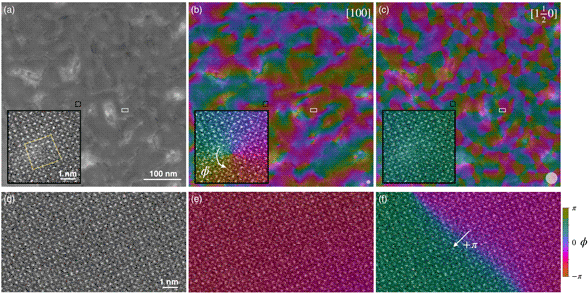
As a real-space technique, atomic-resolution STEM imaging contains both amplitude and geometric phase information about structural order in materials, with the latter encoding important information about local variations and heterogeneities present in crystalline lattices. Such phase information can be extracted using geometric phase analysis (GPA), a method which has generally focused on spatially mapping elastic strain. Here we demonstrate an alternative phase demodulation technique and its application to reveal complex structural phenomena in correlated quantum materials. As with other methods of image phase analysis, the phase lock-in approach can be implemented to extract detailed information about structural order and disorder, including dislocations and compound defects in crystals. Extending the application of this phase analysis to Fourier components that encode periodic modulations of the crystalline lattice, such as superlattice or secondary frequency peaks, we extract the behavior of multiple distinct order parameters within the same image, yielding insights into not only the crystalline heterogeneity but also subtle emergent order parameters such as antipolar displacements. When applied to atomic-resolution images spanning large (~0.5 × 0.5 μm2) fields of view, this approach enables vivid visualizations of the spatial interplay between various structural orders in novel materials.

Type
Software and Instrumentation
Copyright
Copyright © The Author(s), 2022. Published by Cambridge University Press on behalf of the Microscopy Society of America

Access options

Get access to the full version of this content by using one of the access options below. (Log in options will check for institutional or personal access. Content may require purchase if you do not have access.)

Footnotes

Current address: Rowland Institute at Harvard, Cambridge, MA 02142, USA.

Current address: Department of Materials Science and Engineering, University of California Davis, Davis, CA 95616, USA.

References

Ahn, C, Rabe, K & Triscone, JM (2004). Ferroelectricity at the nanoscale: Local polarization in oxide thin films and heterostructures. Science 303, 488491.CrossRefGoogle ScholarPubMed
Catalan, G, Lubk, A, Vlooswijk, A, Snoeck, E, Magen, C, Janssens, A, Rispens, G, Rijnders, G, Blank, DH & Noheda, B (2011). Flexoelectric rotation of polarization in ferroelectric thin films. Nat Mater 10, 963967.CrossRefGoogle ScholarPubMed
Catalano, S, Gibert, M, Fowlie, J, Iniguez, J, Triscone, JM & Kreisel, J (2018). Rare-earth nickelates RNiO3: Thin films and heterostructures. Rep Prog Phys 81, 046501.CrossRefGoogle ScholarPubMed
Chu, MW, Szafraniak, I, Scholz, R, Harnagea, C, Hesse, D, Alexe, M & Gösele, U (2004). Impact of misfit dislocations on the polarization instability of epitaxial nanostructured ferroelectric perovskites. Nat Mater 3, 8790.CrossRefGoogle ScholarPubMed
El Baggari, I, Baek, DJ, Zachman, MJ, Lu, D, Hikita, Y, Hwang, HY, Nowadnick, EA & Kourkoutis, LF (2021). Charge order textures induced by non-linear couplings in a half-doped manganite. Nat Commun 12, 3747.CrossRefGoogle Scholar
El Baggari, I, Savitzky, BH, Admasu, AS, Kim, J, Cheong, SW, Hovden, R & Kourkoutis, LF (2018). Nature and evolution of incommensurate charge order in manganites visualized with cryogenic scanning transmission electron microscopy. Proc Natl Acad Sci USA 115, 14451450.CrossRefGoogle ScholarPubMed
Fan, Z, Ma, T, Wei, J, Yang, T, Zhou, L & Tan, X (2020). TEM investigation of the domain structure in PbHfO3 and PbZrO3 antiferroelectric perovskites. J Mater Sci 55, 49534961.CrossRefGoogle Scholar
Findlay, S, Shibata, N, Sawada, H, Okunishi, E, Kondo, Y, Yamamoto, T & Ikuhara, Y (2009). Robust atomic resolution imaging of light elements using scanning transmission electron microscopy. Appl Phys Lett 95, 191913.CrossRefGoogle Scholar
Gao, P, Kumamoto, A, Ishikawa, R, Lugg, N, Shibata, N & Ikuhara, Y (2018). Picometer-scale atom position analysis in annular bright-field STEM imaging. Ultramicroscopy 184, 177187.CrossRefGoogle ScholarPubMed
Gao, P, Liu, HJ, Huang, YL, Chu, YH, Ishikawa, R, Feng, B, Jiang, Y, Shibata, N, Wang, EG & Ikuhara, Y (2016). Atomic mechanism of polarization-controlled surface reconstruction in ferroelectric thin films. Nat Commun 7, 11318.CrossRefGoogle ScholarPubMed
Guo, H, Wang, Z, Dong, S, Ghosh, S, Saghayezhian, M, Chen, L, Weng, Y, Herklotz, A, Ward, TZ, Jin, R, Pantelides, ST, Zhu, Y, Zhang, J & Plummer, EW (2017). Interface-induced multiferroism by design in complex oxide superlattices. Proc Natl Acad Sci USA 114, E5062E5069.CrossRefGoogle ScholarPubMed
Han, Y, Li, MY, Jung, GS, Marsalis, MA, Qin, Z, Buehler, MJ, Li, LJ & Muller, DA (2018). Sub-nanometre channels embedded in two-dimensional materials. Nat Mater 17, 129133.CrossRefGoogle ScholarPubMed
Hong, SS, Gu, M, Verma, M, Harbola, V, Wang, BY, Lu, D, Vailionis, A, Hikita, Y, Pentcheva, R, Rondinelli, JM & Hwang, HY (2020). Extreme tensile strain states in La0.7Ca0.3MnO3 membranes. Science 368, 7176.CrossRefGoogle ScholarPubMed
Hwang, J, Zhang, JY, D'Alfonso, AJ, Allen, LJ & Stemmer, S (2013). Three-dimensional imaging of individual dopant atoms in SrTiO3. Phys Rev Lett 111, 266101.CrossRefGoogle ScholarPubMed
Hÿtch, M & Gandais, M (1995). Quantitative criteria for the detection and characterization of nanocrystals from high-resolution electron microscopy images. Philos Mag A 72, 619634.CrossRefGoogle Scholar
Hÿtch, M, Snoeck, E & Kilaas, R (1998). Quantitative measurement of displacement and strain fields from HREM micrographs. Ultramicroscopy 74, 131146.CrossRefGoogle Scholar
Hÿtch, MJ (1997). Analysis of variations in structure from high resolution electron microscope images by combining real space and fourier space information. Microsc Microanal Microstruct 8, 4157.CrossRefGoogle Scholar
Hÿtch, MJ, Putaux, JL & Pénisson, JM (2003). Measurement of the displacement field of dislocations to 0.03 Å by electron microscopy. Nature 423, 270273.CrossRefGoogle ScholarPubMed
Jia, CL, Nagarajan, V, He, JQ, Houben, L, Zhao, T, Ramesh, R, Urban, K & Waser, R (2007). Unit-cell scale mapping of ferroelectricity and tetragonality in epitaxial ultrathin ferroelectric films. Nat Mater 6, 6469.CrossRefGoogle ScholarPubMed
Kim, H, Zhang, JY, Raghavan, S & Stemmer, S (2016). Direct observation of Sr vacancies in SrTiO3 by quantitative scanning transmission electron microscopy. Phys Rev X 6, 041063.Google Scholar
Lawler, MJ, Fujita, K, Lee, J, Schmidt, AR, Kohsaka, Y, Kim, CK, Eisaki, H, Uchida, S, Davis, JC, Sethna, JP & Kim, Ea. (2010). Intra-unit-cell electronic nematicity of the high-Tc copper-oxide pseudogap states. Nature 466, 347351.CrossRefGoogle Scholar
Lazić, I, Bosch, EGT & Lazar, S (2016). Phase contrast STEM for thin samples: Integrated differential phase contrast. Ultramicroscopy 160, 265280.CrossRefGoogle ScholarPubMed
LeBeau, JM, Findlay, SD, Allen, LJ & Stemmer, S (2008). Quantitative atomic resolution scanning transmission electron microscopy. Phys Rev Lett 100, 206101.CrossRefGoogle ScholarPubMed
Liao, Z, Sun, W, Zhang, Q, Li, JF & Zhu, J (2019). Microscopic origin of the high piezoelectric response of Sm-doped BiFeO3 near the morphotropic phase boundary. J Appl Phys 125, 175113.CrossRefGoogle Scholar
Loudon, JC, Kourkoutis, LF, Ahn, JS, Zhang, CL, Cheong, SW & Muller, DA (2007). Valence changes and structural distortions in "charge ordered" manganites quantified by atomic-scale scanning transmission electron microscopy. Phys Rev Lett 99, 237205.CrossRefGoogle ScholarPubMed
Lu, D, Baek, DJ, Hong, SS, Kourkoutis, LF, Hikita, Y & Hwang, HY (2016). Synthesis of freestanding single-crystal perovskite films and heterostructures by etching of sacrificial water-soluble layers. Nat Mater 15, 12551260.CrossRefGoogle ScholarPubMed
Mesaros, A, Fujita, K, Eisaki, H, Uchida, S, Davis, J, Sachdev, S, Zaanen, J, Lawler, M & Kim, EA (2011). Topological defects coupling smectic modulations to intra–unit-cell nematicity in cuprates. Science 333, 426430.CrossRefGoogle ScholarPubMed
Milward, GC, Calderon, MJ & Littlewood, P (2005). Electronically soft phases in manganites. Nature 433, 607610.CrossRefGoogle ScholarPubMed
Nelson, CT, Winchester, B, Zhang, Y, Kim, S-J, Melville, A, Adamo, C, Folkman, CM, Baek, S-H, Eom, C-B, Schlom, DG, Chen, L-Q & Pan, X (2011). Spontaneous vortex nanodomain arrays at ferroelectric. Nano Lett 11(2), 828834.CrossRefGoogle ScholarPubMed
Nord, M, Vullum, PE, MacLaren, I, Tybell, T & Holmestad, R (2017). Atomap: A new software tool for the automated analysis of atomic resolution images using two-dimensional gaussian fitting. Adv Struct Chem Imag 3, 9.CrossRefGoogle ScholarPubMed
Peters, JJP, Beanland, R, Alexe, M, Cockburn, JW, Revin, DG, Zhang, SY & Sanchez, AM (2015). Artefacts in geometric phase analysis of compound materials. Ultramicroscopy 157, 9197.CrossRefGoogle ScholarPubMed
Ramesh, R & Spaldin, NA (2007). Multiferroics: Progress and prospects in thin films. Nat Mater 6, 2129.CrossRefGoogle ScholarPubMed
Rouviere, JL & Sarigiannidou, E (2005). Theoretical discussions on the geometrical phase analysis. Ultramicroscopy 106, 117.CrossRefGoogle ScholarPubMed
Savitzky, BH, El, I, Clement, CB, Waite, E, Goodge, BH, Baek, DJ, Sheckelton, JP, Pasco, C, Nair, H, Schreiber, NJ, Hoffman, J, Admasu, AS, Kim, J, Cheong, Sw., Bhattacharya, A, Schlom, DG, Mcqueen, TM, Hovden, R & Kourkoutis, LF (2018). Image registration of low signal-to-noise cryo-STEM data. Ultramicroscopy 191, 5665.CrossRefGoogle ScholarPubMed
Savitzky, BH, El Baggari, I, Admasu, AS, Kim, J, Cheong, SW, Hovden, R & Kourkoutis, LF (2017). Bending and breaking of stripes in a charge ordered manganite. Nat Commun 8, 1883.CrossRefGoogle Scholar
Spaldin, NA & Fiebig, M (2005). The renaissance of magnetoelectric multiferroics. Science 309, 391392.CrossRefGoogle ScholarPubMed
Tokura, Y & Hwang, HY (2008). Complex oxides on fire. Nat Mater 7, 694695.CrossRefGoogle ScholarPubMed
Voyles, PM, Grazul, JL & Muller, DA (2003). Imaging individual atoms inside crystals with ADF-STEM. Ultramicroscopy 96, 251273.CrossRefGoogle ScholarPubMed
Wang, Y, Salzberger, U, Sigle, W, Suyolcu, YE & van Aken, PA (2016). Oxygen octahedra picker: A software tool to extract quantitative information from STEM images. Ultramicroscopy 168, 4652.CrossRefGoogle ScholarPubMed
Wang, Z, Goodge, BH, Baek, DJ, Zachman, MJ, Huang, X, Bai, X, Brooks, CM, Paik, H, Mei, AB, Brock, JD, Maria, JP, Kourkoutis, LF & Schlom, D (2019). Epitaxial SrTiO3 film on silicon with narrow rocking curve despite huge defect density. Phys Rev Mater 3, 073403.CrossRefGoogle Scholar
Wei, XK, Tagantsev, AK, Kvasov, A, Roleder, K, Jia, CL & Setter, N (2014). Ferroelectric translational antiphase boundaries in nonpolar materials. Nat Commun 5, 3031.CrossRefGoogle ScholarPubMed
Xu, C, Chen, Y, Cai, X, Meingast, A, Guo, X, Wang, F, Lin, Z, Lo, TW, Maunders, C, Lazar, S, Wang, N, Lei, D, Chai, Y, Zhai, T, Luo, X & Zhu, Y (2020 a). Two-dimensional antiferroelectricity in nanostripe-ordered In2Se3. Phys Rev Lett 125, 047601.CrossRefGoogle Scholar
Xu, R, Huang, J, Barnard, ES, Hong, SS, Singh, P, Wong, EK, Jansen, T, Harbola, V, Xiao, J, Wang, BY, Crossley, S, Lu, D, Liu, S & Hwang, HY (2020 b). Strain-induced room-temperature ferroelectricity in SrTiO3 membranes. Nat Commun 11, 3141.CrossRefGoogle ScholarPubMed
Yan, S, Iaia, D, Morosan, E, Fradkin, E, Abbamonte, P & Madhavan, V (2017). Influence of domain walls in the incommensurate charge density wave state of Cu-intercalated 1T-TiSe2. Phys Rev Lett 118, 106405.CrossRefGoogle Scholar
Yankovich, AB, Berkels, B, Dahmen, W, Binev, P, Sanchez, SI, Bradley, SA, Li, A, Szlufarska, I & Voyles, PM (2014). Picometre-precision analysis of scanning transmission electron microscopy images of platinum nanocatalysts. Nat Commun 5, 4155.CrossRefGoogle ScholarPubMed
Zheng, Q, Schreiber, NJ, Zheng, H, Yan, J, McGuire, MA, Mitchell, JF, Chi, M & Sales, BC (2018). Real space visualization of competing phases in La0.6Sr2.4Mn2O7 single crystals. Chem Mater 30, 79627969.CrossRefGoogle Scholar
Zhu, Y, Ophus, C, Ciston, J & Wang, H (2013). Interface lattice displacement measurement to 1 pm by geometric phase analysis on aberration-corrected HAADF STEM images. Acta Mater 61, 56465663.CrossRefGoogle Scholar
Supplementary material: PDF

Goodge et al. supplementary material

Goodge et al. supplementary material

Download Goodge et al. supplementary material(PDF)
PDF 17.6 MB