Hostname: page-component-586b7cd67f-t7czq Total loading time: 0 Render date: 2024-11-28T05:00:09.978Z Has data issue: false hasContentIssue false

Determination of the Chemical Compositions of Fine titanium Carbide and Niobium Carbide Precipitates in Isothermally Aged Ferritic Steel by Atom Probe Tomography Analysis

Published online by Cambridge University Press:  07 December 2020

Yukiko Kobayashi*
Affiliation:
Advanced Technology Research Labs., Nippon Steel Corporation, 20-1 Shintomi, Futtsu, Chiba293-8511, Japan Graduate School of Pure and Applied Sciences, University of Tsukuba, Tsukuba305-8577, Japan
Jun Takahashi
Affiliation:
Advanced Technology Research Labs., Nippon Steel Corporation, 20-1 Shintomi, Futtsu, Chiba293-8511, Japan
Kazuto Kawakami
Affiliation:
Nippon Steel Technology Co., Ltd., 20-1 Shintomi, Futtsu, Chiba293-0011, Japan
Kazuhiro Hono
Affiliation:
Graduate School of Pure and Applied Sciences, University of Tsukuba, Tsukuba305-8577, Japan National Institute for Materials Science, 1-2-1 Sengen, Tsukuba305-0047, Japan
*
*Author for correspondence: Yukiko Kobayashi, E-mail: [email protected]
Get access

Abstract

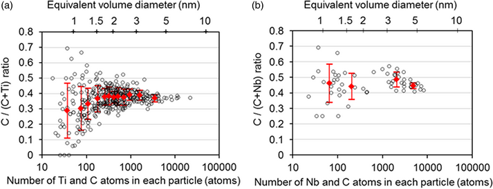
The carbon (C) ratios, namely the atomic ratios of C/(C + M), in nano-sized coherent MC precipitates (M = Ti, Nb) with the NaCl-type (B1) structure in ferritic steels, which had been isothermally aged at 580 °C, were investigated using atom probe tomography (APT). Considering the influences of the trajectory aberration, detection loss, and peak overlap, we determined the C ratios to be ~0.40 and ~0.45 for an equivalent volume diameter of 1.5–5 nm and 1–5 nm for the TiC and NbC precipitates, respectively, suggesting that there is a considerable fraction of C vacancies in both nano-sized precipitates. The apparent C ratios show significant scatter with decreasing particle size, while the apparent mean C ratios of very fine TiC particles, smaller than 1.5 nm, decreased with decreasing particle size. With the use of one of the latest APT instruments with a high detection efficiency, the scattering in the apparent C ratios was reduced because the counting statistics were improved; however, the artificial enrichment of C atoms to particular crystallographic directions of ferrite hindered the determination of the C ratio for very fine TiC particles smaller than 1.5 nm.

Type
Materials Science Applications
Copyright
Copyright © The Author(s), 2020. Published by Cambridge University Press on behalf of the Microscopy Society of America

Access options

Get access to the full version of this content by using one of the access options below. (Log in options will check for institutional or personal access. Content may require purchase if you do not have access.)

References

Albertsen, K & Schaller, H-J (1995). Constitution and thermodynamics of the system Ti-C. Z Metallkd 86, 319325.Google Scholar
Angseryd, J, Liu, F, Andrén, H-O, Gerstl, SSA & Thuvander, M (2011). Quantitative APT analysis of Ti(C,N). Ultramicroscopy 111, 609614.CrossRefGoogle Scholar
Baker, RG & Nutting, J (1959). The tempering of a Cr-Mo-V-W and Mo-V steel, In Precipitation Processes in Steels, Special report no. 64, pp. 122. London: The Iron and Steel Institute.Google Scholar
Breen, AJ, Xie, KY, Moody, MP, Gault, B, Yen, HW, Wong, CC, Cairney, JM & Ringer, SP (2014). Resolving the morphology of niobium carbonitride nano-precipitates in steel using atom probe tomography. Microsc Microanal 20, 11001110.CrossRefGoogle ScholarPubMed
Cahn, JW & Hilliard, JE (1958). Free energy of a nonumiform system. I. Interfacial free energy. J Chem Phys 28, 258267.CrossRefGoogle Scholar
Courtois, E, Epicier, T & Scott, C (2006). EELS study of niobium carbo-nitride nano-precipitates in ferrite. Micron 37, 492502.CrossRefGoogle ScholarPubMed
Danoix, F, Bemont, E, Maugis, P & Blavette, D (2006). Atom probe tomography 1. Early stages of precipitation of NbC and NbN in ferritic steels. Adv Eng Mat 8, 12021205.CrossRefGoogle Scholar
Danoix, F, Grancher, G, Bostel, A & Blavette, D (2007). Standard deviations of composition measurements in atom probe analyses – Part II: 3D atom probe. Ultramicroscopy 107, 739743.CrossRefGoogle ScholarPubMed
Dhara, S, Marceau, RKW, Wood, K, Dorin, T, Timokhina, IB & Hodgson, PD (2018). Atom probe tomography data analysis procedure for precipitate and cluster identification in a Ti-Mo steel. Data in Brief 18, 968982.CrossRefGoogle Scholar
Gault, B, Danoix, F, Hoummada, K, Mangelinck, D & Leitner, H (2012). Impact of directional walk on atom probe microanalysis. Ultramicroscopy 113, 182191.CrossRefGoogle Scholar
Hatzoglou, C, Radiguet, B, Vurpillot, F & Pareige, P (2018). A chemical composition correction model for nanoclusters observed by APT - Application to ODS steel nanoparticles. J Nuc Mater 505, 240248.CrossRefGoogle Scholar
Kapoor, M, O'Malley, R & Thompson, GB (2016). Atom probe tomography study of multi-microalloyed carbide and carbo-nitride precipitates and the precipitation sequence in Nb-Ti HSLA steels. Metal Mater Trans A 47, 1984–1955.CrossRefGoogle Scholar
Kawakami, K & Matsumiya, T (2012). Numerical analysis of hydrogen trap state by TiC and V4C3 in bcc-Fe. ISIJ Int 52, 16931697.CrossRefGoogle Scholar
Kim, YW, Song, SW, Seo, SJ, Hong, SG & Lee, CS (2013). Development of Ti and Mo micro-alloyed hot-rolled high strength sheet steel by controlling thermomechanical controlled processing schedule. Mater Sci Eng A 565, 430438.CrossRefGoogle Scholar
Kobayashi, Y, Takahashi, J & Kawakami, K (2011). Anomalous distribution in atom map of solute carbon in steel. Ultramicroscopy 111, 600603.CrossRefGoogle ScholarPubMed
Kobayashi, Y, Takahashi, J & Kawakami, K (2012). Experimental evaluation of the particle size dependence of the dislocation-particle interaction force in TiC-precipitation-strengthened steel. Scr Mater 67, 854857.CrossRefGoogle Scholar
Kolli, RP & Seidman, DN (2007). Comparison of compositional and morphological atom-probe tomography analyses for a multicomponent Fe-Cu steel. Microsc Microanal 13, 272284.CrossRefGoogle ScholarPubMed
Lipatnikov, VN, Zueva, LV, Gusev, AI & Kottar, A (1998). Disorder-order phase transformations and electrical resistivity of nonstoichiometric titanium carbide. Phys Solid State 40, 12111218.CrossRefGoogle Scholar
Miller, MK (2000). Atom Probe Tomography: Analysis at the Atomic Level. New York, USA: Kluwer Academic/Plenum Publishers.CrossRefGoogle Scholar
Miller, MK, Cerezo, A, Hetherington, MG & Smith, GDW (1996). Atom Probe Field Ion Microscopy. Oxford, UK: Oxford University Press.Google Scholar
Miyamoto, G, Shinbo, K & Furuhara, T (2012). Quantitative measurement of carbon content in Fe–C binary alloys by atom probe tomography. Scr Mater 67, 9991002.CrossRefGoogle Scholar
Mukherjee, S, Timokhina, IB, Zhu, C, Ringer, SP & Hodgson, PD (2013). Three-dimensional atom probe microscopy study of interphase precipitation and nanoclusters in thermomechanically treated titanium–molybdenum steels. Acta Mater 61, 25212530.CrossRefGoogle Scholar
Panayi, P, Clifton, PH, Lloyd, G, Shellswell, G & Cerezo, A (2006). A wide angle achromatic reflectron for the atom probe. In Technical Digest of 50th International Field Emission Symposium, Guillin, China, p. 63.Google Scholar
Prosa, TJ, Geiser, BP, Lawrence, DJ, Olson, JD & Larson, DJ (2014). Developing detection efficiency standards for atom probe tomograpzhy. In Proceedings of SPIE 9173, Instrumentation, Metrology, and Standards for Nanomanufacturing, Optics and Semiconductors VIII, San Diego, USA, p. 917307.Google Scholar
Sha, W, Chang, L, Smith, GDW, Cheng, L & Mittemeijer, EJ (1992). Some aspects of atom-probe analysis of Fe-C and Fe-N systems. Surf Sci 266, 416423.CrossRefGoogle Scholar
Smith, CJ, Weinberger, CR & Thompson, GB (2018). Phase stability and microstructural formations in the niobium carbides. J Eur Ceram Soc 38, 48504866.CrossRefGoogle Scholar
Smith, JF, Carlson, ON & De Avillez, RR (1990). C−Nb, Binary alloy phase diagrams, 2nd ed. In Massalski, TB & Okamoto, H (Eds.), p. 863. Materials Park, Ohio, USA: ASM International.Google Scholar
Stefano, DD, Nazarov, R, Hickel, T, Neugebauer, J, Mrovec, M & Elsasser, C (2016). First-principles investigation of hydrogen interaction with TiC precipitates in α-Fe. Phys Rev B 93, 184108.Google Scholar
Stephenson, LT, Moody, MP, Gault, B & Ringer, SP (2011). Estimating the physical cluster-size distribution within materials using atom-probe. Microsc Res Tech 74, 799803.Google ScholarPubMed
Sun, Z, Ahuja, R & Lowther, JE (2010). Mechanical properties of vanadium carbide and a ternary vanadium tungsten carbide. Solid State Commun 150, 697700.CrossRefGoogle Scholar
Takahashi, J, Kawakami, K & Kobayashi, Y (2012). Consideration of particle-strengthening mechanism of copper-precipitation-strengthened steels by atom probe tomography analysis. Mater Sci Eng A 535, 144152.CrossRefGoogle Scholar
Takahashi, J, Kawakami, K & Kobayashi, Y (2018). Origin of hydrogen trapping site in vanadium carbide precipitation strengthening steel. Acta Mater 153, 193204.CrossRefGoogle Scholar
Takahashi, J, Kawakami, K & Kobayashi, Y (2020). Study on quantitative analysis of carbon and nitrogen in stoichiometric θ-Fe3C and γ-Fe4N by atom probe tomography. Microsc Microanal 26, 185193.CrossRefGoogle Scholar
Takahashi, J, Kawakami, K, Kobayashi, Y & Tarui, T (2010). The first direct observation of hydrogen trapping sites in TiC precipitation-hardening steel through atom probe tomography. Scr Mater 63, 261264.CrossRefGoogle Scholar
Thuvander, M, Kvist, A, Johnson, LJS, Weidow, J & Andrén, H-O (2013). Reduction of multiple hits in atom probe tomography. Ultramicroscopy 132, 8185.CrossRefGoogle ScholarPubMed
Thuvander, M, Shinde, D, Rehan, A, Ejnermark, S & Stiller, K (2019). Improving compositional accuracy in APT analysis of carbides using a decreased detection efficiency. Microsc Microanal 25, 454461.CrossRefGoogle ScholarPubMed
Thuvander, M, Weidow, J, Angseryd, J, Falk, LKL, Liu, F, Sonestedt, M, Stiller, K & Andrén, H-O (2011). Quantitative atom probe analysis of carbides. Ultramicroscopy 111, 604608.CrossRefGoogle ScholarPubMed
Timokhina, IB, Hodgson, PD, Ringer, SP, Zheng, RK & Pereloma, EV (2007). Precipitate characterisation of an advanced high-strength low-alloy (HSLA) steel using atom probe tomography. Scr Mater 56, 601604.CrossRefGoogle Scholar
Tsong, TT (1978). Field ion image formation. Surf Sci 70, 211233.CrossRefGoogle Scholar
Vaumousse, D, Cerezo, A & Warren, PJ (2003). A procedure for quantification of precipitate microstructures from three-dimensional atom probe data. Ultramicroscopy 95, 215221.CrossRefGoogle ScholarPubMed
Vurpillot, F, Bostel, A & Blavette, D (2000). Trajectory overlaps and local magnification in three-dimensional atom probe. Appl Phys Lett 76, 31273129.CrossRefGoogle Scholar
Wang, J, Weyland, M, Bikmukhametov, I, Miller, MK, Hodgson, PD & Timokhina, I (2019). Transformation from cluster to nano-precipitate in microalloyed ferritic steel. Scr Mater 160, 5357.CrossRefGoogle Scholar
Wei, FG, Hara, T & Tsuzaki, K (2004). High-resolution transmission electron microscopy study of crystallography and morphology of TiC precipitates in tempered steel. Phil Mag 11, 17351751.CrossRefGoogle Scholar
Wei, FG & Tsuzaki, K (2006). Quantitative analysis on hydrogen trapping of TiC particles in steel. Metal Mater Trans A 37, 331353.CrossRefGoogle Scholar
Wei, FG & Tsuzaki, K (2007). Nanosized carbides to improve the practical strength of martensitic steels. Ferrum (Bulletin of the Iron and Steel Institute of Japan) 12, 766770.Google Scholar
Yamaguchi, Y, Takahashi, J & Kawakami, K (2009). The study of quantitativeness in atom probe analysis of alloying elements in steel. Ultramicroscopy 109, 541544.CrossRefGoogle Scholar
Zueva, LV & Gusev, I (1999). Effect of nonstoichiometry and ordering on the period of the basis structure of cubic titanium carbide. Phys Solid State 41, 10321038.CrossRefGoogle Scholar