Hostname: page-component-586b7cd67f-rcrh6 Total loading time: 0 Render date: 2024-11-28T03:05:26.408Z Has data issue: false hasContentIssue false

A Comparison of a Direct Electron Detector and a High-Speed Video Camera for a Scanning Precession Electron Diffraction Phase and Orientation Mapping

Published online by Cambridge University Press:  01 September 2020

Ian MacLaren*
Affiliation:
School of Physics and Astronomy, University of Glasgow, Glasgow G12 8QQ, UK
Enrique Frutos-Myro
Affiliation:
School of Engineering, University of Glasgow, Glasgow G12 8QQ, UK
Damien McGrouther
Affiliation:
School of Physics and Astronomy, University of Glasgow, Glasgow G12 8QQ, UK
Sam McFadzean
Affiliation:
School of Physics and Astronomy, University of Glasgow, Glasgow G12 8QQ, UK
Jon Karl Weiss
Affiliation:
NanoMEGAS USA, 1095 W Rio Salado Parkway, Suite 110, Tempe, AZ 85281, USA
Doug Cosart
Affiliation:
NanoMEGAS USA, 1095 W Rio Salado Parkway, Suite 110, Tempe, AZ 85281, USA
Joaquim Portillo
Affiliation:
NanoMEGAS SPRL, Bd.Edmond Machtens 79 bte 22, 1080 Brussels, Belgium Centres Cientifics i Tecnologics, Universitat de Barcelona, Sole i Sabaris, 1-3, Barcelona 08028, Spain
Alan Robins
Affiliation:
NanoMEGAS SPRL, Bd.Edmond Machtens 79 bte 22, 1080 Brussels, Belgium
Stavros Nicolopoulos
Affiliation:
NanoMEGAS SPRL, Bd.Edmond Machtens 79 bte 22, 1080 Brussels, Belgium
Eduardo Nebot del Busto
Affiliation:
Quantum Detectors Ltd., R104, RAL, Harwell, Oxford OX11 0QX, UK
Richard Skogeby
Affiliation:
Quantum Detectors Ltd., R104, RAL, Harwell, Oxford OX11 0QX, UK
*
*Author for correspondence: Ian MacLaren, E-mail: [email protected]
Get access

Abstract

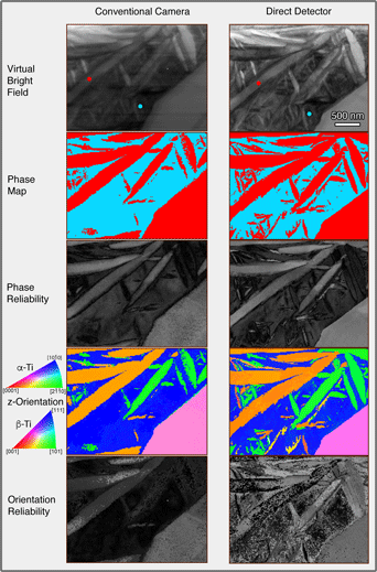
A scanning precession electron diffraction system has been integrated with a direct electron detector to allow the collection of improved quality diffraction patterns. This has been used on a two-phase α–β titanium alloy (Timetal® 575) for phase and orientation mapping using an existing pattern-matching algorithm and has been compared to the commonly used detector system, which consisted of a high-speed video-camera imaging the small phosphor focusing screen. Noise is appreciably lower with the direct electron detector, and this is especially noticeable further from the diffraction pattern center where the real electron scattering is reduced and both diffraction spots and inelastic scattering between spots are weaker. The results for orientation mapping are a significant improvement in phase and orientation indexing reliability, especially of fine nanoscale laths of α-Ti, where the weak diffracted signal is rather lost in the noise for the optically coupled camera. This was done at a dose of ~19 e2, and there is clearly a prospect for reducing the current further while still producing indexable patterns. This opens the way for precession diffraction phase and orientation mapping of radiation-sensitive crystalline materials.

Type
Software and Instrumentation
Copyright
Copyright © Microscopy Society of America 2020

Access options

Get access to the full version of this content by using one of the access options below. (Log in options will check for institutional or personal access. Content may require purchase if you do not have access.)

References

Beacham, R, Mac Raighne, A, Maneuski, D, O'Shea, V, McVitie, S & McGrouther, D (2011). Medipix2/Timepix detector for time resolved transmission electron microscopy. J Instrum 6, C12052.CrossRefGoogle Scholar
Cooper, D, Bernier, N, Rouvière, J-L, Wang, Y-Y, Weng, W, Madan, A, Mochizuki, S & Jagannathan, H (2017). High-precision deformation mapping in finFET transistors with two nanometre spatial resolution by precession electron diffraction. Appl Phys Lett 110(22), 223109.CrossRefGoogle ScholarPubMed
Darbal, AD, Narayan, RD, Vartuli, C, Aoki, T, Mardinly, J, Nicolopoulos, S & Weiss, JK (2014). Applications of automated high resolution strain mapping in TEM on the study of strain distribution in MOSFETs. Microsc Microanal 20(S3), 10661067.CrossRefGoogle Scholar
Darbal, AD, Narayan, RD, Vartuli, C, Lian, G, Graham, R, Shaapur, F, Nicolopoulos, S & Weiss, JK (2013). Automated high precision strain measurement using nanobeam diffraction coupled with precession. Microsc Microanal 19(S2), 702703.CrossRefGoogle Scholar
Doherty, TAS, Winchester, AJ, Macpherson, S, Johnstone, DN, Pareek, V, Tennyson, EM, Kosar, S, Kosasih, FU, Anaya, M, Abdi-Jalebi, M, Andaji-Garmaroudi, Z, Wong, EL, Madéo, J, Chiang, Y-H, Park, J-S, Jung, Y-K, Petoukhoff, CE, Divitini, G, Man, MKL, Ducati, C, Walsh, A, Midgley, PA, Dani, KM & Stranks, SD (2020). Performance-limiting nanoscale trap clusters at grain junctions in halide perovskites. Nature 580(7803), 360366.CrossRefGoogle ScholarPubMed
Eggeman, AS, Krakow, R & Midgley, PA (2015). Scanning precession electron tomography for three-dimensional nanoscale orientation imaging and crystallographic analysis. Nat Commun 6, 7267.CrossRefGoogle ScholarPubMed
Estradé, S, Portillo, J, Mendoza, J, Kosta, I, Serret, M, Müller, C & Peiró, F (2012). Assessment of misorientation in metallic and semiconducting nanowires using precession electron diffraction. Micron 43(8), 910915.CrossRefGoogle ScholarPubMed
Gjønnes, J, Hansen, V, Berg, BS, Runde, P, Cheng, YF, Gjønnes, K, Dorset, DL & Gilmore, CJ (1998). Structure model for the phase AlmFe derived from three-dimensional electron diffraction intensity data collected by a precession technique. Comparison with convergent-beam diffraction. Acta Crystallogr A 54, 306319.CrossRefGoogle Scholar
Gjønnes, K (1997). On the integration of electron diffraction intensities in the Vincent-Midgley precession technique. Ultramicroscopy 69(1), 111.CrossRefGoogle Scholar
Hart, MJ, Bassiri, R, Borisenko, KB, Veron, M, Rauch, EF, Martin, LW, Rowan, S, Fejer, MM & MacLaren, I (2016). Medium range structural order in amorphous tantala spatially resolved with changes to atomic structure by thermal annealing. J Non-Cryst Solids 438, 1017.CrossRefGoogle Scholar
Humbert, M, Wagner, F, Moustahfid, H & Esling, C (1995). Determination of the orientation of a parent [beta] grain from the orientations of the inherited [alpha] plates in the phase transformation from body-centred cubic to hexagonal close packed. J Appl Crystallogr 28(5), 571576.CrossRefGoogle Scholar
Johnstone, DN, Crout, P, Laulainen, J, Høgås, S, Martineau, B, Bergh, T, Smeets, S, Francis, C, Opheim, E, Prestat, E, Collins, S, Danaie, M, Furnival, T, Ånes, HW, Morzy, J, Iqbal, A, Doherty, T, von Lany, M, Ostasevicius, T, Tovey, R, Jacobsen, E & Poon, T (2020 a) pyxem/pyxem: pyxem 0.11.0. Available at https://doi.org/10.5281/zenodo.3831473.CrossRefGoogle Scholar
Johnstone, DN, Firth, FCN, Grey, CP, Midgley, PA, Cliffe, MJ & Collins, SM (2020 b). Direct imaging of correlated defect nanodomains in a metal–organic framework. J Am Chem Soc 142(30), 1308113089.CrossRefGoogle Scholar
Kolb, U, Gorelik, T, Kübel, C, Otten, MT & Hubert, D (2007). Towards automated diffraction tomography: Part I—data acquisition. Ultramicroscopy 107(6), 507513.CrossRefGoogle ScholarPubMed
Krajnak, M, McGrouther, D, Maneuski, D, O'Shea, V & McVitie, S (2016). Pixelated detectors and improved efficiency for magnetic imaging in STEM differential phase contrast. Ultramicroscopy 165, 4250.CrossRefGoogle ScholarPubMed
Mac Raighne, A, Fernandez, GV, Maneuski, D, McGrouther, D & O'Shea, V (2011). Medipix2 as a highly flexible scanning/imaging detector for transmission electron microscopy. J Instrum 6, C01047.Google Scholar
Martineau, B, Johnstone, DN, van Helvoort, ATJ, Midgley, PA & Eggeman, AS (2019). Unsupervised machine learning applied to scanning precession electron diffraction data. Adv Struct Chem Imaging 5, 3.CrossRefGoogle Scholar
Mir, JA, Clough, R, MacInnes, R, Gough, C, Plackett, R, Shipsey, I, Sawada, H, MacLaren, I, Ballabriga, R, Maneuski, D, O'Shea, V, McGrouther, D & Kirkland, AI (2017). Characterisation of the Medipix3 detector for 60 and 80 keV electrons. Ultramicroscopy 182, 4453.CrossRefGoogle Scholar
Moeck, P, Rouvimov, S, Rauch, EF, Véron, M, Kirmse, H, Häusler, I, Neumann, W, Bultreys, D, Maniette, Y & Nicolopoulos, S (2011). High spatial resolution semi-automatic crystallite orientation and phase mapping of nanocrystals in transmission electron microscopes. Crystallogr Res Technol 46(6), 589606.CrossRefGoogle Scholar
Mugnaioli, E, Gorelik, T & Kolb, U (2009). “Ab initio” structure solution from electron diffraction data obtained by a combination of automated diffraction tomography and precession technique. Ultramicroscopy 109(6), 758765.CrossRefGoogle ScholarPubMed
Nord, M, Ross, A, McGrouther, D, Barthel, J, Moreau, M, Hallsteinsen, I, Tybell, T & MacLaren, I (2019 a). Three-dimensional subnanoscale imaging of unit cell doubling due to octahedral tilting and cation modulation in strained perovskite thin films. Phys Rev Mater 3(6), 063605.CrossRefGoogle Scholar
Nord, M, Semisalova, A, Kákay, A, Hlawacek, G, MacLaren, I, Liersch, V, Volkov, OM, Makarov, D, Paterson, GW, Potzger, K, Lindner, J, Fassbender, J, McGrouther, D & Bali, R (2019 b). Strain anisotropy and magnetic domains in embedded nanomagnets. Small 15(52), 1904738.CrossRefGoogle ScholarPubMed
Ophus, C (2019). Four-dimensional scanning transmission electron microscopy (4D-STEM): From scanning nanodiffraction to ptychography and beyond. Microsc Microanal 25(3), 563582.CrossRefGoogle ScholarPubMed
Paterson, GW, Webster, RWH, Ross, A, Paton, KA, Macgregor, TA, McGrouther, D, MacLaren, I & Nord, M (2020). Fast pixelated detectors in scanning transmission electron microscopy. Part II: post-acquisition data processing, visualization, and structural characterization. arXiv e-prints, pp. arXiv:2004.02777, and Microscopy and Microanalysis, 120. doi:10.1017/S1431927620024307.CrossRefGoogle Scholar
Pennycook, TJ, Lupini, AR, Yang, H, Murfitt, MF, Jones, L & Nellist, PD (2015). Efficient phase contrast imaging in STEM using a pixelated detector. Part 1: Experimental demonstration at atomic resolution. Ultramicroscopy 151, 160167.CrossRefGoogle ScholarPubMed
Ramachandra, R, Bouwer, JC, Mackey, MR, Bushong, E, Peltier, ST, Xuong, NH & Ellisman, MH (2014). Improving signal to noise in labeled biological specimens using energy-filtered TEM of sections with a drift correction strategy and a direct detection device. Microsc Microanal 20(3), 706714.CrossRefGoogle Scholar
Rauch, EF, Portillo, J, Nicolopoulos, S, Bultreys, D, Rouvimov, S & Moeck, P (2010). Automated nanocrystal orientation and phase mapping in the transmission electron microscope on the basis of precession electron diffraction. Z Kristallogr-Cryst Mater 225(2-3), 103109.CrossRefGoogle Scholar
Reisinger, M, Zalesak, J, Daniel, R, Tomberger, M, Weiss, JK, Darbal, AD, Petrenec, M, Zechner, J, Daumiller, I, Ecker, W, Sartory, B & Keckes, J (2016). Cross-sectional stress distribution in AlxGa1-xN heterostructure on Si(111) substrate characterized by ion beam layer removal method and precession electron diffraction. Mater Design 106, 476481.CrossRefGoogle Scholar
Rouvimov, S, Rauch, EF, Moeck, P & Nicolopoulos, S (2009). Automated crystal orientation and phase mapping of iron oxide nano-crystals in a transmission electron microscope. Microsc Microanal 15(S2), 12901291.CrossRefGoogle Scholar
Ruskin, RS, Yu, ZH & Grigorieff, N (2013). Quantitative characterization of electron detectors for transmission electron microscopy. J Struct Biol 184(3), 385393.CrossRefGoogle ScholarPubMed
Ryll, H, Simson, M, Hartmann, R, Holl, P, Huth, M, Ihle, S, Kondo, Y, Kotula, P, Liebel, A, Muller-Caspary, K, Rosenauer, A, Sagawa, R, Schmidt, J, Soltau, H & Struder, L (2016). A pnCCD-based, fast direct single electron imaging camera for TEM and STEM. J Instrum 11(19), 04006.CrossRefGoogle Scholar
Sunde, JK, Johnstone, DN, Wenner, S, van Helvoort, ATJ, Midgley, PA & Holmestad, R (2019). Crystallographic relationships of T-/S-phase aggregates in an Al-Cu-Mg-Ag alloy. Acta Mater 166, 587596.CrossRefGoogle Scholar
Sunde, JK, Marioara, CD, van Helvoort, ATJ & Holmestad, R (2018). The evolution of precipitate crystal structures in an Al-Mg-Si(-Cu) alloy studied by a combined HAADF-STEM and SPED approach. Mater Charact 142, 458469.CrossRefGoogle Scholar
Tate, MW, Purohit, P, Chamberlain, D, Nguyen, KX, Hovden, R, Chang, CS, Deb, P, Turgut, E, Heron, JT, Schlom, DG, Ralph, DC, Fuchs, GD, Shanks, KS, Philipp, HT, Muller, DA & Gruner, SM (2016). High dynamic range pixel array detector for scanning transmission electron microscopy. Microsc Microanal 22(1), 237249.CrossRefGoogle ScholarPubMed
Valery, A, Rauch, EF, Clément, L & Lorut, F (2017). Retrieving overlapping crystals information from TEM nano-beam electron diffraction patterns. J Microsc-Oxford 268(2), 208218.CrossRefGoogle ScholarPubMed
Vincent, R & Midgley, PA (1994). Double conical beam-rocking system for measurement of integrated electron diffraction intensities. Ultramicroscopy 53(3), 271282.CrossRefGoogle Scholar
Yang, H, Jones, L, Ryll, H, Simson, M, Soltau, H, Kondo, Y, Sagawa, R, Banba, H, Maclaren, I & Nellist, PD (2015 a). 4D STEM: High efficiency phase contrast imaging using a fast pixelated detector. J Phys Conf Ser 644, 012032.CrossRefGoogle Scholar
Yang, H, MacLaren, I, Jones, L, Martinez, GT, Simson, M, Huth, M, Ryll, H, Soltau, H, Sagawa, R, Kondo, Y, Ophus, C, Ercius, P, Jin, L, Kovacs, A & Nellist, PD (2017). Electron ptychographic phase imaging of light elements in crystalline materials using Wigner distribution deconvolution. Ultramicroscopy 180, 173179.CrossRefGoogle ScholarPubMed
Yang, H, Pennycook, TJ & Nellist, PD (2015 b). Efficient phase contrast imaging in STEM using a pixelated detector. Part II: Optimisation of imaging conditions. Ultramicroscopy 151, 232239.CrossRefGoogle ScholarPubMed