Hostname: page-component-586b7cd67f-vdxz6 Total loading time: 0 Render date: 2024-11-30T20:34:14.708Z Has data issue: false hasContentIssue false

Atomic Resolution Imaging of Light Elements in a Crystalline Environment using Dynamic Hollow-Cone Illumination Transmission Electron Microscopy

Published online by Cambridge University Press:  10 June 2020

Hamish G. Brown*
Affiliation:
National Center for Electron Microscopy, Molecular Foundry, Lawrence Berkeley National Laboratory, Berkeley, CA, USA
Jim Ciston
Affiliation:
National Center for Electron Microscopy, Molecular Foundry, Lawrence Berkeley National Laboratory, Berkeley, CA, USA
*
*Author for correspondence: Hamish G. Brown, E-mail: [email protected]
Get access

Abstract

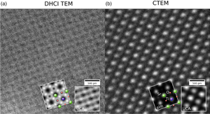
Multiple electron scattering and the nonintuitive nature of image formation with coherent radiation complicate the interpretation of conventional transmission electron microscopy images. Precession of the illuminating beam in transmission electron microscopy (TEM) can lead to more robust and interpretable images with some penalty to image contrast, a technique known as dynamic hollow-cone illumination TEM. We demonstrate direct and robust imaging of light and heavy atoms in a crystalline environment with this technique. This method is similar to the annular bright-field technique in scanning transmission electron microscopy, via the principle of reciprocity. Dynamic hollow-cone illumination TEM is challenging in practice due to sensitivity to the misalignment of the precession axis, microscope objective aperture, and crystal zone axis.

Type
Software and Instrumentation
Copyright
Copyright © Microscopy Society of America 2020

Access options

Get access to the full version of this content by using one of the access options below. (Log in options will check for institutional or personal access. Content may require purchase if you do not have access.)

References

Avilov, A, Kuligin, K, Nicolopoulos, S, Nickolskiy, M, Boulahya, K, Portillo, J, Lepeshov, G, Sobolev, B, Collette, JP, Martin, N, Robins, AC & Fischione, P (2007). Precession technique and electron diffractometry as new tools for crystal structure analysis and chemical bonding determination. Ultramicroscopy 107, 431444.CrossRefGoogle ScholarPubMed
Barnum, A (2019). k-scanning: An alternative to 4D-STEM. Microsc Microanal 25, 19321933.CrossRefGoogle Scholar
Brown, HG, Ishikawa, R, Sánchez-Santolino, G, Lugg, NR, Ikuhara, Y, Allen, LJ & Shibata, N (2017). A new method to detect and correct sample tilt in scanning transmission electron microscopy bright-field imaging. Ultramicroscopy 173, 7683.CrossRefGoogle ScholarPubMed
Ciston, J, Deng, B, Marks, LD, Own, CS & Sinkler, W (2008). A quantitative analysis of the cone-angle dependence in precession electron diffraction. Ultramicroscopy 108, 514522.CrossRefGoogle ScholarPubMed
Cowley, JM (1969). Image contrast in a transmission scanning electron microscope. Appl Phys Lett 15, 5859.CrossRefGoogle Scholar
Cowley, JM & Moodie, AF (1957). Fourier images: I-the point source. Proc Phys Soc B 70, 486.CrossRefGoogle Scholar
Cullis, A & Maher, D (1975). Topographical contrast in the transmission electron microscope. Ultramicroscopy 1, 97112.CrossRefGoogle ScholarPubMed
Findlay, SD, Shibata, N, Sawada, H, Okunishi, E, Kondo, Y & Ikuhara, Y (2010). Dynamics of annular bright field imaging in scanning transmission electron microscopy. Ultramicroscopy 110, 903923.CrossRefGoogle ScholarPubMed
Findlay, SD, Shibata, N, Sawada, H, Okunishi, E, Kondo, Y, Yamamoto, T & Ikuhara, Y (2009). Robust atomic resolution imaging of light elements using scanning transmission electron microscopy. Appl Phys Lett 95, 191913.CrossRefGoogle Scholar
Geipel, T & Mader, W (1996). Practical aspects of hollow-cone imaging. Ultramicroscopy 63, 6574.CrossRefGoogle Scholar
Henderson, R (1995). The potential and limitations of neutrons, electrons and X-rays for atomic resolution microscopy of unstained biological molecules. Q Rev Biophys 28, 171193.CrossRefGoogle ScholarPubMed
Ishikawa, R, Okunishi, E, Sawada, H, Kondo, Y, Hosokawa, F & Abe, E (2011). Direct imaging of hydrogen-atom columns in a crystal by annular bright-field electron microscopy. Nat Mater 10, 278281.CrossRefGoogle Scholar
Kunath, W, Zemlin, F & Weiss, K (1985). Apodization in phase-contrast electron microscopy realized with hollow-cone illumination. Ultramicroscopy 16, 123138.CrossRefGoogle Scholar
Nagata, F, Matsuda, T, Komoda, T & Hama, K (1976). High resolution observation of biological specimens by an incoherent illumination method. Microscopy 25, 237243.Google ScholarPubMed
Nagayama, K, Danev, R, Shigematsu, H, Hosogi, N, Fukuda, Y, Nitta, K & Kaneko, Y (2010). Phase contrast enhancement with phase plates in biological electron microscopy. Microsc Today 18, 1013.CrossRefGoogle Scholar
Own, C, Marks, L & Sinkler, W (2005). Electron precession: A guide for implementation. Rev Sci Instrum 76, 033703.CrossRefGoogle Scholar
Pennycook, SJ & Boatner, LA (1988). Chemically sensitive structure-imaging with a scanning transmission electron microscope. Nature 336, 565.CrossRefGoogle Scholar
Pennycook, TJ, Lupini, AR, Yang, H, Murfitt, MF, Jones, L & Nellist, PD (2015). Efficient phase contrast imaging in stem using a pixelated detector. Part 1: Experimental demonstration at atomic resolution. Ultramicroscopy 151, 160167.CrossRefGoogle ScholarPubMed
Pogany, AP & Turner, PS (1968). Reciprocity in electron diffraction and microscopy. Acta Crystallogr A 24, 103109.CrossRefGoogle Scholar
Rebled, JM, Yedra, Ll., Estrade, S, Portillo, J & Peiro, F (2011). A new approach for 3D reconstruction from bright field TEM imaging: Beam precession assisted electron tomography. Ultramicroscopy 111, 15041511.CrossRefGoogle ScholarPubMed
Rez, P (2003). Comparison of phase contrast transmission electron microscopy with optimized scanning transmission annular dark field imaging for protein imaging. Ultramicroscopy 96, 117124.CrossRefGoogle ScholarPubMed
Rosenauer, A, Krause, FF, Müller, K, Schowalter, M & Mehrtens, T (2014). Conventional transmission electron microscopy imaging beyond the diffraction and information limits. Phys Rev Lett 113, 096101.CrossRefGoogle ScholarPubMed
Saxton, WO, Jenkins, WK, Freeman, LA & Smith, D (1978). TEM observations using bright field hollow cone illumination. Optik (Jena) 49, 505510.Google Scholar
Seki, T, Takanashi, N & Abe, E (2018). Integrated contrast-transfer-function for aberration-corrected phase-contrast stem. Ultramicroscopy 194, 193198.CrossRefGoogle ScholarPubMed
Sidorov, MV, McCartney, MR & Smith, DJ (2000). High resolution TEM imaging with hollow-cone illumination. Microsc Microanal 3, 11911192.CrossRefGoogle Scholar
Spence, JCH (2013). High-Resolution Electron Microscopy, Chapter 5. Oxford: OUP, pp. 973977.CrossRefGoogle Scholar
Vincent, R & Midgley, PA (1994). Double conical beam-rocking system for measurement of integrated electron diffraction intensities. Ultramicroscopy 53, 271282.CrossRefGoogle Scholar
Zhou, D, Müller-Caspary, K, Sigle, W, Krause, FF, Rosenauer, A & van Aken, PA (2016). Sample tilt effects on atom column position determination in ABF–STEM imaging. Ultramicroscopy 160, 110117.CrossRefGoogle ScholarPubMed
Supplementary material: File

Brown and Ciston supplementary material

Brown and Ciston supplementary material 1

Download Brown and Ciston supplementary material(File)
File 71.5 KB
Supplementary material: PDF

Brown and Ciston supplementary material

Brown and Ciston supplementary material 2

Download Brown and Ciston supplementary material(PDF)
PDF 951.1 KB