Hostname: page-component-78c5997874-dh8gc Total loading time: 0 Render date: 2024-11-14T07:27:14.491Z Has data issue: false hasContentIssue false

Novel 3D-printed methacrylated chitosan-laponite nanosilicate composite scaffolds enhance cell growth and biomineral formation in MC3T3 pre-osteoblasts

Published online by Cambridge University Press:  23 August 2018

Tugba Cebe
Affiliation:
Department of Materials Science and Engineering, University of Texas at Arlington, Arlington, Texas 76019, USA
Neelam Ahuja
Affiliation:
Department of Graduate Nursing, College of Nursing and Health Innovation, University of Texas at Arlington, Arlington, Texas 76019, USA
Felipe Monte
Affiliation:
Department of Graduate Nursing, College of Nursing and Health Innovation, University of Texas at Arlington, Arlington, Texas 76019, USA
Kamal Awad
Affiliation:
Department of Materials Science and Engineering, University of Texas at Arlington, Arlington, Texas 76019, USA; and Department of Refractories and Ceramics, National Research Centre, Giza 12622, Egypt
Kimaya Vyavhare
Affiliation:
Department of Materials Science and Engineering, University of Texas at Arlington, Arlington, Texas 76019, USA
Pranesh Aswath
Affiliation:
Department of Materials Science and Engineering, University of Texas at Arlington, Arlington, Texas 76019, USA
Jian Huang
Affiliation:
Department of Graduate Nursing, College of Nursing and Health Innovation, University of Texas at Arlington, Arlington, Texas 76019, USA
Marco Brotto
Affiliation:
Department of Graduate Nursing, College of Nursing and Health Innovation, University of Texas at Arlington, Arlington, Texas 76019, USA
Venu Varanasi*
Affiliation:
Department of Materials Science and Engineering, University of Texas at Arlington, Arlington, Texas 76019, USA; and Department of Graduate Nursing, College of Nursing and Health Innovation, University of Texas at Arlington, Arlington, Texas 76019, USA
*
a)Address all correspondence to this author. e-mail: [email protected]
Get access

Abstract

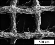
This study compared the effect of gelatin- and chitosan-based scaffolds on osteoblast biomineralization. These scaffolds have been modified using methacrylate and laponite nanosilicates to improve their mechanical strength and support osteoblast function. Scaffold materials were prepared to have the same compressive strength (14–15 MPa) such that differences in cell response would be isolated to differences in biopolymer chemistry. The materials were tested for rheological properties to optimize the bio-ink for successful 3D printing using a robocast-assisted deposition system. Osteoblasts were cultured on the surface of 3D-printed methacrylated chitosan-laponite (MAC-Lp), methacrylated gelatin-laponite (MAG-Lp), MAC, and MAG scaffolds. MAC-Lp scaffolds showed increased cell viability, cell growth, and biomineral formation as compared to MAG-Lp scaffolds. FTIR results showed the presence of higher biomineral phosphate and extracellular matrix (ECM) collagen-like amide formation on MAC-Lp scaffolds as compared to MAG-Lp scaffolds. MAC-Lp scaffolds showed increased density of ECM-like tissue from SEM analysis, stained mineral nodules from Alizarin staining, and the existence of Ca–P species evident by X-ray absorbance near edge structure analysis. In conclusion, MAC-Lp scaffolds enhanced osteoblast growth and biomineral formation as compared to MAG-Lp scaffolds.

Keywords

Type
Article
Copyright
Copyright © Materials Research Society 2018 

Access options

Get access to the full version of this content by using one of the access options below. (Log in options will check for institutional or personal access. Content may require purchase if you do not have access.)

Footnotes

Since the original publication of this article, the Journal of Materials Research has moved to a new design layout. This article has been republished in the new design, and no changes have been made to the article content.

References

Saxena, S., Ray, A., Kapil, A., Pavon Djavid, G., Letourneur, D., Gupta, B., and Meddahi-Pellé, A.: Development of a new polypropylene-based suture: Plasma grafting, surface treatment, characterization, and biocompatibility studies. Macromolecular bioscience. 11, 373 (2011).CrossRefGoogle ScholarPubMed
Chiarello, E., Cadossi, M., Tedesco, G., Capra, P., Calamelli, C., Shehu, A., and Giannini, S.: Autograft, allograft and bone substitutes in reconstructive orthopedic surgery. Aging: Clin. Exp. Res. 25, 101 (2013).Google ScholarPubMed
Grove, J.R.: Autograft, allograft and xenograft options in the treatment of neglected Achilles tendon ruptures: a historical review with illustration of surgical repair. Surgeon. 15, 47 (2008).Google Scholar
Peppas, N.A. and Langer, R.: New challenges in biomaterials. Science 263, 1715 (1994).CrossRefGoogle ScholarPubMed
Censi, R., Schuurman, W., Malda, J., di Dato, G., Burgisser, P.E., Dhert, W.J.A., van Nostrum, C.F., di Martino, P., Vermonden, T., and Hennink, W.E.: A printable photopolymerizable thermosensitive p(HPMAm-lactate)-PEG hydrogel for tissue engineering. Adv. Funct. Mater. 21, 1833 (2011).CrossRefGoogle Scholar
Wangtueai, S. and Noomhorm, A.: Processing optimization and characterization of gelatin from lizardfish (Saurida spp.) scales. LWT–Food Sci. Technol. 42, 825 (2009).CrossRefGoogle Scholar
Schwenke, K.D.: The Science and Technology of Gelatin. Herausgegeben von A. G. Ward u. A. Courts, XVI und 564 Seiten mit zahlreichen Abb. u. Tab., Academic Press London, New York, San Francisco 1977. Preis: 18,00 £; 39,50 $. Food/Nahrung. 22, 444 (1978).CrossRefGoogle Scholar
Maurer, P.H.: II. Antigenicity of gelatin in rabbits and other species. J. Exp. Med. 100, 515 (1954).CrossRefGoogle ScholarPubMed
Zhu, J. and Marchant, R.: Design properties of hydrogel tissue-engineering scaffolds. Expert Rev. Med. Devices 8, 607 (2011).CrossRefGoogle ScholarPubMed
Haraguchi, K. and Li, H-J.: Mechanical properties and structure of polymer−clay nanocomposite gels with high clay content. Macromolecules 39, 1898 (2006).CrossRefGoogle Scholar
Tian, W.M., Hou, S.P., Ma, J., Zhang, C.L., Xu, Q.Y., Lee, I.S., Li, H.D., Spector, M., and Cui, F.Z.: Hyaluronic acid–poly-D-lysine-based three-dimensional hydrogel for traumatic brain injury. Tissue Eng. 11, 513 (2005).CrossRefGoogle ScholarPubMed
Clark, A.H., Richardson, R.K., Ross-Murphy, S.B., and Stubbs, J.M.: Structural and mechanical properties of agar/gelatin co-gels. Small-deformation studies. Macromolecules 16, 1367 (1983).CrossRefGoogle Scholar
Yasuda, K., Ping Gong, J., Katsuyama, Y., Nakayama, A., Tanabe, Y., Kondo, E., Ueno, M., and Osada, Y.: Biomechanical properties of high-toughness double network hydrogels. Biomaterials 26, 4468 (2005).CrossRefGoogle ScholarPubMed
Iyer, P., Walker, K., and Madihally, S.: Increased matrix synthesis by fibroblasts with decreased proliferation on synthetic chitosan-gelatin porous structures. Biotechnol. Bioeng. 109, 1314 (2012).CrossRefGoogle ScholarPubMed
Shigemasa, Y., Saito, K., Sashiwa, H., and Saimoto, H.: Enzymatic degradation of chitins and partially deacetylated chitins. Int. J. Biol. Macromol. 16, 43 (1994).CrossRefGoogle ScholarPubMed
Khor, E. and Lim, L.: Implantable applications of chitin and chitosan. Biomaterials 24, 2339 (2003).CrossRefGoogle ScholarPubMed
Madihally, S.V. and Matthew, H.W.T.: Porous chitosan scaffolds for tissue engineering. Biomaterials 20, 1133 (1999).CrossRefGoogle ScholarPubMed
Suh, J.K. and Matthew, H.W.: Application of chitosan-based polysaccharide biomaterials in cartilage tissue engineering: A review. Biomaterials 21, 2589 (2000).Google ScholarPubMed
Mi, F-L., Tan, Y-C., Liang, H-F., and Sung, H-W.: In vivo biocompatibility and degradability of a novel injectable-chitosan-based implant. Biomaterials 23, 181 (2002).CrossRefGoogle ScholarPubMed
Aiba, S., Minoura, N., Taguchi, K., and Fujiwara, Y.: Covalent immobilization of chitosan derivatives onto polymeric film surfaces with the use of a photosensitive hetero-bifunctional crosslinking reagent. Biomaterials 8, 481 (1987).CrossRefGoogle ScholarPubMed
Cai, K., Yao, K., Cui, Y., Lin, S., Yang, Z., Li, X., Xie, H., Qing, T., and Luo, J.: Surface modification of poly(D,L-lactic acid) with chitosan and its effects on the culture of osteoblasts in vitro. J. Biomed. Mater. Res. 60, 398 (2002).CrossRefGoogle ScholarPubMed
Lahiji, A., Sohrabi, A., Hungerford, D.S., and Frondoza, C.G.: Chitosan supports the expression of extracellular matrix proteins in human osteoblasts and chondrocytes. J. Biomed. Mater. Res. 51, 586 (2000).3.0.CO;2-S>CrossRefGoogle ScholarPubMed
Arpornmaeklong, P., Pripatnanont, P., and Suwatwirote, N.: Properties of chitosan-collagen sponges and osteogenic differentiation of rat-bone-marrow stromal cells. Int. J. Oral Maxillofac. Surg. 37, 357 (2008).CrossRefGoogle ScholarPubMed
Li, B., Wang, Y., Jia, D., and Zhou, Y.: Gradient structural bone-like apatite induced by chitosan hydrogel via ion assembly. J. Biomater. Sci., Polym. Ed. 22, 505 (2011).CrossRefGoogle ScholarPubMed
Jiang, T., Abdel Fattah, W., and Laurencin, C.: In vitro evaluation of chitosan/poly(lactic acid-glycolic acid) sintered microsphere scaffolds for bone tissue engineering. Biomaterials 27, 4894 (2006).CrossRefGoogle ScholarPubMed
Kim, I-Y., Seo, S-J., Moon, H-S., Yoo, M-K., Park, I-Y., Kim, B-C., and Cho, C-S.: Chitosan and its derivatives for tissue engineering applications. Biotechnol. Adv. 26, 1 (2008).CrossRefGoogle ScholarPubMed
Harward, M.E.: An introduction to clay colloid chemistry. For clay technologists, geologists, and soil scientists. H. van Olphen, ed. Interscience (Wiley), New York, 1963. xvi + 301 pp. Illus. $10. Science 143, 1023 (1964).CrossRefGoogle Scholar
Wu, C-J., Gaharwar, A.K., Schexnailder, P.J., and Schmidt, G.: Development of biomedical polymer-silicate nanocomposites: A materials science perspective. Materials 3, 2986 (2010).CrossRefGoogle Scholar
Bordes, P., Pollet, E., and Avérous, L.: Nano-biocomposites: Biodegradable polyester/nanoclay systems. Prog. Polym. Sci. 34, 125 (2009).CrossRefGoogle Scholar
Xavier, J., Thakur, T., Desai, P., Jaiswal, M., Sears, N., Cosgriff Hernandez, E., Kaunas, R., and Gaharwar, A.: Bioactive nanoengineered hydrogels for bone tissue engineering: A growth-factor-free approach. ACS Nano 9, 3109 (2015).CrossRefGoogle ScholarPubMed
Gaharwar, A., Mihaila, S., Swami, A., Patel, A., Sant, S., Reis, R., Marques, A., Gomes, M., and Khademhosseini, A.: Bioactive silicate nanoplatelets for osteogenic differentiation of human mesenchymal stem cells. Adv. Mater. 25, 3329 (2013).CrossRefGoogle ScholarPubMed
Cesarano, J. III, and Calvert, P.D.: Freeforming objects with low-binder slurry. U.S. Patent 6,027, 326 (2000).Google Scholar
Lewis, J.A., Smay, J.E., Stuecker, J., and Cesarano, J.: Direct ink writing of three-dimensional ceramic structures. J. Am. Ceram. Soc. 89, 3599 (2006).CrossRefGoogle Scholar
Nichol, J., Koshy, S., Bae, H., Hwang, C., Yamanlar, S., and Khademhosseini, A.: Cell-laden microengineered gelatin methacrylate hydrogels. Biomaterials 31, 5536 (2010).CrossRefGoogle ScholarPubMed
Lu, S., Song, X., Cao, D., Chen, Y., and Yao, K.: Preparation of water-soluble chitosan. J. Appl. Polym. Sci. 91, 3497 (2004).CrossRefGoogle Scholar
Varanasi, V.G., Saiz, E., Loomer, P.M., Ancheta, B., Uritani, N., Ho, S.P., Tomsia, A.P., Marshall, S.J., and Marshall, G.W.: Enhanced osteocalcin expression by osteoblast-like cells (MC3T3-E1) exposed to bioactive coating glass (SiO2–CaO–P2O5–MgO–K2O–Na2O system) ions. Acta Biomater. 5, 3536 (2009).CrossRefGoogle ScholarPubMed
Demirkiran, H., Hu, Y., Zuin, L., Appathurai, N., and Aswath, P.B.: XANES analysis of calcium and sodium phosphates and silicates and hydroxyapatite–Bioglass®45S5 co-sintered bioceramics. Mater. Sci. Eng., C 31, 134 (2011).CrossRefGoogle Scholar
Rajendran, J., Gialanella, S., and Aswath, P.B.: XANES analysis of dried and calcined bones. Mater. Sci. Eng., C 33, 3968 (2013).CrossRefGoogle ScholarPubMed
Aruwajoye, O.O., Kim, H.K., and Aswath, P.B.: Bone apatite composition of necrotic trabecular bone in the femoral head of immature piglets. Calcif. Tissue Int. 96, 324 (2015).CrossRefGoogle ScholarPubMed
Kruse, J., Leinweber, P., Eckhardt, K-U., Godlinski, F., Hu, Y., and Zuin, L.: Phosphorus L2, 3-edge XANES: Overview of reference compounds. J. Synchrotron Radiat. 16, 247 (2009).CrossRefGoogle Scholar
Mano, N., Mao, F., and Heller, A.: Characteristics of a miniature compartment-less glucose-O2 biofuel cell and its operation in a living plant. J. Am. Chem. Soc. 125, 6588 (2003).CrossRefGoogle Scholar
Ravenelle, F., and Rahmouni, M.: Contramid®: High-amylose starch for controlled drug delivery. Polysaccharides for drug delivery and pharmaceutical applications 934, 79104 (2006).CrossRefGoogle Scholar
Darder, A., Baltodano, M., and Torres, R.D.: Critical pedagogy: An introduction. In: The Critical Pedagogy Reader (Routledge, Abingdon, United Kingdom, 2003), pp. 121.Google Scholar
Saraiva, S.M., Miguel, S.P., Ribeiro, M.P., Coutinho, P., and Correia, I.J.: Synthesis and characterization of a photocrosslinkable chitosan-gelatin hydrogel aimed for tissue regeneration. RSC Adv. 5, 63478 (2015).CrossRefGoogle Scholar
Han, J., Lei, T., and Wu, Q.: High-water-content mouldable polyvinyl alcohol-borax hydrogels reinforced by well-dispersed cellulose nanoparticles: Dynamic rheological properties and hydrogel formation mechanism. Carbohydr. Polym. 102, 306 (2014).CrossRefGoogle ScholarPubMed
Shen, M., Li, L., Sun, Y., Xu, J., Guo, X., and Prud’homme, R.K.: Rheology and adhesion of poly(acrylic acid)/laponite nanocomposite hydrogels as biocompatible adhesives. Langmuir 30, 1636 (2014).CrossRefGoogle ScholarPubMed
Gaharwar, A., Kishore, V., Rivera, C., Bullock, W., Wu, C-J., Akkus, O., and Schmidt, G.: Physically crosslinked nanocomposites from silicate-crosslinked PEO: Mechanical properties and osteogenic differentiation of human mesenchymal stem cells. Macromolecular bioscience. 12, 779 (2012).CrossRefGoogle ScholarPubMed
Peter, M., Ganesh, N., Selvamurugan, N., Nair, S.V., Furuike, T., Tamura, H., and Jayakumar, R.: Preparation and characterization of chitosan–gelatin/nanohydroxyapatite composite scaffolds for tissue engineering applications. Carbohydr. Polym. 80, 687 (2010).CrossRefGoogle Scholar
Pleshko, N., Boskey, A., and Mendelsohn, R.: Novel infrared spectroscopic method for the determination of crystallinity of hydroxyapatite minerals. Biophys. J. 60, 786 (1991).10.1016/S0006-3495(91)82113-0CrossRefGoogle ScholarPubMed
Chen, P-Y. and McKittrick, J.: Compressive mechanical properties of demineralized and deproteinized cancellous bone. J. Mech. Behav. Biomed. Mater. 4, 961 (2011).CrossRefGoogle ScholarPubMed
Xiaoyu, Z., Payal, B., Melissa, O., and Zanello, L.: 1α,25(OH)2-vitamin D3 membrane-initiated calcium signaling modulates exocytosis and cell survival. J. Steroid Biochem. Mol. Biol. 103, 457 (2007).CrossRefGoogle ScholarPubMed
Gaharwar, A., Schexnailder, P., Jin, Q., Wu, C-J., and Schmidt, G.: Addition of chitosan to silicate cross-linked PEO for tuning osteoblast cell adhesion and mineralization. ACS Appl. Mater. Interfaces 2, 3119 (2010).CrossRefGoogle ScholarPubMed
Reffitt, D.M., Ogston, N., Jugdaohsingh, R., Cheung, H.F.J., Evans, B.A.J., Thompson, R.P.H., Powell, J.J., and Hampson, G.N.: Orthosilicic acid stimulates collagen type 1 synthesis and osteoblastic differentiation in human osteoblast-like cells in vitro. Bone 32, 127 (2003).10.1016/S8756-3282(02)00950-XCrossRefGoogle ScholarPubMed
Varanasi, V., Owyoung, J., Saiz, E., Marshall, S., Marshall, G., and Loomer, P.: The ionic products of bioactive glass particle dissolution enhance periodontal ligament fibroblast osteocalcin expression and enhance early mineralized tissue development. J. Biomed. Mater. Res., Part A 98, 177 (2011).CrossRefGoogle ScholarPubMed
Hoppe, A., Güldal, N.S., and Boccaccini, A.: A review of the biological response to ionic dissolution products from bioactive glasses and glass-ceramics. Biomaterials 32, 2757 (2011).CrossRefGoogle ScholarPubMed
Maehira, F., Iinuma, Y., Eguchi, Y., Miyagi, I., and Teruya, S.: Effects of soluble silicon compound and deep-sea water on biochemical and mechanical properties of bone and the related gene expression in mice. J. Bone Miner. Metab. 26, 446 (2008).CrossRefGoogle ScholarPubMed
Izu, A., Kumai, T., Tohno, Y., Tohno, S., Minami, T., Yamada, G., and Yamada, M-O.: Silicon intake to vertebral columns of mice after dietary supply. Biol. Trace Elem. Res. 113, 297 (2006).CrossRefGoogle ScholarPubMed
Yang Huixia, H.S., Wenbo, W., and Aiqin, W.: Composite hydrogel beads based on chitosan and laponite: Preparation, swelling, and drug release behaviour. Iran. Polym. J. 20, 479–490 (2011).Google Scholar
Murphy, C.M., Haugh, M.G., and O’Brien, F.J.: The effect of mean pore size on cell attachment, proliferation and migration in collagen–glycosaminoglycan scaffolds for bone tissue engineering. Biomaterials 31, 461 (2010).CrossRefGoogle ScholarPubMed
Raja, I.S. and Fathima, N.N.: Porosity and dielectric properties as tools to predict drug release trends from hydrogels. SpringerPlus 3, 393 (2014).CrossRefGoogle ScholarPubMed
Supplementary material: File

Cebe et al. supplementary material

Figures S1-S3

Download Cebe et al. supplementary material(File)
File 9.6 MB